原 著
高齢者大動脈弁狭窄症例における大動脈弁石灰化度と
頸動脈動脈硬化病変との関連に関する検討
長崎 治能
Correlation Between Calcific Aortic Valvular Changes and Atherosclerosis in Carotid Arteries in Elderly Patients with Aortic Stenosis
Harutaka Nagasaki (Division of Cardiology, Saitama Medical School, Moroyama, Iruma-gun, Saitama 350 -0495, Japan)
To examine the association among the sclerotic and calcified changes in the aortic valve, atherosclerotic changes in carotid arteries and cardiovascular risk factors in the elderly patients(>60 years old) with aortic stenosis(AS) < 1.5 cm2(aortic valve area), 49 consecutive patients(average 75.5 years old, 23 male and 26 female) were
prospectively studied. The degree of calcification of the aortic valve was classified into four groups as follows, group I; no calcification, group II; mildly calcified(small isolated spots), group III; moderately calcified(multiple larger spots), and group IV; heavily calcified(extensive thickening and calcification of all cusp). We quantitatively evaluated the carotid atherosclerosis by using carotid ultrasonography. The upper limit of normal for the IMCT (intima - media complex thickening) was defined as 1.0 mm, and lesions with an IMCT≧1.1 mm were defined as atheromatous plaques. The traditional cardiovascular risk factors(smoking, hypertension, total cholesterol and HDL - cholesterol, diabetes) and high - sensitivity CRP and fibrin were simultaneously assessed. All patients had the calcified aortic valve. The patients in group III(n=22) and IV(n=18) were 81% of patients studied. Twenty-three (66%) of 35 patients who underwent carotid ultrasonography had atheromatous lesions. There was no significant correlation between calcific aortic valve changes and stenotic severity in this series. Although the increased LDL - cholesterol level, decreased HDL - cholesterol level and increased HbA1c% were significantly associated with severity of atherosclerotic changes in carotid arteries, they were not correlated with the severity of calcific aortic valvular changes. In conclusion, severity of calcific valvular changes in the elderly patients with aortic stenosis were not associated with cardiovascular risk factors, but carotid atherosclerosis was significantly correlated with them. These results suggest the difference in pathogenesis and development between calcific changes in aortic stenosis and carotid atherosclerosis in the elderly populations.
Keywords: calcific aortic valvular changes, atherosclerosis in carotid arteries, aortic stenosis, cardiovascular risk factors
J Saitama Med School 2004;31:67-72
(Received November 28, 2003) I.はじめに 大動脈弁狭窄症の主な原因として,先天性,リウマ チ性,硬化石灰化がある.65歳以上の大動脈弁狭窄症 の90%は,三弁性の弁硬化石灰化によるものであり, 高齢者の増加に伴いその絶対数も増えつつある1).高齢 者の大動脈弁狭窄症の原因は,大動脈硬化病変が大動 脈側の弁側に起こり,進行して大動脈弁の硬化石灰化 が生じながら進行していくとの説もあるが,その機序 は不明である2 - 4).一方,最近の研究により,動脈硬化 は炎症の関与が大であることが明らかにされた5). 本研究では,弁の硬化石灰化の重症度は,全身の動 脈硬化病変と相関するとの仮説を立て,大動脈弁の硬 化石灰化度および血行動態上の重症度と,中動脈であ る頸動脈(総頸動脈の分岐部を含む遠位部から近位部 6 cm)の硬化病変との関連を検討した.同時に,動脈 硬化危険因子と大動脈弁硬化石灰化の重症度および頸 動脈動脈硬化重症度との関連も解析した. 埼玉医科大学循環器内科学教室 〔平成 15 年 11 月 28 日 受付〕
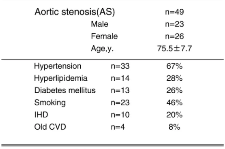
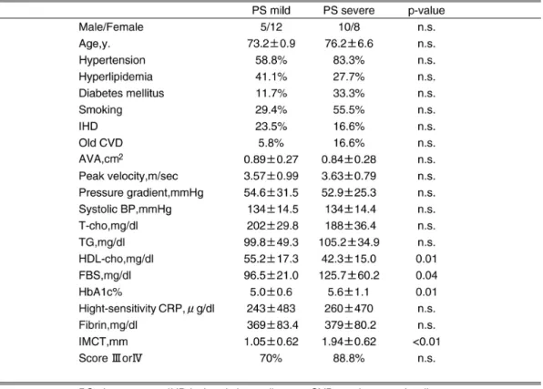
II.対象と方法 対象者:60才以上で,大動脈弁口面積1.5 cm2以下 である,外来通院中の患者49例(男性23例,女性26例, 平均年齢75.5歳)を対象とした.先天性の2尖弁によ る大動脈弁狭窄症は除外した.対象例の中には,症状 はあるが高齢のため手術適応でない例,他の合併症の ため手術できない例,本人や家族が手術を拒否してい る例も含まれた.これらの例において,NYHA分類, 既往歴,心臓超音波検査所見,頸動脈超音波検査所 見,動脈硬化の危険因子(総コレステロール値,中性 脂肪値,HDL-コレステロール値,LDL-コレステロー ル値[Friedewaldの式から算出した],空腹時血糖値, HbA1c値,血清fibrin値,高感度CRP値),投与薬につ いて調べた. 心臓超音波検査:断層超音波検査法およびドプラ心 エコー法をおこなった.使用装置として,HEWLETT PACKARD SONOS5500を使用した.検査中にvideoを 撮影し,そのvideoを2人の経験のある観察者による 合議性で弁の石灰化を4段階に評価(Ⅰ群;石灰化な し,Ⅱ群;小さく孤立する石灰化,Ⅲ群;点在する 石灰化,Ⅳ群;弁全体が肥厚し全体の石灰化)した6). 連続波ドプラー法から,最大流速値を測定し,簡易 ベルヌーイの式から大動脈弁口前後の圧較差を計算 した7).また,パルス・ドプラー法による連続の式か ら弁口面積を求めた. 頸動脈超音波検査:頸動脈,特に総頸動脈分岐部 は動脈硬化の好発部位であり,同部の動脈硬化度 の評価は全身の動脈硬化度推定の指標となる.7.5 MHz のプローブを用いると,体表に近い血管の内膜 中 膜 複 合 体 厚(intima - media complex thickening ; IMCT)や0.1 - 0.3 mm程度の局所的なプラークも描出 可能である.IMCTが1.0 mm以下までを正常とし,1.1 mm 以上を呈する箇所を動脈硬化性病変(プラーク) と定義した8).またプラークを有さない頸動脈におい ては,IMCTの最も厚い部位を測定した.血管エコー の結果よりplaque score (PS)を算出し,動脈硬化の 指標として用いた.これは分岐部を含む遠位部から 近位部にかけての6 cmに渡って左右頸動脈を評価す ることにより,頸動脈分岐部近辺の動脈硬化度を総 合的に定量化する評価法である(Fig.1).IMCTが 1.1 mm 以上の部位を異常肥厚と評価し,厚み(mm) を計測した.それらを左右の血管で総計し,PSを算 出し個人の動脈硬化度の値とした.頸動脈硬化度を, PS 値5未満を軽度,5以上を高度の2段階に分類した 8 - 11). 統計学的解析は,併存率や頻度の比較をχ2検定, 平均値の比較をunpaired t - test,相関関係の検定には Pearsonの相関係数を用いて,p<0.05を有意とした. III.結 果 1.患者背景 患者背景をTable 1に示した.平均年齢は75.5歳,男 女比は1:1で,多くの例が,動脈硬化危険因子を有し ていた.これら症例にみられた主な危険因子の併存率 は,高血圧症67%,高脂血症28%,糖尿病26%,喫 煙歴46%,虚血性心疾患20%,脳血管障害の既往8% であった.このうち,動脈硬化危険因子を,3因子有 するものが9例,4因子以上を有する例が7例認めら れた. 2.大動脈弁硬化石灰化度と弁狭窄重症度 本研究の対象症例は,大動脈弁口面積1.5 cm2以下 の症例であるが,これらの例における弁の硬化石灰化 度と大動脈弁口最大流速には関連が認められなかった (Table 2).弁硬化石灰化度が高度例では,弁口面積が 減少する傾向がみられたが,統計学的には有意でな かった. 3.大動脈弁の硬化石灰化度と動脈硬化危険因子の関連 心臓超音波検査の結果,全例において弁の石灰化 が確認された.その程度から,I群はなし,II群9例 (18.3 %),III群22例(44.8 %),IV群18例(36.7 %)に 分類された.Ⅱ群とⅢ+Ⅳ群の2群に分類し,動脈硬 化危険因子との関連を検討した(Table 2).Ⅲ+Ⅳ群
Fig. 1. Diagram of evaluation of carotid atherosclerosis.
The cervical carotid artery was evaluated using a 7.5 -MHz duplex type probe. Plaque score was computed by summing maximum thickness in millimeters of plaques in each segment on both sides (a+b+c+contralateral plaques). S1, region of internal carotid artery (ICA)<15 mm distal to its bifurcation from common carotid artery(CCA);S2, region of ICA and CCA<15 mm proximal to bifurcation;S3, region of CCA>15 mm and<30 mm proximal to bifurcation;S4, region of CCA> 30 mm proximal to bifurcation below flow divider. Length of individual plaques was not considered in determining plaque score. Modified from the references 8-10).
では,年齢,収縮期血圧,大動脈弁圧較差,空腹時血 糖値, 高感度CRP値,fibrin値,IMCTがⅡ群より高い 傾向があったが,中性脂肪値のみが有意に高値であっ た.Ⅱ群で,大動脈弁口面積および最大流速値,総コ レステロール値,HDL-コレステロール値がⅢ+Ⅳ群 より高い傾向がみられたが,統計学的には有意ではな かった.LDL-コレステロール値は,Ⅲ+Ⅳ群でII群よ り高値であったが,有意差は認められなかった. 4.大動脈弁狭窄度と動脈硬化危険因子との関連 大動脈弁口最大流速および大動脈弁口面積と総コ レステロール値,HDL-コレステロール値,糖尿病 の有無等の動脈硬化危険因子との関連は認められな かった. 5.大動脈弁硬化石灰化度と頸動脈動脈硬化度との関連 頸動脈に1.1 mm以上のプラーク病変を認めた症 例は,頸動脈超音波検査を施行できた35例中23例 (66%)で,平均最大IMCTは1.5±0.8 mmであった. PS値から,頸動脈硬化軽度群17例,重症群18例に分 けた.大動脈弁硬化石灰化が高度なⅢ+Ⅳ群の28例中 16例(57.3%),Ⅱ群の7例中2例(28.5%)に,重症の 頸動脈硬化病変を認めたが,両者間には有意な関係は みられなかった(Table 3).また,大動脈弁狭窄の血行 動態上の重症度の指標である大動脈弁口面積,大動脈 弁圧較差および弁硬化石灰化度の重症度(群分類)に も,両群間で差は認めなかった. 6.大動脈弁狭窄度と頸動脈動脈硬化度との関連 大動脈弁口最大流速および大動脈弁口面積で評価し た大動脈弁狭窄度の評価と頸動脈IMCTとPSとの関連 は認められなかった. 7.頸動脈硬化度と動脈硬化危険因子との関連 頸動脈硬化高度群は軽度群に比して,空腹時血糖値, HbA1c値は有意に高値で,HDL-コレステロール値 は有意に低値で,両群間で統計学的差が認められた.
Table 2. Clinical Characteristics of the Patients with Aortic Valve Calcification in Group II and Group III, IV Table 1. Clinical Characteristics of the Patients
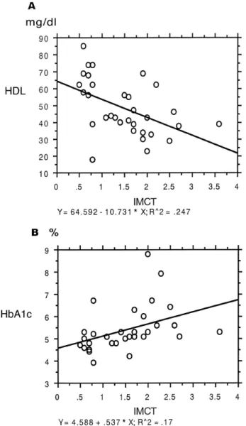
高感度CRP値,fibrin値,収縮期血圧値には差はみら れなかった(Table 3).他の頸動脈硬化度の指標であ る最大IMCTとHbA1cとHDL-コレステロール値との 間に,弱いながら有意な相関が認められた(Fig. 2). IV.考 察 本研究の対象である,大動脈弁口面積1.5 cm2以下 の中等度以上の大動脈弁狭窄症の全例に弁硬化石灰化 病変が認められた.しかし,弁の硬化石灰化と弁口面 積との間には相関はみられなかった.大動脈弁硬化石 灰化度および狭窄重症度と動脈硬化危険因子との関連 を検討したが,統計学的には,大動脈弁硬化石灰化度 と空腹時中性脂肪値のみが有意であった. 本研究では,大動脈弁の硬化石灰化の指標として, 心臓超音波検査で評価した石灰化scoreを用いたが, その程度と総コレステロール値等の動脈硬化危険因 子には関連が見られなかった.心臓超音波法を用いて 弁硬化石灰化の有無を評価し,病変の有無と臨床因 子との関連を5200人の多数で検討したCardiovascular Health Study では,大動脈弁石灰化の危険因子は,年 齢,男性,低身長,喫煙歴,高血圧歴,リポ蛋白(a) (Lipoprotein(a);Lp(a))高値が主たるもので,他に LDL - コレステロール高値が有意なものであったとし ている12).また,Helsinki Aging 研究では,年齢,高血
圧,低body mass indexが危険因子であり,糖尿病,喫 煙,高コレステロール血症は関連がみられなかったこ とが報告されている13).本邦では,Lp(a)高値が関連し たとの研究結果が示されている14).これらの研究は, 主として大動脈弁の病変の有無と臨床因子との関連 を解析しており,本研究とは対象例が異なると考えら れる. 最近の研究では,動脈硬化の重症度と進展のマー カーとして,高感度CRPが注目されているが15),本研 究では,高感度CRP値および血中fibrin値と大動脈弁 の硬化石灰化度との関連はみられなかった. 重症大動脈弁狭窄症であるが無症状例を対象に して,平均約2年間,弁狭窄進行に関与する要因を検 討した研究から,大動脈弁の中等度以上の石灰化が 唯一の予測因子であることが報告されている6).この 研究では,弁狭窄が進行して手術適応となる経過と 年齢,高血圧,高コレステロール血症等の臨床因子と は関連がなかったとしている.この結果は,我々の研 究と同様に,その重症度に関与する要因が認められな いものであった.その共通点は研究対象例が,弁硬化 のためにすでに大動脈弁狭窄症を発症した例であった ことである.
本研究からは,大動脈弁の硬化石灰化度および狭 窄重症度と頸動脈動脈硬化には,関連は認められな かった.大動脈弁硬化石灰化度と頸動脈動脈硬化の 程度とを比較した研究は少なく,重症大動脈弁狭窄 症例では,冠動脈疾患例よりも頸動脈動脈硬化は軽 度であるとの報告がある16).その原因として血圧低下 がむしろ防御的に作用することが推測されている.ま た,大動脈弁狭窄症例の弁置換術前と術1年後に,頸 動脈のIMCTを比較した研究では,術後では血管のリ モデリングが起こり,IMCT値は増加する結果が示さ れている.これは術後には血圧が上昇し,頸動脈内皮 にかかるずり応力が増加するためIMCTが増加すると 考えられている.本研究では,このような流体力学的 要因を,分析に加えていないが,頸動脈動脈硬化の初 期病変に影響を与えている可能性があり,今後検討す べき点であると考えられる. 本研究から,頸動脈動脈硬化の重症度と,HDL-コ レステロール低値,空腹時血糖高値,HbA1c高値とに 関連がみられた.Handaらは,同様の検討から,頸部 動脈硬化病変と,年齢,性別,高脂血症,糖尿病との 関連を認めたが,高血圧,喫煙歴との関連性は無かっ たことを報告している8).本研究の結果も,高齢者にお いても,脂質異常,耐糖能異常が,頸動脈硬化の危険 因子であることを確認するものであった. Pohleら は, 石 灰 化 を 伴 う 大 動 脈 弁 狭 窄 症 例 で は,LDL-コレステロール値が130 mg/dlを越えると, 病変が進行しやすいことを報告した2).本研究の対象 例のLDL-コレステロールの平均値は121±33.9 mg/dl であり,彼らの報告にくらべると低値であった.本研 究の治療的な意義を考察すると,大動脈弁狭窄症に対 する有効な内科治療法がないため,やや低めのコレス テロール値の例であっても,進行阻止のためのコレス テロール低下療法は検討に値する治療法であると考え られる. 本研究の限界は次のような点にあると考えられる. 本研究では,高齢者ですでに大動脈弁に変化が生じ 弁口面積が中等度以上に減少した例を対象に,重症 度と動脈硬化危険因子との関連を検討したため,重症 度に影響する因子の解析となった点である.さらに, 大動脈弁硬化症発症と動脈硬化危険因子との関連を 検討するためには,年齢,性別を一致させた健常者を コントロールとする方法が必要である.また,大動脈 弁硬化石灰化の評価を,心エコー検査を用いて石灰 化度を指標として行ったが,解像力に限界があり定 性的評価にとどまった.今後,治療による効果判定を 行うためには,心臓超音波法を用いた弁石灰化の定 量的方法,あるいは定量化が容易なEBCT(electron beam computed tomography)を用いるのがよいと考え られる1). V.結 語 高齢者の大動脈弁狭窄症における頸動脈の動脈 硬化病変の併存率は高率であったが,大動脈弁の硬 化石灰化の重症度と頸動脈病変の重症度には関連は 認められなかった.大動脈弁硬化石灰化重症度には 中性脂肪値が,頸動脈硬化病変の重症度には耐糖能 障害と,高LDL-コレステロール値,低HDL-コレステ ロール値が,関連する結果が得られた.高感度CRP値 は,大動脈弁硬化石灰化,頸動脈硬化の重症度と相関 しなかった.本研究から,大動脈弁硬化石灰化の重症 度には,伝統的な動脈硬化危険因子との関連は認め られず,頸動脈のような中動脈動脈硬化とは異なる臨 床因子の影響と機序が推測された.
Fig. 2. A, Scatterplot of intima-media complex thickening:
IMCT(x - axis) and HDL cholesterol level in mg/dl(y - axis). Bold line indicates regression line. The corresponding correlation coefficient was r=0.24(p<0.01). B, Scatterplot of IMCT(x - axis) and HbA1c%(y -axis). Bold line indicates regression line.� The corresponding correlation coefficient was r=0.17(p=0.01).
謝 辞 稿を終えるにあたり,本研究のために終始ご懇篤 なる御指導を賜りました,埼玉医科大学循環器内科学 教室 西村重敬教授に深甚なる謝意を捧げますと共に, 御協力いただいた循環器内科学教室,心臓病センター 心エコー室の皆様に感謝いたします.なお,本論文の 要旨の一部は第51回日本心臓病学会において発表さ れた. 文 献
1) Otto CM, Lind BK, Kitzman DW, Gersh BJ, Siscovick DS. Association of aortic - valve sclerosis with cardiovascular mortality and morbidity in the elderly. N Engl J Med 1999;341:142 - 7.
2) Pohle K, Maf fer t R, Ropers D, Moshage W, Stilianakis N, Daniel WG, et al. Progression of aortic valve calcification. Circulation 2001;104:1927 - 32. 3) Otto CM, Kuusisto J, Reichenbach DD, Gown AM,
Obrien KD. Characterization of the early lesion of degenerative valvular aortic stenosis. Circulation 1994;90:844 - 53.
4) Palta S, Pai AM, Gill KS, Pai RG. New insights into the progression of aortic stenosis. Circulation 2000; 101:2497 - 502.
5) Libby P, Ridker PM, Maseri A. Inflammation and atherosclerosis. Circulation 2002;105:1135 - 43. 6) Rosenhek R, Binder T, Porenta G, Lang I, Christ G,
Schemper M, et al. Predictors of outcome in severe, asymptomatic aortic stenosis. N Engl J Med 2000; 343:611 - 7.
7) Laskey WK, Kussmaul WG. Subvalvular gradients in patients with valvular aortic stenosis. Circulation 2001;104:1019 - 22.
8) Handa N, Matsumoto M, Maeda H, Hougaku H,
Ogawa S, Fukunaga R, et al. Ultrasonic evaluation of early carotid atherosclerosis. Stroke 1990;21: 1567 - 72.
9) Handa N, Matsumoto M, Maeda H, Hougaku H, Kamada T. Ischemic stroke events and carotid atherosclerosis. Stroke 1995;26:1781 - 6.
10) Nagao T, Sadoshima S, Ibayashi S, Takeya Y, Fujishima M. Increase in extracranial atherosclerotic carotid lesion in patients with brain ischemia in Japan. Stroke 1994;25:766 - 70.
11) 福島洋行,伊藤幹彦,石丸新,岩本俊彦,高崎優. 閉塞性動脈硬化症に併存する頸動脈病変. 脈管学 2001;41:37 - 41.
12) Stewart BF, Siscovick D, Lind BK, Gardin JM, Gottdiener JS, Smith VE, et al. Clinical factors associated with calcific aortic valve disease. J Am Coll Cardiol 1997;29:630 - 4.
13) Aronow WS, Ahn C, Kronzon I, Goldman ME. Association of coronar y risk factors and use of statins with progression of mild valvular aortic stenosis in older persons. Am J Cardiol 2001;88: 693 - 5.
14) Ridker PM, Cushman M, Stampfer MJ, Tracy RP, Hennekens CH. Inflammation, aspirin, and the risk of cardiovascular disease in apparently healthy men. N Engl Med 1997;336:973 - 9.
15) Galante A, Pietroiusi A, Vellini M, Piccolo P, Possati G, Bonis MD, et al. C - reactive protein is increased in patients with degenerative aortic valvular stenosis. J Am Coll Cardiol 2001;38:1078 - 82.
16) Irace C, Gnasso A, Cirillo F, Leonardo G, Ciamei M, Crivaro A, et al. Arterial remodeling of the common carotid arter y after aortic valve replacement in patients with aor tic stenosis. Stroke 2002;33: 2446 - 50.