Suppression of LPS-induced Cyclooxygenase-2 (cox-2)
Gene Expression in Mouse Macrophages by
Excretory/Secretory Products of
Spirometra erinaceieuropaei Plerocercoids
Soji Fukumoto, Paramasari Dirgahayu*, Kozue Nunomura, Haruyo Matsuura and Kazumitsu Hirai
Division of Molecular Medical Zoology, Department of Microbiology and Pathology, School of Medicine, Tottori University Faculty of Medicine, Yonago 683-8503 Japan
The escape mechanism of parasites from inflammatory processes has been thought to be one of the most important tools for their survival in infected tissues. On the other hand, inducible cyclooxygenase-2 (cox-2) has been shown to play a major role in the process of inflammation. We investigated the effect of excretory/secretory (ES) products from the plerocercoids of Spirometra erinaceieuropaei on cox-2 gene expression in a murine mac-rophage cell line (RAW 264.7). Preincubation of the macmac-rophages with the ES products inhibited LPS-induced cox-2 mRNA expression by 75% as well as its protein production. Dibutyryl cAMP enhances LPS-induced cox-2 mRNA expression. The ES products also inhibited this enhanced expression by 46%. Inhibition of p38 mitogen-activated protein kinase (MAPK) with SB203580 or that of extracellular signal-regulated protein kinase with PD98059 reduced LPS-induced cox-2 mRNA expression by 75% or 52%, respectively, along with its protein production. This evidence suggests that both MAPK pathways are crucial for full induction of cox-2 gene expression. One experiment using a transcriptional inhibitor, actinomycin-D, showed that the ES products destabilized LPS-induced cox-2 mRNA in RAW 264.7 macrophages. These results show that ES prod-ucts from the plerocercoids of Spirometra erinaceieuropaei suppress LPS-induced cox-2 mRNA expression in murine RAW 264.7 macrophages, and may attenuate inflammation around the plerocercoids of S. erinaceieuropaei.
Key words: Spirometra erinaceieuropaei; lipopolysaccharide; macrophage; cyclooxygenase-2; mitogen-activated protein kinase
Cyclooxygenase (cox) catalyzes the synthesis of prostaglandins (PGs) from arachidonic acid. Two isozymes, cox-1 and cox-2, have been identified, and they are encoded by separate genes. The cox-1 isozyme is constitutively present in most tis-sues and seems to catalyze the synthesis of PGs
for normal physiological functions (O’Neill and Ford-Hutchinson, 1993). In contrast, cox-2 is not or is scarcely detectable in most tissues under bas-al conditions, but is dramaticbas-ally induced by bac-terial lipopolysaccharide (LPS), cytokines, growth factors or carcinogens (Kujubu et al., 1991).
* Present address: Department of Parasitology, Faculty of Medicine, Sebelas Maret University, Surakarta, Indonesia Abbreviations: cox, cyclooxygenase;dbcAMP, dibutyryl cAMP; DMEM, Dulbecco’s modified Eagle’s medium; ERK,
The cox-2 isozyme is responsible for high levels of PG production, and secreted PGs are involved in various physiological events including progression of inflammation, immunomodula-tion, and transmission of pain (Vane and Boot-ing, 1998; Kim et al., 2004). The central role of PGs in inflammation was firmly established after the discovery that the anti-inflammatory action of nonsteroidal anti-inflammatory drugs, such as acetylsalicylic acid, was mediated by the inhibi-tion of cox. It has been widely accepted that mac-rophages play an important role in the regulation of inflammation and immune response. In in-flammatory reactions, macrophages are the main producers of large quantities of PGE2 (Giroux and
Descoteaux, 2000).
Parasite survival may depend on the ability of the parasite to modulate host immune response by the release of immunomodulatory molecules that protect the organism (Maizels et al., 2001; Harnett et al., 2004). When larval plerocercoids of S. erinaceieuropaei are taken orally in many mammals including humans and rodents, these plerocercoids invade the peritoneal cavity from the intestine, and can survive in the tissues of hosts. In humans, infection with larval plero-cercoids causes “sparganosis” in various tissues (Kudesia et al., 1998). Experimentally infected plerocercoids do not induce the strong inflamma-tory responses around the parasites and they can survive for long periods in the tissues of mice; hence, we hypothesized that larval plerocercoids of S. erinaceieuropaei secrete an immunosuppres-sive factor(s). We previously showed that excre-tory/secretory (ES) products from plerocercoids suppress LPS-induced expressions of chemokines KC and JE, the murine homologues of neutro-phil chemoattractant growth related oncogene α (CXCL1/GRO-α) and monocyte chemoattractant protein-1 (CCL2/MCP-1), respectively (Fukumoto et al., 1997), tumor necrosis factor (TNF)-α (Mi-ura et al., 2001; Dirgahayu et al., 2002), and IL-1β (Dirgahayu et al., 2004) in murine macrophages. We also found that these suppressions are caused by the reduction of phosphorylation of
mitogen-activated protein kinase (MAPK), particularly extracellular signal-regulated kinase 1/2 (ERK1/2) and p38 MAPK (Dirgahayu et al., 2002; Dirga-hayu et al., 2004).
Studies using MAPK inhibitors, SB203580 and PD98059 revealed that MAPK is involved in LPS-induced cox-2 transcription in RAW 264.7 macrophages (Chanmugam et al., 1995; Hwang et al., 1997). SB203580 inhibits the p38 MAPK pathway (Ajizian et al., 1999), and inhibits the promoter activities of cox-2 (Ho et al., 2004), while PD98059 is a specific inhibitor for the ERK1/2 pathway (Alessi et al., 1995). In addition to the transcriptional effects of MAPK, AgC10, a mucin from Trypanosoma cruzi destabilized
cox-2 mRNA by inhibiting the activation of p38 MAPK (Alcaide and Fresno, 2004).
Besides MAPKs, the importance of NF-κB for the induction of cox-2 in LPS-activated mono-cyte-macrophage lineage has been emphasized (D’ Acquisto et al., 1998). However, we found that ES products do not affect the LPS-induced nuclear translocation of NF-κB (Dirgahayu et al., 2002).
Since ES products have the ability to sup-press the phosphorylation of ERK1/2 and p38 MAPK in LPS-activated macrophages, we hy-pothesized that ES products could suppress LPS-induced cox-2 gene expression in macrophages. We have shown in this paper that ES products suppress LPS-induced cox-2 mRNA expression and cox-2 protein in RAW 264.7 cells, and ES products destabilize cox-2 mRNA. This is the first report demonstrating that ES products from the parasitic helminth reduce cox-2 mRNA and cox-2 protein in macrophages.
Materials and Methods
Plerocercoids of Spirometra erinaceieuro-paei and their ES products
Plerocercoids of Spirometra erinaceieuropaei were collected from two species of snakes (Elaphe
Prefecture, Japan and stored for 6 to 10 months in the subcutaneous tissues of golden hamsters, which were housed and maintained according to the guidelines for proper treatment of animals at the Research Center for Bioscience and Technol-ogy, Tottori University, Japan. ES products were obtained as described previously (Miura et al., 2001). To obtain ES products, 25 plerocercoids aseptically removed from hamsters were incubat-ed for 24 h in serum-free 25 mL Dulbecco’s mod-ified Eagle’s medium (DMEM) in a 10 cm dish, and the incubation medium was dialyzed against distilled water and lyophilized. The protein con-centration of this sample dissolved in DMEM was assayed using a protein assay kit (Bio-Rad, Melville, NY) and adjusted to 50 µg/mL. The av-erage amount of total protein secreted from each plerocercoid for 24 h was approximately 30 µg in the present experiment.
Culture of macrophages
A murine macrophage cell line RAW 264.7 (RCB0535) was purchased from RIKEN Cell Bank (Wako, Japan). The cells were cultured in DMEM supplemented with 5% heat-inactivated fetal bovine serum (FBS) (Invitrogen, Carlsbad, CA) containing 100 U/mL penicillin G (Banyu Pharmaceutical, Tokyo, Japan) and 100 µg/mL streptomycin (Meiji Seika, Tokyo, Japan), plated in tissue culture dishes (Greiner Bio-One, Tokyo, Japan) and finally incubated at 37˚C in an atmo-sphere of 5% CO2 in a humidified incubator. In
the present study, 5 × 10⁶ cells were preincubated
in a 10 cm culture dish for 3 h or 6 h in the pres-ence or abspres-ence of ES products (5 µg/mL). The cells were washed with PBS and stimulated with LPS (100 ng/mL) in DMEM supplemented with 5% FBS. After 3 h of incubation with LPS, large amounts of cox-2 mRNA were expressed in a time-course experiment. Then, the cells were incubated with LPS for 3 h in the present experi-ments. A concentration of 5 µg/mL ES products was used, because this concentration had been effective in TNF-α gene expression (Miura et al.,
2001), and similar activation pathways such as NF-κB and MAPK are involved in LPS-induced
cox-2 and TNF-α gene expressions.
Reverse transcription-PCR analysis
Total RNA was prepared using an ISOGEN Kit (Nippon Gene, Tokyo, Japan) according to the manufacturer’s protocol. Total RNA (1 µg) was reverse-transcribed, and PCR reactions were per-formed on a Perkin Elmer DNA Thermal Cycler (Takara Bio, Kusatsu, Japan). Primers used were as follows: cox-2: 517 bp, 5'-CTT CCT GAT CAA AAG AAG TGC TG-3' (sense) and 5'-ACT TGA GTT TGA AGT GGT AAC CGC-3' (antisense); and β-actin: 349 bp, 5'-TGG AAT CCT GTG GCA TCC ATG AA-3' (sense) and 5'-TAA AAC GCA GCT CAG TAA CAG TCC-3' (antisense). The PCR cycles consisted of denaturation at 94˚C for 30 s, annealing at 60˚C for 1 min and exten-sion at 72˚C for 1 min. The number of cycles used was 29 for semi-quantitative analysis of cox-2 or 25 for β-actin. A 15-µL aliquot of each PCR product was separated by electrophoresis on a 1.5% agarose gel, stained with ethidium bromide and photographed. For quantification, photo-graphs showing PCR products were scanned and analyzed using the software Densitograph version 4.0 (ATTO, Tokyo, Japan). The amount of cox-2 mRNA was normalized to that of β-actin.
Preparation of total RNA and Northern blot analysis
Northern hybridization analysis was performed as previously described (Fukumoto et al., 1997). Briefly, 10 µg of total RNA was separated on an 1% agarose gel containing 2.2 M formaldehyde, and subsequently blotted onto a MagnaGraph Nylon transfer membrane (Micron Separation, Westboro, MA) and then cross-linked to the mem-brane using an UV cross-linker (Funa-UV-Linker Fs 1500, Funakoshi, Tokyo, Japan). After prehy-bridization, the blots were hybridized with cDNA plasmid probes radiolabelled by [α-32P]dCTP
(MP Biomedicals, Irvine, CA) using a Takara BcaBEST Labelling Kit (Takara Bio), and then autoradiography was performed. The murine-517 bp of the cox-2 cDNA fragment was prepared by RT-PCR, followed by confirmation with sequenc-ing. A plasmid encoding the glyceraldehyde-3-phosphate dehydrogenase (gapdh) cDNA, used as a control, was kindly provided by Dr. David Stern (Columbia University). Relative cox-2 mRNA expressions were analyzed using a Molecular Im-ager, Model GS-535 (Bio-Rad) with reference to the expression of gapdh mRNA.
Preparation of cell lysate and Western blot
After the treatment, cell lysates were prepared as previously described (Dirgahayu et al., 2002). Briefly, the adherent cells were disrupted in cold lysis buffer (20 mM Tris, pH 7.5, 120 mM EDTA, 10% glycerol, 1% Triton X-100, 1 mM sodium orthovanadate, 1 mM phenylmethylsulfonyl fluo-ride, 5 µg/mL aprotinin, 5 µg/mL pepstatin and 5 µg/mL leupeptin), homogenized for 5 s using a microtube homogenizer, and then kept on ice
for 30 min. Insoluble material was removed by centrifugation at 4˚C, 12,500 rpm for 10 min and protein concentration in the clear supernatant was determined using a protein assay kit (Bio-Rad). The solubilized proteins (80 µg) were boiled in SDS-loading buffer, then separated on a 10% SDS-polyacrylamide gel with a running buffer containing 25 mM Tris, 192 mM glycine, pH 8.3, and 0.1% SDS. After electrophoresis, the sepa-rating gel was soaked in transfer buffer (25 mM Tris-HCl, 0.2 M glycine, 20% (v/v) methanol, pH 8.5) for 15 min and electrophoretically transferred to Hybond-ECL nitrocellulose (Amersham Bio-sciences, Piscataway, NJ) using a semidry blotting apparatus (Novablot, Amersham Biosciences), and then blocked overnight in Tris-buffered sa-line/tween-20 (0.05%) supplemented with 3% non-fat milk. The membranes were washed, incubated overnight with phospho-specific an-tibody recognizing cox-2 (1:300) detected with anti-rabbit horseradish peroxidase-conjugated antibodies (1:3,000) and subsequently visualized by an enhanced chemiluminescence reagent (Am-ersham Biosciences). The gel was stained with
Fig. 1. Suppression of LPS-induced cox-2 mRNA expression in RAW 264.7 macrophages by ES products. RAW
264.7 cells were left untreated or treated with ES products (5 µg/mL) for 3 h or 6 h, and the medium was replaced prior to stimulation with LPS (100 ng/mL) for 3 h.
A: Total RNA was obtained from the cells, and the expression of cox-2 and gapdh mRNA was assessed by
North-ern blot analysis.
B: The cox-2 mRNA level was quantified and normalized to gapdh. The relative cox-2 mRNA levels are
present-ed as percentages of the LPS-inducpresent-ed control in the absence of ES products.
C: The cox-2 protein in each cell lysate was analyzed by Western blot analysis. The same volume of each cell
lysate was resolved by SDS-PAGE, and the gel was stained with Coomassie Brilliant Blue to show that total ��
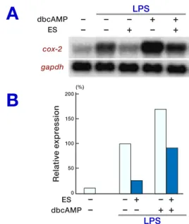
Fig. 2. The suppressive effect of ES products on cox-2
mRNA expression induced by dbcAMP and LPS. RAW 264.7 cells were left untreated or preincubated with ES products for 6 h prior to medium change. The cells were then either left untreated or treated with dbcAMP (0.1 mM) for 15 min and were then stimulated with LPS (100 ng/mL) for 3 h. Total RNA was obtained from the cells and cox-2 and gapdh mRNA expression was assessed by Northern blot analysis (A). The cox-2 mRNA level was
quantified and was normalized to gapdh. The relative
cox-2 mRNA levels are presented as percentages of the LPS-induced control in the absence of ES products and dbcAMP (100%) (B). The figure is representative of 3
independent experiments.
Coomassie Brilliant Blue to demonstrate that an equal amount of proteins was loaded in each lane.
Results
Suppression of LPS-induced cox-2 mRNA expression by ES products
No appreciable band was observed in controls without LPS stimulation. Firstly, the effect of ES products on cox-2 mRNA expression and pro-tein production were investigated. RAW 264.7 cells were either left untreated or preincubated with ES products (5 µg/mL) for 3 h or 6 h. The cells were stimulated with 100 ng/mL LPS for 3 h. Total RNA or total protein was subjected to Northern blot or Western blot, respectively. The result shows that ES products strongly suppressed the levels of LPS-induced cox-2 mRNA (Figs. 1A and B) and cox-2 protein (Fig. 1C) to a similar de-gree. The level of constitutively expressed gapdh mRNA was unaltered by exposure to ES products. Since preincubation for 3 h or 6 h showed no sig-nificant differences on cox-2 mRNA expression or protein production, a 6 h pretreatment was used for the following experiments.
Suppressive effect of ES products on cox-2 mRNA induced by dbcAMP and LPS
The plerocercoids release PGE2 (Fukushima et
al., 1993; Gao et al., 1998), which enhances the induction of LPS-induced cox-2 mRNA by an ad-enyl cyclase/cAMP-dependent pathway (Hinz et al., 2000). We tested the possibility of ES prod-ucts on LPS-induced cox-2 mRNA expression in the presence of dibutyryl cAMP (dbcAMP). As seen in Fig. 2, LPS-induced cox-2 mRNA expres-sion (Lane 2) was further enhanced by treatment with dbcAMP 1.7 times (Lane 4). Pretreatment of macrophages with ES products prior to stimula-tion with LPS (Lane 3) or with LPS and dbcAMP (Lane 5) strongly suppressed cox-2 mRNA ex-pression by 75% and 46%, respectively. No
ap-preciable band was detected in the untreated con-trol (Lane 1), nor in cells treated with dbcAMP or ES products without LPS-induction (data not shown).
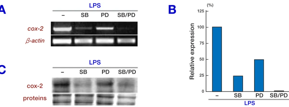
Involvement of MAPK in LPS-induced cox-2 mRNA
We examined the effects of MAPK inhibitors for the ERK1/2 pathway (PD98059) and for p38 MAPK (SB203580) in LPS-induced cox-2 mRNA expression. The macrophages were pretreated for 1 h with SB203580 (10 µM), PD98059 (50 µM) or both, and these cells were then stimulated with LPS for 3 h. The total RNA or protein was
sub-jected to semi-quantitative RT-PCR or Western blot analysis, respectively. SB203580 (Fig. 3A, Lane 2) and PD98059 (Lane 3) suppressed cox-2 mRNA expression by 75% or 52%, respectively, and cox-2 protein production (Fig. 3C, Lanes 2, 3, respectively). The combination of both inhibi-tors reduced the cox-2 mRNA (Fig. 3A, Lane 4) and cox-2 protein (Fig. 3C, Lane 4) to nearly null. Figure 3B shows the quantitative relations.
Next we examined the effect of ES products in combination with MAPK inhibitor(s) on cox-2 mRNA expression by Northern blot analysis. ES products or MAPK inhibitors reduced LPS-in-duced cox-2 mRNA expression, and the combina-tion with ES products and each MAPK inhibitor additively suppressed cox-2 mRNA expression (Fig. 4).
Destabilization of LPS-induced cox-2 mRNA by ES products
The level of cox-2 mRNA is regulated both tran-scriptionally and post-trantran-scriptionally (Wadleigh et al., 2000; Alcaide and Fresno, 2004). There-fore, we examined the effect of ES products on cox-2 mRNA stability in LPS-stimulated macrophages using a transcriptional inhibitor,
Fig. 3. Effect of MAPK inhibitors on LPS-induced cox-2 mRNA expression. RAW 264.7 cells were treated with
PD98059 (50 µM), SB203580 (10 µM) or a combination of both for 1 h and were stimulated with LPS (100 ng/mL) for 3 h. Total RNA and total protein were obtained from the cells and cox-2 and β-actin mRNA expressions were as-sessed by semi-quantitative RT-PCR (A). The cox-2 mRNA level was quantified and was normalized to β-actin. The relative cox-2 mRNA levels are presented as percentages of the LPS-induced control in the absence of MAPK inhibi-tor (B). The cox-2 protein expression was assessed by Western blot analysis (C). One representative of 2 independent
experiments is shown.
Fig. 4. Additive suppression of LPS-induced cox-2
mRNA expression by a combination of MAPK inhibitor and ES products. RAW 264.7 cells were left untreated or preincubated with ES products (5 µg/mL) for 6 h prior to medium change. The cells were then left untreated or treated either with PD98059, SB203580 or a combination of both for 1 h and were then stimulated with LPS (100 ng/mL) for 3 h. Total RNA was obtained from the cells and was subjected to Northern blot analysis (A). The cox-2 mRNA level was quantified and was normalized to
gapdh. The relative cox-2 mRNA levels are presented as percentages of the LPS-induced control in the absence of ES products and MAPK inhibitor (100%) (B). The figure
Fig. 5. Destabilization of LPS-induced cox-2 mRNA by ES products. RAW 264.7 cells were left untreated or
pre-incubted with ES products (5 µg/mL) prior to medium change. The cells were then stimulated with LPS (100 ng/mL) for 3 h and treated with actinomycin D (10 µg/mL). Total RNA was obtained at different time points (0, 15, 30, 60, 90 and 120 min) after the addition of actinomycin D. Total RNA was obtained from the cells and was subjected to Northern blot analysis. The cox-2 mRNA expression was quantified and was normalized to gapdh. One representa-tive experiment of three is shown. Act D, actinomycin D.
actinomycin-D. As shown previously, the mac-rophages were treated with ES products for 6 h, washed, and stimulated with LPS for 3 h. Then the cells were further treated with actinomycin D (10 µg/mL) at varying intervals. Total RNA was subjected to Northern blot hybridization to exam-ine the remaining cox-2 mRNA. As seen in Fig. 5, cox-2 mRNA was degraded rapidly in macro-phages treated with ES products, in comparison to cells without treatment of ES products. The cox-2 mRNA levels were normalized to gapdh mRNA levels. The remaining cox-2 mRNA level at 90 min was 43% in the absence of ES products or 17% in the presence of ES products. The level at 120 min was 25% in the absence of ES products or 3% in the presence of ES products. ES prod-ucts reduced the stability of cox-2 mRNA.
Discussion
Some parasites such as filarial nematodes secrete immunomodulatory molecules that protect the or-ganism (Maizels et al., 2001; Harnett e al., 2004). The cox-2 isozyme has been known as one of the potent inflammatory factors of host inflamma-tion through PGs (Vane and Booting, 1998; Kim et al., 2004). We found that ES products from plerocercoids of S. erinaceieuropaei inhibit cox-2 gene expression in LPS-activated macrophages. Although AgC10, a mucin from Trypanosoma
cruzi, a protozoan parasite, is reported to desta-bilize cox-2 mRNA (Alcaide and Fresno, 2004), this is the first report that shows that the parasitic helminth suppresses cox-2 gene expression.
It is well known that PGE2 potentiates
LPS-induced cox-2 mRNA expression via an adenyryl cyclase/cAMP-dependent pathway (Hinz et al., 2000). As the plerocercoids released PGE2 in the
supernatants of the in vitro culture (Fukushima et al., 1993; Gao et al., 1998), we also examined the effect of ES products on cox-2 mRNA expres-sion stimulated by the cAMP-dependent pathway. Stimulation by dbcAMP, a cell-permeable cAMP analogue, could not induce cox-2 gene expres-sion, but enhanced the induction of cox-2 mRNA by LPS, and the ES products suppressed the en-hanced cox-2 mRNA expression. These results suggest that ES products may inhibit the autocrine feed-forward loop of PGE2 in LPS-stimulated
macrophages around the plerocercoids of S.
eri-naceieuropaei.
In agreement with other studies (Chanmugam et al., 1995; Hwang et al., 1997), we showed that ERK1/2 and p38 MAPK pathways are crucial for
cox-2 gene expression and protein production in LPS-activated RAW 264.7 macrophages. Of note, PD98059 (an inhibitor for the ERK1/2 pathway) and SB203580 (an inhibitor of the p38 MAPK pathway) additively suppressed gene expression (Figs. 3 and 4). The ES products and both of MAPKs inhibitors also additively affected cox-2
gene expression (Fig. 4). Recently, we reported that ES products suppress TNF-α mRNA expres-sion by inhibiting ERK1/2 and p38 MAPK path-ways in RAW 264.7 macrophages stimulated with LPS (Dirgahayu et al., 2002). We also showed that ES products do not affect LPS-induced nu-clear translocation of NF-κB by electrophoretic mobility shift assay (Dirgahayu et al., 2002), while the activation of NF-κB is important for the induction of cox-2 mRNA in the LPS-activated monocyte-macrophage lineage (D’Acquisto et al., 1998). Although the mechanism of suppression by ES products needs to be investigated further, these lines of data suggest the possibility that ES products suppress LPS-induced cox-2 gene ex-pression by inhibiting ERK1/2 and p38 MAPK pathways.
Recently, p38 MAP kinase has been re-ported to play a major role in mRNA stabilization through AU-rich elements in the 3'-untranslated regions (Winzen et al., 1999; Lasa et al., 2000 and 2001). The 3'-untranslated region of cox-2 con-tains 22 AUUUA motifs that are recognized to be important determinants of mRNA instability (Chen and Shyu, 1995; Zubiaga et al., 1995). We observed that ES products reduce cox-2 mRNA stability (Fig. 5), and suppose that ES products destabilize cox-2 mRNA by inhibiting the phos-phorylation of p38 MAPK.
The present investigation showed that ES products suppress cox-2 mRNA expression and cox-2 protein production, which may attenuate in-flammation around the plerocercoids.
Acknowledgments: The authors are grateful to Dr. Kazutoyo Miura, Division of Molecular Medical Zool-ogy, Department of Microbiology and PatholZool-ogy, School of Medicine, Tottori University Faculty of Medicine, for his useful comments on the manuscript.
This work was supported by Grants-in-Aid for Sci-entific Research (numbers 12670231 and 16590339) from the Ministry of Education, Culture, Sports, Science and Technology of Japan.
References
1 Ajizian SJ, English BK, Meals EA. Specific inhibi-tors of p38 and extracellular signal-regulated kinase mitogen-activated protein kinase pathways block in-ducible nitric oxide synthase and tumor necrosis fac-tor accumulation in murine macrophages stimulated with lipopolysaccharide and interferon-γ. J Infect Dis 1999;179:939–944.
2 Alcaide P, Fresno M. AgC10, a mucin from
Try-panosoma cruzi, destabilizes TNF and cyclooxygen-ase-2 mRNA by inhibiting mitogen-activated protein kinase p38. Eur J Immunol 2004;34:1695–1704. 3 Alessi DR, Cuenda A, Cohen P, Dudley DT, Saltiel
AR. PD 098059 is a specific inhibitor of the activa-tion of mitogen-activated protein kinase kinase in vi-tro and in vivo. J Biol Chem 1995;270:27489–27494. 4 Chanmugam P, Feng L, Liou S, Jang BC, Boudreau
M, Yu G, et al. Radicicol, a protein tyrosine kinase inhibitor, suppresses the expression of mitogen-inducible cyclooxygenase in macrophages stimulated with lipopolysaccharide and in experimental glo-merulonephritis. J Biol Chem 1995;270:5418–5426. 5 Chen CY, Shyu AB. AU-rich elements:
charac-terization and importance in mRNA degradation. Trends Biochem Sci 1995;20:465–470.
6 D’Acquisto F, Sautebin L, Iuvone T, Di Rosa M, Carnuccio R. Prostaglandins prevent inducible ni-tric oxide synthase protein expression by inhibiting nuclear factor-κB activation in J774 macrophages. FEBS Lett 1998;440:76–80.
7 Dirgahayu P, Fukumoto S, Miura K, Hirai K. Ex-cretory/secretory products from plerocercoids of
Spi-rometra erinaceieuropaei suppress the TNF-α gene expression by reducing phosphorylation of ERK1/2 and p38 MAPK in macrophages. Int J Parasitol 2002;32:1155–1162.
8 Dirgahayu P, Fukumoto S, Tademoto S, Kina Y, Hirai K. Excretory/secretory products from plero-cercoids of Spirometra erinaceieuropaei suppress interleukin-1β gene expression in murine macro-phages. Int J Parasitol 2004;34:577–584.
9 Fukumoto S, Hirai K, Tanihata T, Ohmori Y, Stuehr DJ, Hamilton TA. Excretory/secretory products from plerocercoids of Spirometra erinacei reduce iNOS and chemokine mRNA levels in peritoneal macrophages stimulated with cytokines and/or LPS. Parasite Immunol 1997;19:325–332.
10 Fukushima T, Isobe A, Hojo N, Shiwaku K, Yamane Y, Torii M. The metabolism of arachidonic acid to prostaglandin E2 in plerocercoids of Spirometra
eri-nacei. Parasitol Res 1993;79:634–638.
11 Gao T, Fukushima T, Isobe A, Hojo N, Shiwaku K, Yamane Y. Arachidonic acid metabolism to prostaglandin E2, D2, F2α, and I2 in the
plerocer-coids of Spirometra erinaceieuropaei. J Parasitol 1998;84:1107–1111.
12 Giroux M, Descoteaux A. Cyclooxygenase-2 ex-pression in macrophages: modulation by protein ki-nase C-α. J Immunol 2000;165:3985–3991.
13 Harnett W, McInnes IB, Harnett MM. ES-62, a filarial nematode-derived immunomodulator with anti-inflammatory potential. Immunol Lett 2004;94: 27–33.
14 Hinz B, Brune K, Pahl A. Prostaglandin E2 upregu-lates cyclooxygenase-2 expression in lipopolysaccha-ride-stimulated RAW 264.7 macrophages. Biochem Biophys Res Commun 2000;272:744–748.
15 Ho FM, Lai CC, Huang LJ, Kuo TC, Chao CM, Lin WW. The anti-inflammatory carbazole, LCY-2-CHO, inhibits lipopolysaccharide-induced inflam-matory mediator expression through inhibition of the p38 mitogen-activated protein kinase signaling pathway in macrophages. Br J Pharmacol 2004;141: 1037–1047.
16 Hwang D, Jang BC, Yu G, Boudreau M. Expres-sion of mitogen-inducible cyclooxygenase induced by lipopolysaccharide: mediation through both mitogen-activated protein kinase and NF-κB signal-ing pathways in macrophages. Biochem Pharmacol 1997;54:87–96.
17 Kim BH, Kang KS, Lee YS. Effect of retinoids on LPS-induced COX-2 expression and COX-2 associ-ated PGE2 release from mouse peritoneal macro-phages and TNF-α release from rat peripheral blood mononuclear cells. Toxicol Lett 2004;150:191–201. 18 Kudesia S, Indira DB, Sarala D, Vani S, Yasha TC,
Jayakumar PN, et al. Sparganosis of brain and spi-nal cord: unusual tapeworm infestation (report of two cases). Clin Neurol Neurosurg 1998;100:148– 152.
19 Kujubu DA, Fletcher BS, Varnum BC, Lim RW, Her-schman HR. TIS10, a phorbol ester tumor promoter-inducible mRNA from Swiss 3T3 cells, encodes a novel prostaglandin synthase/cyclooxygenase homo-logue. J Biol Chem 1991;266:12866–12872.
20 Lasa M, Mahtani KR, Finch A, Brewer G, Saklatvala J,
Clark AR. Regulation of cyclooxygenase 2 mRNA stability by the mitogen-activated protein kinase p38 signaling cascade. Mol Cell Biol 2000;20:4265– 4274.
21 Lasa, M, Brook M, Saklatvala J, Clark AR. Dexa-methasone destabilizes cyclooxygenase 2 mRNA by inhibiting mitogen-activated protein kinase p38. Mol Cell Biol 2001;21:771–780.
22 Maizels RM, Gomez-Escobar N, Gregory WF, Murray J, Zang X. Immune evasion genes from filarial nema-todes. Int J Parasitol 2001;31:889–898.
23 Miura K, Fukumoto S, Digahayu P, Hirai K, Excre-tory/secretory products from plerocercoids of
Spiro-metra erinaceieuropaei suppress gene expressions and production of tumor necrosis factor-α in murine macrophages stimulated with lipopolysaccharide or lipoteichoic acid. Int J Parasitol 2001;31:39–47. 24 O’Neill GP, Ford-Hutchinson AW. Expression of
mRNA for cyclooxygenase-1 and cyclooxygenase-2 in human tissues. FEBS Lett 1993;330:156–160. 25 Vane JR, Botting RM. Anti-inflammatory drugs and
their mechanism of action. Inflamm Res 1998;47 Suppl 2:S78–87.
26 Wadleigh DJ, Reddy ST, Kopp E, Ghosh S, Her-schman HR. Transcriptional activation of the cyclo-oxygenase-2 gene in endotoxin-treated RAW 264.7 macrophages. J Biol Chem 2000;275:6259-6266. 27 Winzen R, Kracht M, Ritter B, Wilhelm A, Chen
CY, Shyu AB, et al. The p38 MAPK pathway signals for cytokine-induced mRNA stabilization via MAP kinase-activated protein kinase 2 and AU-rich region-targeted mechanism. EMBO J 1999;18:4969-4980.
28 Zubiaga AM, Belasco JG, Greenberg ME. The no-namer UUAUUUAUU is the key AU-rich sequence motif that mediates mRNA degradation. Mol Cell Biol 1995;15:2219–2230.
Received November 17, 2005; accepted November 25, 2005 Corresonding author: Soji Fukumoto, MD, PhD