113
Vascular Leiomyoma of the Nasal Cavity: Case Report and Review of
the Literature
Mari Osaki, Mitsuhiko Osaki, Isamu Kodani, Hironobu Adachi, Itoji Shibata* and Hisao Ito
Division of Organ Pathology, Department of Microbiology and Pathology, School of Medi-cine, Tottori University Faculty of MediMedi-cine, Yonago 683-8503 and *Shibata Otorhino-laryngoly Clinic, Tottori 680-0862 Japan
We reported a rare case of nasal vascular leiomyoma that developed in a 67-year-old man. The tumor showed characteristic histological findings with proliferation of smooth muscle cells intermingled with dilated venous vessels. Review of the literature indicated that nasal vascular leiomyoma is characterized by female predominance, occurrence in aged patients and nasal obstruction or pain as the primary symptom.
Key words: immunohistochemistry; nasal cavity; vascular leiomyoma Yonago Acta medica 2002;45:113–116
Leiomyomas are benign myogenic neoplasms that may occur wherever smooth muscle is present. According to Enzinger and Weiss (1995), who analyzed a total of 7,748 leiomyo-mas, 95% of the tumors occurred in the female genitalia (uterus), 3% in the skin, 0.9% in the gastrointestinal tract and the remainder in vari-ous sites. Leiomyomas are classified into 3 his-tological subtypes: vascular, nonvascular and epithelioid or leiomyoblastoma (Batsakis, 1979). Vascular leiomyoma is a relatively un-common smooth muscle tumor rarely found in the neck and head area. Herein, we present a rare case of vascular leiomyoma developed in the nasal septum.
Patient Report
A 67-year-old man visited an otorhinolaryngoly clinic in June 1999, with a 2-week history of left-sided nasal obstruction and intermittent bloody discharge. He had no other symptoms such as headache, pain or epistaxis. Physical examination revealed a round wide-based mass measuring 8 mm in diameter on the left nasal sep-tum, covered with a dark reddish nasal mucous membrane. No teleangiectasis or bleeding was noted. He was followed up for about 6 months.
Thereafter, the tumor was completely excised with marginal normal nasal mucosa. Post-operative recovery was uneventful. The patient has shown no recurrence of the tumor and was well 15 months postoperatively.
Pathological findings
The resected specimen measured about 10 mm in diameter, including peripheral normal muco-sa. The cut surface showed a homogeneous, pale tan tissue without obvious hemorrhage or necrosis.
Histologically, the squamous epithelia covering the tumor showed slight hyperkera-tosis, but not cellular atypia (Fig. 1). The tumor was composed of elongated blunt-shaped spin-dle cells intermingled with numerous irregular-ly dilated blood vessels, which had a thin single layer of endothelial cells (Fig. 2). Mitoses were not noted. The margin of the tumor was cir-cumscribed, despite the absence of a fibrous capsule.
Formalin-fixed, paraffin-embedded sections were immunohistologically stained with the avidin-biotin peroxidase complex method, using commercially available antibodies for desmin, factor VIII, CD-34, α-smooth muscle actin, muscle-specific actin-HHF-35 and myoglobin.
M. Osaki et al.
114
Fig. 1. Solid-type tumor showing compacted vascular vessels and muscles measuring about 8 mm in diameter. This tumor is thickly encapsulated by squamous cells with slight hyperkeratosis (hematoxylin and eosin stain; original magnification × 50).
The tumor cells as well as the walls of the blood vessels showed marked immunoreactivity for desmin, α-smooth muscle actin (Fig. 3) and muscle-specific actin-HHF-35, but not for myoglobin. The lining endothelial cells of the dilated vessels showed positive immunostain-ing for factor VIII and CD-34.
Fig. 2. Thick muscular walled-vessels and intervascular smooth muscle bundles (hematoxylin and eosin stain; original magnification × 50).
Discussion
Leiomyomas developing in the nasal cavity and paranasal sinuses are extremely rare. Maesaka et al. (1966) initially reported an intranasal leio-myoma in 1966. To the best of our knowledge,
Vascular leiomyoma of nasal cavity
115
Fig. 3. Most of the spindle cells showing strong positive cytoplasmic reactivity for α-smooth muscle actin on the left side of the lesion (peroxidase anti-peroxidase method for α-smooth muscle actin antigen; original magnification × 50).
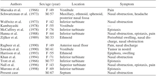
35 nasal leiomyomas have been subsequently reported until now. Of these, only 5 tumors developed in the nasal septum (Timiryaleev, 1973; Barr et al., 1990; Trott et al., 1994; Llorente et al., 1996), and 16 cases including the present tumor were histologically classified as vascular type (Table 1), which involved 11 females (mean age: 54.4 years, ranging from 25 to 76) and 5 males (mean age: 55.0 years, rang-ing from 33 to 77). Prominent clinical sym-ptoms are nasal obstruction and pain, and nasal bleeding is rather less frequent. The tumor de-veloped most frequently in the lower extrem-ities, followed by the upper extremities and less frequently in the nasal septum (Hachisuga et al., 1984). Thus, nasal vascular leiomyoma might be characterized by female predominance, occur-rence in aged patients, and nasal obstruction or pain as the primary symptom.
According to the report written by Morimoto (1973), vascular leiomyomas were classified into 3 histological subtypes: solid or capillary, cavernous and venous. The venous type devel-oped most frequently in the head and neck. His-tologically, the venous type has vascular chan-nels with thick muscular walls that are easily discernible from smooth muscle bundles, as ob-served in the present case. Although we con-ducted immunohistochemical analysis for
Table 1. Reported cases of vascular leiomyoma of the nasal cavity
Authors Sex/age (year) Location Symptom
Maesaka et al. (1966) F /49 Vestibule Pain
Schwartzman et al. (1973) M /57 Maxillary, ethmoid, sphenoid, Nasal obstruction, headache posterior nasal fossa
Wolfwitz et al. (1973) F /42 Inferior turbinate Nasal obstruction
Kambayashi (1978) F /55 Vestibule —
McCaffrey et al. (1978) F /76 Inferior turbinate Epistaxis
Hanna et al. (1988) F /64 Inferior turbinate Nasal obstruction, epistaxis, pain Zijlker et al. (1989) M /33 Ethmoid Periorbital swelling, nasal
dis-charge, nasal obstruction Ragbeer et al. (1990) F /49 Anterior nasal floor Pain, nasal discharge
Sawada et al. (1990) M /41 Vestibule Tumor in nostril
Harrison et al. (1993) F /55 Ethmoid Epiphora, swelling
Khan et al. (1994) F /71 Inferior turbinate Nasal obstruction Trott et al. (1966) M /77 Inferior turbinate Epistaxis
Nall et al. (1996) F /43 Superior turbinate Nasal obstruction, epistaxis, pain Murono et al. (1998) F /69 Inferior turbinate Epistaxis
Present case M /67 Septum Nasal obstruction
M. Osaki et al.
116
myoma of the nasal septum: report of a case and review of the literature. J Laryngol Otol 1996; 110:65–68.
10 Maesaka A, Keyaki Y, Nakanishi T. Nasal an-gioleiomyoma and leiomyosarcoma: report of 2 cases. Otologia (Fukuoka) 1966;12:42.
11 McCaffrey TV, McDonald TJ, Unni KK. Leio-myoma of the nasal cavity: report of a case. J Laryngol Otol 1978;92:817–819.
12 Morimoto N. Angiomyoma (vascular leiomyoma): a clinicopathologic study. Med J Kagoshima Univ 1973;24:663–683.
13 Murono S, Ohmura T, Sugamori S, Furukawa M. Vascular leiomyoma with abundant adipose cells of the nasal cavity. Am J Otolaryngol 1998;19: 50–53.
14 Nall AV, Stringer SP, Baughman RA. Vascular leiomyoma of the superior turbinate: first report case. Head Neck 1996;19:63–67.
15 Ragbeer MS, Stone J. Vascular leiomyoma of the nasal cavity: report of a case and review of lit-erature. J Oral Maxillofac Surg 1990;48:1113– 1117.
16 Sawada Y. Angioleiomyoma of the nasal cavity. J Oral Maxillofac Surg 1990;48:1100–1101. 17 Schwartzman J, Schwartzman J.
Leiomyoangio-ma of paranasal sinuses: case report. Laryngo-scope 1973;83:1856–1858.
18 Timiryaleev, MKH. Angioleiomyoma of the nasal septum. Vestn Otolaryngol 1973;35:106. 19 Trott MS, Gewirtz A, Lavertu P, Wood BG,
Sebek BA. Sinonasal leiomyomas. Otolaryngol Head Neck Surg 1994;111:660–664.
20 Wolfwitz BL, Schmaman A. Smooth muscle tu-mours of the upper respiratory tract. S Afr Med J 1973;47:1189–1191.
21 Zijlker TD, Visser R. A vascular leiomyoma of the ethmoid. Report of case. Rhinology 1989; 27:129–135.
Received September 25, 2002; accepted October 7, 2002 Corresponding author: Mitsuhiko Osaki academic interest, histopathological diagnosis
is easily done with routinely stained hematoxy-lin and eosin sections. Malignant transform-ation of vascular leiomyoma has not been re-ported, in spite of local recurrence in a few cases (2 of 562 cases) (Hachisuga, 1984). It should be stressed that otorhinolaryngologists should have an awareness of the possible exis-tence of vascular leiomyoma in the nasal cavity.
References
1 Barr GD, More IAR, McCallum HM. Leiomyo-ma of the nasal septum. J Laryngol Otol 1990; 104:891–893.
2 Batsakis JG (ed). Tumors of the head and neck: clinical and pathological considerations. 2nd ed. Baltimore, MD: Williams & Wilkins; 1979. p. 354–356.
3 Enzinger FM, Weiss SW. Soft tissue tumors. 3rd ed. St Louis: Mosby; 1995. p. 467–489. 4 Hachisuga T, Hashimoto H, Enjoji M.
Angio-leiomyoma: a clinicopathologic reappraisal of 562 cases. Cancer 1984;54:126–130.
5 Hanna GS, Akosa AB, Ali MH. Vascular leio-myoma of the inferior turbinate: report of a case and review of the literature. J Laryngol Otol 1988;102:1159–1160.
6 Harrison D, Lund VJ. Tumors of muscle origin. In: Harrison D, Lund VJ, eds. Tumours of the up-per jaw. Edinburgh, United Kingdom: Churchill Livingstone; 1993. p.187–197.
7 Kambayashi J. Vascular leiomyoma of the nasal vestibule. J Jpn Otolaryngol (Tokyo) 1976;50: 313–316.
8 Khan MHZ, Jones AS, Haqqani MT. Angioleio-myoma of the nasal cavity: report of a case and review of the literature. J Laryngol Otol 1994; 108:244–246.