Yonago Acta Medica 2019;62:085–093 Original Article
Corresponding author: Hiroki Chikumi, MD, PhD chikumi@med.tottori-u.ac.jp
Received 2018 December 17 Accepted 2019 January 4
Abbreviations: Akt, v-akt murine thymoma viral oncogene; ANOVA, analysis of variance; ATCC, American Type Culture Collection; D-MEM, Dulbecco’s modified Eagle’s medium; EGF, epidermal growth factor; EGFR, epidermal growth factor receptor; ERK, extracellular signal-regulated kinase; FBS, fetal bovine serum; FDA, Food and Drug Administration; HRP, horseradish peroxidase; KRAS, v-ki-ras2 kirsten rat sarcoma viral oncogene homolog; LKB1, liver kinase B1; MEK, mitogen-activated protein kinase/extracellular signal-regulated kinase; mTOR, mammalian target of rapamycin; NSCLC, non-small cell lung cancer; PBS, phosphate-buffered saline; PCR, polymerase chain reaction; PI3K, phosphoinositol-3-kinase; PTEN, phosphatase and tensin homolog deleted from chromosome 10; PVDF, PolyVinylidene DiFluoride; RAF, v-raf murine sarcoma viral oncogene homolog; RNAi, RNA interference; RPMI, Roswell Park Memorial Institute; SCLC, small cell lung cancer; SD, standard deviation; SDS-PAGE, sodium dodecyl sulfate-polyacrylamide gel electrophoresis; siRNA, small interfering RNA; SOS, son of sevenless; TKIs, tyrosine kinase inhibi-tors; TP53, tumor protein p53; WST-8, water-soluble tetrazolium salt
Effect of Cetuximab and EGFR Small Interfering RNA Combination Treatment in
NSCLC Cell Lines with Wild Type EGFR and Use of KRAS as a Possible Biomarker
for Treatment Responsiveness
Naomi Miyake,* Hiroki Chikumi,† Kosuke Yamaguchi,* Miyako Takata,‡ Miki Takata,* Kensaku Okada,† Tsuyoshi Kitaura,† Masaki Nakamoto† and Akira Yamasaki*
*Division of Medical Oncology and Molecular Respirology, Department of Multidisciplinary Internal Medicine, School of Medicine, Tottori University Faculty of Medicine, Yonago 683-8503, Japan, †Division of Infectious Diseases, Tottori University Hospital, Yonago 683-8504, Japan, and ‡Department of Pathobiological Science and Technology, School of Health Science, Tottori University Faculty of Medicine, Yonago 683-8503, Japan
ABSTRACT
Background The epidermal growth factor receptor (EGFR) is a therapeutic target for patients with non-small cell lung cancer (NSCLC). Cetuximab is an anti-EGFR monoclonal antibody that inhibits EGFR signaling and proliferation of colorectal cancer and head and neck cancers. Since only few NSCLC patients ben-efit from cetuximab therapy, we evaluated a novel com-bination treatment using cetuximab and EGFR small interfering RNA (siRNA) to strongly suppress EGFR signaling and searched for a biomarker in NSCLC cell lines harboring wild-type EGFR.
Methods Alterations in EGFR and its downstream genes in five NSCLC cell lines (A549, Lu99, 86-2, Sq19 and Ma10) were assessed through sequencing. The pro-tein expression levels of these molecules were assessed through western blotting. The effect of combination treatment was determined through cell proliferation assay, caspase-3/7 assay, invasion assay, and migration assay.
Results All cell lines were harboring wild-type
EGFR, whereas KRAS, PTEN, TP53 and LKB1 were
mutated in A549 and Lu99; Lu99 and Sq19; Lu99, 86-2, Sq19 and Ma10; and A549, 86-2, and Sq19 cell lines, re-spectively. PTEN was not expressed in Sq19, and LKB1 was not expressed in both A549 and Sq19. TP53 was not expressed in both A549 and Lu99. The combination of cetuximab and EGFR siRNA significantly suppressed cell proliferation in 86-2, Sq19 and Ma10, which express wild-type KRAS. It induced apoptosis in A549, 86-2 and Ma10 cells, which express wild type PTEN. The combi-nation treatment had no effect either on cell invasion nor migration in all cell lines.
Conclusion EGFR targeted therapy using the com-bination of cetuximab and EGFR siRNA is effective in NSCLC cell lines harboring wild-type EGFR. Wild-type
KRAS may act as a potential biomarker for response to
combination treatment by the induction of apoptosis in cells with wild-type PTEN.
Key words cetuximab; EGFR siRNA; KRAS; non-small cell lung cancer
Human lung cancer is the leading cause of cancer deaths worldwide. Lung cancer is classified into small cell lung cancer (SCLC) and NSCLC, accounting for approximately 15–20% and 80–85%, respectively. In addition, NSCLC is further classified into adenocarcino-ma, squamous cell carcinoma and large cell carcinoma. NSCLC is difficult to treat compared with SCLC.1 EGFR is transmembrane protein that regulates cell pro-liferation, apoptosis, angiogenesis and metastasis. EGFR is activated by EGF binding, which further activates the EGFR downstream signaling pathway.2 In lung cancer, overexpression and mutation of EGFR was observed in NSCLC patients, which makes it an important therapeu-tic target. Studies reveal that EGFR mutations occur in 30–40% of NSCLC patients.3
Currently, two classes of EGFR targeted drugs are used to treat cancers. One of them is EGFR-tyrosine
N. Miyake et al. kinase inhibitors (TKIs) and the other is anti-EGFR monoclonal antibodies. In lung cancer, EGFR-TKIs are the predominantly used EGFR-targeted drugs,4 and first-generation EGFR-TKIs such as gefitinib and erlotinib are recommended for first-line treatment in NSCLC patients with EGFR mutations.5 However, anti-EGFR monoclonal antibodies, such as cetuximab, are not used in NSCLC patients, although they are broadly used in patients with colorectal cancer and head and neck cancers.6 A randomized, multicenter, phase III study [First-Line ErbituX (FLEX)] of cetuximab re-vealed improvements in the overall survival of NSCLC patients.7 However, survival upon addition of cetuximab to conventional chemotherapy was only prolonged by one month and was considered insignificant compared with the cost of treatment. Therefore, novel combination therapies with cetuximab and biomarkers to identify patients who would benefit from the therapy have been extensively sought.8
RNA interference (RNAi) is a novel strategy that degrades the target mRNA by small interfering RNA (siRNA) consisting of 19–25 base pair, which leads to the down regulation of protein expression.9 In various fields such as medicine, biology and engineering, RNAi has been widely used as a tool for gene functional analysis.10–12 Recently, Food and Drug Administration (FDA) approved the first-ever therapeutic drug based on siRNA.13 Therefore, inhibition of specific molecules by siRNA is now a promising therapy in the cancer.
In the previous study, we found that NSCLC cell lines with EGFR mutation and lack of AKT activation were sensitive for cetuximab monotherapy.14 One pos-sible mechanism of this phenomenon is that these cells might become physiologically dependent on EGFR signaling pathway for their growth,15 therefore they are sensitive for the inhibition of EGFR function by cetux-imab. There is no report on overcoming the resistance of NSCLC cells with wild type EGFR to cetuximab.
In this study, we developed a novel combination treatment using cetuximab and EGFR siRNA to strong-ly suppress EGFR signaling pathways and studied its effect on NSCLC cell lines harboring wild-type EGFR. Additionally, we explored candidate gene alterations in cells showing response to treatment to find possible biomarkers for this combination treatment.
MATERIALS AND METHODS Cell lines
Human NSCLC cell lines including the adenocarcinoma cell lines A549 and Ma10, the squamous cell carcinoma cell line Sq19, the giant cell carcinoma cell line Lu99, and the large cell carcinoma cell line 86-2 were used
in this study. A549 was purchased from the American Type Culture Collection (ATCC, Manassas, VA). Sq19, Lu99, RERF-LC-AI and 86-2 were provided by the Riken Cell Bank (Riken, Tsukuba, Japan). The cell lines were cultured in Roswell Park Memorial Institute (RPMI) 1640 medium or Dulbecco’s modified Eagle’s medium (D-MEM) (Wako, Osaka, Japan) supplemented with 10% fetal bovine serum(FBS), 100 U/ml penicillin G, and 0.1 mg/ml streptomycin at 37℃ in a humidified incubator containing 5% CO2. The gene status of cell lines that were not published in literature was deter-mined using polymerase chain reaction (PCR) and direct sequencing.16
EGFR siRNA transfection
The EGFR siRNA (sense: 5’-CUCUGGAGGAAAAGAAAGU-3’ and antisense: 5’-ACUUUCUUUUCCUCCAGAG-5’-CUCUGGAGGAAAAGAAAGU-3’) and the negative control siRNA (no information disclo-sure) were purchased from Santa Cruz Biotechnology (Santa Cruz Biotechnology, Dallas, TX). The cell lines were transfected with 10 nM siRNA using Lipofectamine 3000 (Invitrogen, Carlsbad, CA). Transfection was performed according to the manufacturer’s protocol. Western blotting
After the cell lines were transfected with siRNA, cells were collected and washed with phosphate-buffered saline (PBS) (-), and subsequently lysed in a lysis buffer. The sample was subjected to 8–10% sodium dodecyl sulfate-polyacrylamide gel electrophoresis (SDS-PAGE) and separated proteins were transferred to Immobilon-P PolyVinylidene DiFluoride (PVDF) membranes (Merck Millipore, Billerica, MA). The membranes were incubated with EGFR, anti-PTEN, anti-Akt, anti-KRAS, anti-TP53, anti-LKB1 antibodies (1:1,000 dilution, Cell signaling technology, Beverly, MA) and β-actin antibody (1:2,000 dilution, Sigma-Aldrich, St. Louis, MO). Primary antibodies were detected using horseradish peroxidase (HRP)-conjugated secondary antibodies (1:1,000 dilution, anti-mouse IgG and anti-Rabbit IgG respectively; GE Healthcare Bio-sciences Amersham, Diegem, Belgium, UK). The mem-branes were subjected to chemiluminescence detection assay using ECL Prime Western Blotting Detection Reagents (GE Healthcare Bio-sciences Amersham). Cell proliferation assay
Cell proliferation after treatment with 0, 0.01, 0.1 and 1.0 µM cetuximab for 6 days was detected by water-soluble tetrazolium salt (WST-8) for colorimetric cell viability assay (Dojindo Molecular Technologies, Kumamoto, Japan). The procedure was performed according to the
Cetuximab and EGFR siRNA combination treatment in NSCLC
manufacture’s instruction. The absorbance at 450 nm with a reference wave length of 655 nm was measured by a microplate reader, Sunrise RAINBOW (Tecan Trading AG, Männedorf Switzerland).
Active caspase-3/7 assay
Active caspase-3/7 was measured using a homogeneous luminescent method with Caspase-3/7 Glo assay kit (Promega, Madison, WI). The operating procedure was performed according to the manufacture’s instruction. After the cell lines were transfected with EGFR siRNA and treated with 0, 0.01, 0.1 and 1.0 µM cetuximab for 24 h, the luminescence was measured by using a multifunctional microplate reader, Infi nite F500 (Tecan Trading AG).
Migration and invasion assay
The in vitro migration assay used an HTS Transwell-96 permeable support with 8 µm pore polyester membrane (Corning Incorporated, NY). The in vitro invasion as-says were performed using HTS Transwell-96 well plate with Matrigel-coated filters (Corning Incorporated). Cell lines were seeded into the upper 96 well of the plate and exposed to 0, 0.01, 0.1 and 1.0 µM cetuximab, whereas 10% FBS medium was added into the lower well followed by incubation for 42 h. Non-migratory and non-invasive cells stayed in the upper membrane and migratory and invasive cells passed through basement membrane layer and cling to the bottom of the insert membrane. Migratory and invasive cells were dissociat-ed from membrane by the detachment buffer and were lysed by the lysis buffer. Finally, the lysates were stained with CyQuant GR Dye (Invitrogen, Carlsbad) and the fluorescence produced were measured (excitation: 485 nm/emission: 525 nm) using multifunctional microplate reader, Infi nite F500 (Tecan Trading AG).
Statistical analysis
All experiments were performed in triplicate. The values
were represented as mean ± SD (standard deviation). Statistical analyses were performed by one-way ANOVA (analysis of variance) with Bonferroni’s test using SPSS Statistics 25 (IBM Japan, Tokyo, Japan). The result was considered statistically signifi cant if P < 0.05.
RESULTS
Gene status of cell line
The gene status of EGFR, PTEN, KRAS, TP53 and
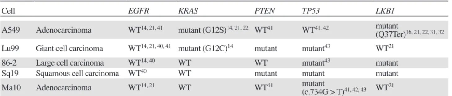
LKB1 in all NSCLC cell lines used in this study are Table 1. Gene atatus of NSCLC cell lines
Cell EGFR KRAS PTEN TP53 LKB1
A549 Adenocarcinoma WT14, 21, 41 mutant (G12S)14, 21, 22 WT41 WT41, 42 mutant
(Q37Ter)16, 21, 22, 31, 32 Lu99 Giant cell carcinoma WT14, 21, 40, 41 mutant (G12C)14 mutant mutant43 WT21
86-2 Large cell carcinoma WT14, 40 WT WT mutant43 mutant
Sq19 Squamous cell carcinoma WT40 WT mutant mutant mutant
Ma10 Adenocarcinoma WT14, 21 WT WT41 mutant
(c.734G > T)41, 42, 43 WT21
EGFR, epidermal growth factor receptor; KRAS,v-ki-ras2 kirsten rat sarcoma viral oncogene homolog; LKB1, liver kinase B1; NSCLC, non-small cell lung cancer; PTEN, phosphatase and tensin homolog deleted from chromosome 10; TP53, tumor protein p53; WT, wild type
LKB1 PTEN EGFR Akt β-actin KRAS TP53
N. Miyake et al., p. 27
Fig. 1. Protein expression in NSCLC cell lines. Protein
expres-sion of EGFR, PTEN, KRAS, Akt, LKB1, TP53 and β-actin in NSCLC cell lines were analyzed by western blotting. Akt, v-akt murine thymoma viral oncogene; EGFR, epidermal growth factor receptor; KRAS, v-ki-ras2 kirsten rat sarcoma viral oncogene homolog; LKB1, liver kinase B1; NSCLC, non-small cell lung cancer; PTEN, phosphatase and tensin homolog deleted from chromosome 10; TP53, tumor protein p53.
N. Miyake et al. β-actin EGFR β-actin EGFR A549 Lu99 β-actin EGFR Ma10 Sq19 β-actin EGFR β-actin EGFR 86-2
N. Miyake et al., p. 28
Fig. 2. EGFR expression in NSCLC cell lines after EGFR siRNA
treatment. Down regulation of EGFR expression was observed by western blotting. EGFR, epidermal growth factor receptor; NSCLC, non-small cell lung cancer; siRNA, small interfering RNA. 0 20 40 60 80 100 120 0.00 0.01 0.10 1.00 0 20 40 60 80 100 120 0.00 0.01 0.10 1.00 0 20 40 60 80 100 120 0.00 0.01 0.10 1.00 Cetuximab (µM) 0 20 40 60 80 100 120 0.00 0.01 0.10 1.00 0 20 40 60 80 100 120 0.00 0.01 0.10 1.00 Cell proliferation (%)
A549 Lu99 86-2 Sq19 Ma10
N. Miyake et al., p. 29
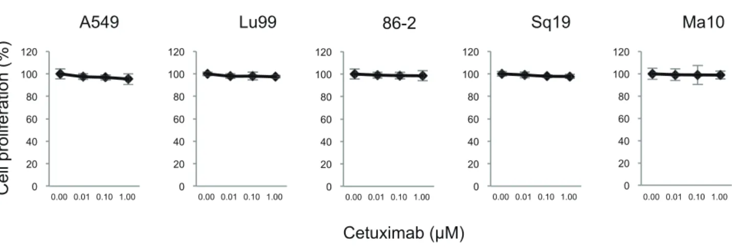
Fig. 3. Effect of cetuximab on cell proliferation of NSCLC cell lines. NSCLC cell lines were treated with 0, 0.01, 0.1 and 1.0 µM
cetux-imab for 6 days, and cell proliferation of was assessed using 8 cell proliferation assay kit. NSCLC, non-small cell lung cancer; WST-8, water-soluble tetrazolium salt.
listed in Table 1. We examined KRAS gene status in 86-2, Sq19 and Ma10, PTEN gene status in Lu99, 86-2 and Sq19, TP53 gene status in Sq19, and LKB1 gene status in 86-2 and Sq19 using PCR and direct sequencing with sequence specifi c primers published elsewhere.16–18 The remaining gene status of the cell lines were cited from a published data as indicated in the table 1. All cell lines exhibited wild-type EGFR, whereas KRAS, PTEN, TP53 and LKB1 were mutated in A549 and Lu99; Lu99 and Sq19; Lu99, 86-2, Sq19 and Ma10; and A549, 86-2 and Sq19, respectively.
Protein expression of NSCLC cell lines
The expression of EGFR, PTEN, Akt, KRAS, TP53 and LKB1 proteins in all NSCLC cell lines was examined using western blotting (Fig.1). EGFR, KRAS and Akt were equally expressed in all cell lines. However, PTEN was not expressed in Sq19 cells, and the expression of Akt and KRAS were comparable with other cell lines. LKB1 was not expressed in A549 and Sq19 cells, and TP53 was not expressed in A549 and Lu99 cells.
Effect of EGFR siRNA on EGFR expression Next, we tested the effect of EGFR specifi c siRNA on the expression of EGFR in all cell lines. As shown in Fig. 2, the expression of EGFR was almost suppressed by EGFR specific siRNA, whereas negative control siRNA that consists of non-targeting 20–25 bp nucleotide strands had no effect on the expression of EGFR. The proliferation of the cells transfected with EGFR siRNA was not different from those of the cells transfected with negative control siRNA (data not shown).
Cetuximab and EGFR siRNA combination treatment in NSCLC
Effect of cetuximab on cell proliferation in NSCLC cell lines
We evaluated the effect of cetuximab on the cell growth in NSCLC cell lines. The number of NSCLC cells after cetuximab treatment (0, 0.01, 0.1 and 1.0 µM) was measured for 6 days using a colorimetric WST-8 assay. As shown in Fig. 3, cetuximab has no effect on the pro-liferation in all NSCLC cell lines. Therefore, it was con-fi rmed that all the cell lines studied here are
cetuximab-N. Miyake et al., p. 30
Cell proliferation (% of control)
Cetuximab Cetuximab-Negative control Cetuximab-EGFR siRNA Cetuximab (µM) 0 20 40 60 80 100 120 0.00 0.01 0.10 1.00 0 20 40 60 80 100 120 0.00 0.01 0.10 1.00 0 20 40 60 80 100 120 140 0.00 0.01 0.10 1.00 0 20 40 60 80 100 120 140 0.00 0.01 0.10 1.00 0 20 40 60 80 100 120 0.00 0.01 0.10 1.00 * * *
A549 Lu99 86-2 Sq19 Ma10
*
Ratio of active caspase-3/7 (ratio 0f control)
0.0 0.2 0.4 0.6 0.8 1.0 1.2 0.00 0.01 0.10 1.00 0.0 0.2 0.4 0.6 0.8 1.0 1.2 1.4 0.00 0.01 0.10 1.00 0.0 0.2 0.4 0.6 0.8 1.0 1.2 1.4 1.6 0.00 0.01 0.10 1.00 0.0 0.2 0.4 0.6 0.8 1.0 1.2 1.4 1.6 0.00 0.01 0.10 1.00 Cetuximab (µM) 0.0 0.2 0.4 0.6 0.8 1.0 1.2 1.4 0.00 0.01 0.10 1.00
A549 Lu99 86-2 Sq19 Ma10
* Cetuximab Cetuximab-Negative control Cetuximab-EGFR siRNA * *
N. Miyake et al., p. 31
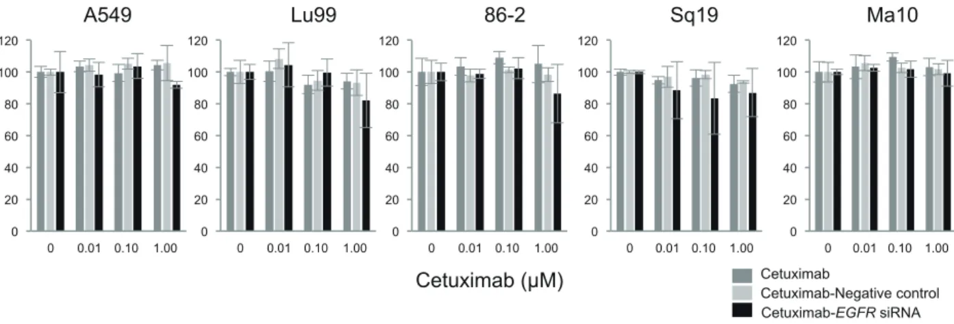
Fig. 4. Inhibition of cell proliferation by the combination treatment of cetuximab with EGFR siRNA. Cells were treated with the
combi-nation of 0, 0.01, 0.1 and 1.0 µM cetuximab and EGFR siRNA for 6 days. Cell proliferation was assessed using WST-8 cell proliferation assay kit. *P < 0.05 compared the combination of cetuximab and negative control siRNA by one-way ANOVA with Bonferroni’s multiple comparison test using SPSS Statistics 25. ANOVA, analysis of variance; EGFR, epidermal growth factor receptor; NSCLC, non-small cell lung cancer; siRNA, small interfering RNA; WST-8, water-soluble tetrazolium salt.
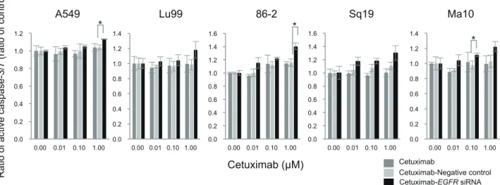
Fig. 5. Apoptosis induction by the combination treatment of cetuximab and EGFR siRNA. Cells were treated with the combination of
cetuximab (0, 0.01, 0.1 and 1.0 µM) and EGFR siRNA for 24 h. The production of active caspase 3/7 as a surrogate marker of apoptosis was assessed using Caspase-3/7 Glo assay kit. *P < 0.05 compared the combination of cetuximab and negative control siRNA by one-way ANOVA with Bonferroni’s multiple comparison test using SPSS Statistics 25. ANOVA, analysis of variance; EGFR, epidermal growth factor receptor; NSCLC, non-small cell lung cancer; siRNA, small interfering RNA.
resistant cell lines.
Effect of combination treatment of cetuximab and EGFR siRNA on cell proliferation
Furthermore, to test the effect of EGFR downregulation on the cetuximab-resistant cells, we evaluated the effect of cetuximab on NSCLC cell proliferation in the absence or presence of EGFR specifi c siRNA. As shown in Fig. 4, the combination of cetuximab with EGFR siRNA showed
N. Miyake et al.
Ratio of migration cells to untreated cells(% 0f control)
0 20 40 60 80 100 120 0 0.01 0.10 1.00 0 20 40 60 80 100 120 0 0.01 0.10 1.00 0 20 40 60 80 100 120 0 0.01 0.10 1.00 0 20 40 60 80 100 120 0 0.01 0.10 1.00 0 20 40 60 80 100 120 0 0.01 0.10 1.00 Cetuximab (µM) Cetuximab Cetuximab-Negative control Cetuximab-EGFR siRNA
A549 Lu99 86-2 Sq19 Ma10
N. Miyake et al., p. 32
N. Miyake et al., p. 33
0 20 40 60 80 100 120 0 0.01 0.10 1.00 0 20 40 60 80 100 120 0 0.01 0.10 1.00 0 20 40 60 80 100 120 0 0.01 0.10 1.00 0 20 40 60 80 100 120 0 0.01 0.10 1.00 0 20 40 60 80 100 120 0 0.01 0.10 1.00Ratio of Invasion cells to untreated cells(% 0f control) Cetuximab (µM) Cetuximab Cetuximab-Negative control
Cetuximab-EGFR siRNA
A549 Lu99 86-2 Sq19 Ma10
*
Fig. 6. Alteration of migration abilities of NSCLC cells by the combination treatment of cetuximab and EGFR siRNA. The NSCLC cell
lines were treated with cetuximab (0, 0.01, 0.1 and 1.0 µM) and EGFR siRNA for 42 h. Cell migration was evaluated after the combi-nation treatment in vitro. No significant alteration of migration was observed compared with the treatment with cetuximab and negative control siRNA. EGFR, epidermal growth factor receptor; NSCLC, non-small cell lung cancer; siRNA, small interfering RNA.
Fig. 7. Alteration of invasion abilities of NSCLC cells by the combination treatment of cetuximab and EGFR siRNA. The NSCLC cell
lines were treated with (0, 0.01, 0.1 and 1.0 µM) cetuximab and EGFR siRNA for 42 h. Cell invasion was evaluated after the combination treatment in vitro. *P < 0.05 compared the combination of cetuximab and negative control siRNA by one-way ANOVA with Bonferroni’s multiple comparison test using SPSS Statistics 25. ANOVA, analysis of variance; EGFR, epidermal growth factor receptor; NSCLC, non-small cell lung cancer; siRNA, non-small interfering RNA.
significant suppression of cell proliferation in 86-2, Sq19 and Ma10 NSCLC cell lines. These cell lines have wild-type KRAS (Table 1). The cell lines that have mutated
KRAS were found to be insensitive to the combination
treatment. These data suggested that the KRAS status is a possible biomarker for the combination treatment of cetuximab with EGFR siRNA.
Effect of combination treatment of cetuximab and
EGFR siRNA on apoptosis
After exploring the mechanism underlying the inhibition
of cell proliferation by the combination treatment of cetuximab and EGFR siRNA, we tested theses effect on apoptosis in each cell line. In this assay, we measured the production of active caspase-3/7 as a surrogate mark-er of apoptosis. As shown in Fig. 5, the combination treatment induced significant apoptosis in A549, 86-2 and Ma10 cell lines. These cell lines harbor wild-type
PTEN, whereas the cell lines in which apoptosis was
not induced by the combination treatment have mutated
PTEN. As wild-type PTEN was reported to promote
Cetuximab and EGFR siRNA combination treatment in NSCLC the combination treatment might facilitate the induction
of apoptosis. Moreover, induction of apoptosis by the combination treatment is a possible mechanism for the inhibition of cell proliferation in 86-2 and Ma10 cell lines.
Effect of combination treatment of cetuximab and EGFR siRNA on migration and invasion. Since cell migration and invasion are the hallmarks of cancer cells,20 we evaluated the effect of the combina-tion treatment of cetuximab with EGFR siRNA on cell migration and invasion. Cell migration was not affected by the combination treatment (Fig. 6). Cell invasion was suppressed by the combination treatment of 0.01 μM cetuximab and EGFR siRNA; however, it was not affected by the combination treatment of the other concentrations of cetuximab and EGFR siRNA (Fig. 7). Therefore, we concluded that the combination treatment mostly affects EGFR pathways that relate to cell prolif-eration and apoptosis.
DISCUSSION
In this study, we explored the effect of the combination treatment of cetuximab with EGFR siRNA in NSCLC cell lines with wild-type EGFR. We found that this combination treatment suppressed the proliferation of cells that carried wild-type KRAS. Therefore, the muta-tional status of KRAS might be a possible biomarker of this combination treatment. In addition, we also showed that combination treatment induces apoptosis, which suppresses the proliferation of cells that carry wild-type
PTEN. These observations suggest that the combination
therapy of cetuximab with EGFR siRNA is promising for the treatment of NSCLC using KRAS and PTEN gene status as biomarkers.
A number of signaling molecules are involved in cancer development. These genes are classified into two categories; oncogenes that promote cancer development and tumor suppressor genes that inhibit cancer develop-ment. EGFR pathways, which play a vital role in cancer development, mainly regulate cell proliferation signals; many genes downstream of EGFR are known oncogenes and tumor suppressor genes. KRAS is the oncogene and
PTEN, TP53, and LKB1 are the tumor suppressor genes
involved in this pathway (Fig. 8). Dysregulation of these molecules by gene mutation or alternation of protein expression is reported to promote cancer development. For example, KRAS mutation constitutively activates the EGFR signaling pathway and promote cell prolif-eration.21–25 PTEN is the tumor suppressor molecule of PI3K-Akt pathway that regulates cell proliferation, cell growth, and inhibition of apoptosis.26 Mutation or loss
of PTEN is reported to inhibit apoptosis, which leads to cell proliferation.18, 27, 28 TP53 is a multifunctional protein that is activated owing to cell-physiologic stress and prevents cancer development via the induction of apop-tosis, cell cycle arrest, DNA repair, and the inhibition of angiogenesis.29 Indeed, TP53 mutation is reported to reduce apoptosis in cancer cells.30 LKB1 inhibits activa-tion of mTOR signal funcactiva-tioning in the downstream of PI3K-Akt pathway, and functional loss of LKB1 leads to cell proliferation.22, 31–33
In this study, we examined five NSCLC cell lines. Since, these cell lines have wild-type EGFR, the aber-rant proliferative activity of these cancer cells is depen-dent on the EGFR downstream molecules. KRAS is ab-errantly activated by mutations in A549 and Lu99 cells and is the cause of the abnormal activity of the growth regulating function of these cancer cell lines. Moreover, we showed that TP53 was inactivated in all cell lines by defective expression (A549) or gene mutation (Lu99, 86-2, Sq19 and Ma10), and one or two additional tumor suppressor genes are inactivated by mutation or defec-tive expression in each cell line. These diverse variety of inactivation of tumor suppressor genes may strongly
Fig. 8. EGFR downstream pathways tested in this study. Tested
molecules that activate EGFR pathway are circled with a contin-uous line, and tested molecules that negatively regulate this path-way are circled with a dashed line. Akt, v-akt murine thymoma viral oncogene; EGFR, epidermal growth factor receptor; ERK, extracellular signal-regulated kinase; KRAS, v-ki-ras2 kirsten rat sarcoma viral oncogene homolog; LKB1, liver kinase B1; MEK, mitogen-activated protein kinase/extracellular signal-regulated ki-nase; mTOR, mammalian target of rapamycin; PI3K, phosphoinositol-3-kinase; PTEN, phosphatase and tensin homolog deleted from chromosome 10; RAF, v-raf murine sarcoma viral oncogene homolog; siRNA, small interfering RNA; SOS, son of sevenless; TP53, tumor protein p53.
N. Miyake et al. contribute to the cancer development.
Dysregulation of oncogenes and tumor suppressor genes are considered as the biomarkers of molecular targeted therapy in cancer. In the anti-EGFR therapy, somatic mutation of tyrosine kinase domain of EGFR that renders aberrant activation was revealed as potent predictors of response to EGFR-TKIs.34, 35 In addition, increased EGFR copy number36 and KRAS mutation37 were reported as negative predictors of EGFR-TKI. For cetuximab, wild-type KRAS was reported to be a predictor of colorectal cancer responsiveness,38 however, it was reported that the KRAS mutation status was not useful in predicting the responsiveness for cetuximab in NSCLC patient.39 In the NSCLC cell lines with wild-type EGFR, we showed that wild-type KRAS would be a common characteristic of the cells that are sensitive for the combination treatment of cetuximab with EGFR siRNA. To our knowledge, this is among the first studies that identified a biomarker of this combination treatment for NSCLC cells with wild-type EGFR.
There are several limitations in this study. First, we have presented data showing that one of the mechanisms of the susceptibility to the combination treatment of cetuximab and EGFR siRNA was the induction of apoptosis in two of three sensitive cell lines (86-2, Ma10). However, in the other sensitive cell line (Sq19), the mechanism was unclear, and a more extensive study of the other signaling molecules affected by this combi-nation treatment is needed. Second, this was an in vitro study using NSCLC cell lines, and its results need to be validated using animal experiments and clinical testing to enable the practical application of the combination treatment of cetuximab with EGFR siRNA.
In conclusion, EGFR targeted treatment using the combination of cetuximab and EGFR siRNA is effec-tive even in the NSCLC cell lines harboring wild-type
EGFR. A possible biomarker of response to this
com-bination treatment is wild-type KRAS, and one of the mechanisms of action is induction of apoptosis in cells with wild-type PTEN. The combination treatment of cetuximab and EGFR siRNA may be a new therapeutic potential in NSCLC patients.
The authors declare no conflict of interest. REFERENCES
1 Pfister DG, Johnson DH, Azzoli CG, Sause W, Smith TJ, Baker S, Jr., et al. American Society of Clinical Oncology treatment of unresectable non-small-cell lung cancer guideline: update 2003. J Clin Oncol. 2004;22:330-53. DOI: 10.1200/JCO.2004.09.053. PMID: 14691125.
2 Scaltriti M, Baselga J. The epidermal growth factor receptor pathway: a model for targeted therapy. Clin Cancer Res.
2006;12:5268-72. DOI: 10.1158/1078-0432.CCR-05-1554. PMID: 17000658.
3 Sharma SV, Bell DW, Settleman J, Haber DA. Epidermal growth factor receptor mutations in lung cancer. Nat Rev Cancer. 2007;7:169-81. DOI: 10.1038/nrc2088. PMID: 17318210.
4 Singh M, Jadhav HR. Targeting non-small cell lung cancer with small-molecule EGFR tyrosine kinase inhibitors. Drug Discov Today. 2018;23:745-53. DOI: 10.1016/j.dru-dis.2017.10.004. PMID: 29031620.
5 Liao BC, Lin CC, Lee JH, Yang JC. Optimal management of EGFR-mutant non-small cell lung cancer with disease progression on first-line tyrosine kinase inhibitor therapy. lung cancer. 2017;110:7-13. DOI: 10.1016/j.lungcan.2017.05.009. PMID: 28676222.
6 Xu MJ, Johnson DE, Grandis JR. EGFR-targeted therapies in the post-genomic era. Cancer Metastasis Rev. 2017;36:463-73. DOI: 10.1007/s10555-017-9687-8. PMID: 28866730.
7 Pirker R, Pereira JR, Szczesna A, von Pawel J, Krzakowski M, Ramlau R, et al. Cetuximab plus chemotherapy in patients with advanced non-small-cell lung cancer (FLEX): an open-label randomised phase III trial. Lancet (London, England). 2009;373:1525-31. DOI: 10.1016/S0140-6736(09)60569-9. PMID: 19410716.
8 Pirker R. EGFR-directed monoclonal antibodies in non-small cell lung cancer. Targeted oncology. 2013;8:47-53. DOI: 10.1007/s11523-012-0244-7. PMID: 23300028.
9 Reynolds A, Leake D, Boese Q, Scaringe S, Marshall WS, Khvorova A. Rational siRNA design for RNA interference. Nat Biotechnol. 2004;22:326-30. DOI: 10.1038/nbt936. PMID: 14758366.
10 Rothenberg SM, Engelman JA, Le S, Riese DJ, 2nd, Haber DA, Settleman J. Modeling oncogene addiction using RNA interference. Proc Natl Acad Sci U S A. 2008;105:12480-4. DOI: 10.1073/pnas.0803217105. PMID: 18711136.
11 Burnett JC, Rossi JJ, Tiemann K. Current progress of siRNA/shRNA therapeutics in clinical trials. Biotechnol J. 2011;6:1130-46. DOI: 10.1002/biot.201100054. PMID: 21744502.
12 Yamanaka S, Gu Z, Sato M, Fujisaki R, Inomata K, Sakurada A, et al. siRNA targeting against EGFR, a promising candi-date for a novel therapeutic application to lung adenocarci-noma. Pathobiology. 2008;75:2-8. DOI: 10.1159/000113789. PMID: 18334834.
13 Adams D, Gonzalez-Duarte A, O’Riordan WD, Yang CC, Ueda M, Kristen AV, et al. Patisiran, an RNAi Therapeutic, for Hereditary Transthyretin Amyloidosis. N Engl J Med. 2018;379:11-21. DOI: 10.1056/NEJMoa1716153. PMID: 29972753.
14 Takata M, Chikumi H, Miyake N, Adachi K, Kanamori Y, Yamasaki A, et al. Lack of AKT activation in lung cancer cells with EGFR mutation is a novel marker of cetuximab sensitivity. Cancer Biology & Therapy. 2012;13:369-78. DOI: 10.4161/cbt.13.6.19238. PMID: WOS:000302786400003. 15 Gazdar AF, Shigematsu H, Herz J, Minna JD. Mutations
and addiction to EGFR: the Achilles ‘heal’ of lung can-cers? Trends Mol Med. 2004;10:481-6. DOI: 10.1016/ j.molmed.2004.08.008. PMID: 15464447.
16 Onozato R, Kosaka T, Achiwa H, Kuwano H, Takahashi T, Yatabe Y, et al. LKB1 gene mutations in Japanese lung cancer patients. Cancer Sci. 2007;98:1747-51. DOI: 10.1111/j.1349-7006.2007.00585.x. PMID: 17711506.
Cetuximab and EGFR siRNA combination treatment in NSCLC Fujisawa T, et al. Role of the PI3K/Akt, mTOR, and STK11/
LKB1 pathways in the tumorigenesis of sclerosing heman-gioma of the lung. Pathol Int. 2008;58:38-44. DOI: 10.1111/ j.1440-1827.2007.02186.x. PMID: 18067639.
18 Jin G, Kim MJ, Jeon HS, Choi JE, Kim DS, Lee EB, et al. PTEN mutations and relationship to EGFR, ERBB2, KRAS, and TP53 mutations in non-small cell lung cancers. lung cancer. 2010;69:279-83. DOI: 10.1016/j.lungcan.2009.11.012. PMID: 20018398.
19 Weng L, Brown J, Eng C. PTEN induces apoptosis and cell cycle arrest through phosphoinositol-3-kinase/Akt-dependent and -independent pathways. Hum Mol Genet. 2001;10:237-42. DOI: 10.1093/hmg/10.3.237. PMID: 11159942.
20 Hanahan D, Weinberg RA. Hallmarks of cancer: the next gen-eration. Cell. 2011;144:646-74. DOI: 10.1016/j.cell.2011.02.013. PMID: 21376230.
21 Uekita T, Fujii S, Miyazawa Y, Iwakawa R, Narisawa-Saito M, Nakashima K, et al. Oncogenic Ras/ERK signaling activates CDCP1 to promote tumor invasion and metastasis. Mol Cancer Res. 2014;12:1449-59. DOI: 10.1158/1541-7786.MCR-13-0587. PMID: 24939643.
22 Mahoney CL, Choudhury B, Davies H, Edkins S, Greenman C, Haaften G, et al. LKB1/KRAS mutant lung cancers con-stitute a genetic subset of NSCLC with increased sensitivity to MAPK and mTOR signalling inhibition. Br J Cancer. 2009;100:370-5. DOI: 10.1038/sj.bjc.6604886. PMID: 19165201.
23 Massarelli E, Varella-Garcia M, Tang X, Xavier AC, Ozburn NC, Liu DD, et al. KRAS mutation is an important predictor of resistance to therapy with epidermal growth factor receptor tyrosine kinase inhibitors in non–small-cell lung cancer. Clin Cancer Res. 2007;13:2890-6. DOI: 10.1158/1078-0432.CCR-06-3043. PMID: 17504988.
24 Riely GJ, Marks J, Pao W. KRAS mutations in non-small cell lung cancer. Proc Am Thorac Soc. 2009;6:201-5. DOI: 10.1513/pats.200809-107LC. PMID: 19349489.
25 D’Arcangelo M, Cappuzzo F. K-Ras Mutations in Non-Small-Cell Lung Cancer: Prognostic and Predictive Value. ISRN Mol Biol. 2012;2012:837306. DOI: 10.5402/2012/837306. PMID: 27398239.
26 Simpson L, Parsons R. PTEN: life as a tumor suppressor. Exp Cell Res. 2001;264:29-41. DOI: 10.1006/excr.2000.5130. PMID: 11237521.
27 Soria JC, Lee HY, Lee JI, Wang L, Issa JP, Kemp BL, et al. Lack of PTEN expression in non-small cell lung cancer could be related to promoter methylation. Clin Cancer Res. 2002;8:1178-84. DOI: Published May 2002. PMID: WOS:000175547700032.
28 Zhao H, Dupont J, Yakar S, Karas M, LeRoith D. PTEN inhibits cell proliferation and induces apoptosis by downregu-lating cell surface IGF-IR expression in prostate cancer cells. Oncogene. 2004;23:786-94. DOI: 10.1038/sj.onc.1207162. PMID: 14737113.
29 Marcel V, Nguyen Van Long F, Diaz JJ. 40 Years of Research Put p53 in Translation. Cancers. 2018;10. DOI: 10.3390/can-cers10050152. PMID: 29883412.
30 Fridman JS, Lowe SW. Control of apoptosis by p53. Oncogene. 2003;22:9030-40. DOI: 10.1038/sj.onc.1207116. PMID: 14663481.
31 Sanchez-Cespedes M. The role of LKB1 in lung cancer. Fam
Cancer. 2011;10:447-53. DOI: 10.1007/s10689-011-9443-0. PMID: 21516316.
32 Matsumoto S, Iwakawa R, Takahashi K, Kohno T, Nakanishi Y, Matsuno Y, et al. Prevalence and specificity of LKB1 genetic alterations in lung cancers. Oncogene. 2007;26:5911-8. DOI: 10.1038/sj.onc.1210418. PMID: 17384680.
33 Zhong D, Guo L, de Aguirre I, Liu X, Lamb N, Sun SY, et al. LKB1 mutation in large cell carcinoma of the lung. lung cancer. 2006;53:285-94. DOI: 10.1016/j.lungcan.2006.05.018. PMID: 16822578.
34 Lynch TJ, Bell DW, Sordella R, Gurubhagavatula S, Okimoto RA, Brannigan BW, et al. Activating mutations in the epi-dermal growth factor receptor underlying responsiveness of non-small-cell lung cancer to gefitinib. N Engl J Med. 2004;350:2129-39. DOI: 10.1056/NEJMoa040938. PMID: 15118073.
35 Paez JG, Janne PA, Lee JC, Tracy S, Greulich H, Gabriel S, et al. EGFR mutations in lung cancer: correlation with clinical response to gefitinib therapy. Science. 2004;304:1497-500. DOI: 10.1126/science.1099314. PMID: 15118125.
36 Cappuzzo F, Hirsch FR, Rossi E, Bartolini S, Ceresoli GL, Bemis L, et al. Epidermal growth factor receptor gene and protein and gefitinib sensitivity in non-small-cell lung cancer. J Natl Cancer Inst. 2005;97:643-55. DOI: 10.1093/jnci/dji112. PMID: 15870435.
37 Pao W, Wang TY, Riely GJ, Miller VA, Pan Q, Ladanyi M, et al. KRAS mutations and primary resistance of lung adenocar-cinomas to gefitinib or erlotinib. PLoS Med. 2005;2:e17. DOI: 10.1371/journal.pmed.0020017. PMID: 15696205.
38 Russo A, Rizzo S, Bronte G, Silvestris N, Colucci G, Gebbia N, et al. The long and winding road to useful predictive factors for anti-EGFR therapy in metastatic colorectal carcinoma: the KRAS/BRAF pathway. Oncology. 2009;77 Suppl 1:57-68. DOI: 10.1159/000258497. PMID: 20130433.
39 Khambata-Ford S, Harbison CT, Hart LL, Awad M, Xu LA, Horak CE, et al. Analysis of potential predictive markers of cetuximab benefit in BMS099, a phase III study of cetuximab and first-line taxane/carboplatin in advanced non-small-cell lung cancer. J Clin Oncol. 2010;28:918-27. DOI: 10.1200/ JCO.2009.25.2890. PMID: 20100958.
40 Nagai Y, Miyazawa H, Huqun, Tanaka T, Udagawa K, Kato M, et al. Genetic heterogeneity of the epidermal growth factor receptor in non-small cell lung cancer cell lines revealed by a rapid and sensitive detection system, the peptide nucleic acid-locked nucleic acid PCR clamp. Cancer Res. 2005;65:7276-82. DOI: 10.1158/0008-5472.CAN-05-0331. PMID: 16105816. 41 Yokota J, Kohno T. Molecular footprints of human lung
cancer progression. Cancer Sci. 2004;95:197-204. DOI: org/10.1111/j.1349-7006.2004.tb02203.x. PMID: 15016317. 42 Iwakawa R, Kohno T, Enari M, Kiyono T, Yokota J.
Prevalence of human papillomavirus 16/18/33 infection and p53 mutation in lung adenocarcinoma. Cancer Sci. 2010;101:1891-6. DOI: 10.1111/j.1349-7006.2010.01622.x. PMID: 20557307.
43 Miyake N, Chikumi H, Takata M, Nakamoto M, Igishi T, Shimizu E. Rapamycin induces p53-independent apoptosis through the mitochondrial pathway in non-small cell lung cancer cells. Oncol Rep. 2012;28:848-54. DOI: 10.3892/ or.2012.1855. PMID: 22710790.