ORIGINAL ARTICLE
Prevalence and associated characteristics of aponeurotic ptosis among a general
population in Japan
Keiichiro Iida1),Shigeyuki Nakaji2),Makoto Mikami1),Katsunori Yokoi1),
Yuriko Saito1),Naoko Wada1),Sayaka Kasai1),Manabu Fukushima1),
Ayako Higuchi1),and Satoshi Urushidate1)
Abstract
Background: Ptosis is caused mainly by age-related degeneration of the aponeurosis caused by lifestyle-related diseases. Some patients with ptosis complain of stiff shoulders and headaches. There have been few actual epidemiological investigations of aponeurotic ptosis and these symptoms. We aimed to find their prevalence and associated characteristics of ptosis in Japanese general population.
Methods: The target population consisted of 1004 volunteers who took part in the 2016 Iwaki Health Promotion Project. We administered a questionnaire, took blood samples, took physical measurements, and measured marginal reflex distance (MRD-1) in each participant. The relationship between survey items (age, gender, difficulty in raising eyelids, headache, stiff shoulders, hypertension, diabetes, dyslipidemia, and body mass index [BMI]) and eyelid droop was examined statistically.
Results: The prevalence of ptosis in this study was 15.4%. Ptosis prevalence correlated with elderly people 65 years or older (odds ratio. 3.07, 95% CI: 2.02–4.66), male sex (odds ratio. 0.29, 95% CI: 0.20–0.43), hypertension (odds ratio. 1.68, 95% CI: 1.11–2.54) and dyslipidemia (odds ratio. 1.51, 95% CI: 1.02–2.23).
Conclusions: The present results suggested that hypertension, dyslipidemia and male sex were thought to be the risk factors of aponeurotic ptosis in Japanese general population.
Hirosaki Med.J. 71:131―137,2021
Key words: Aponeurotic ptosis; Lifestyle; risk factors; general population; logistic regression.
1) Department of Plastic and Reconstructive Surgery, Hirosaki University Graduate School of Medicine, Hirosaki, Aomori, Japan
2) Department of Social Medicine, Hirosaki University Graduate School of Medicine, Hirosaki, Aomori, Japan
Correspondence: K. Iida, MD
Received for publication, December 2, 2020 Accepted for publication, December 18, 2020
Introduction
The most common form of acquired ptosis is aponeurotic ptosis. Aponeurotic ptosis has various causes; in general, it is degeneration of the aponeurosis with age1). Fat infiltration has
been seen in the aponeurosis during the intraoperative findings of ptosis surgery2). In
cases of aponeurosis with fewer fatty deposits, the levator aponeurosis does not need to be surgically advanced very far, and more favorable surgical results are achieved3). Some authors
have pointed out that microcirculatory
distur-bances that result from lifestyle-related diseases such as hypertension, dyslipidemia, and diabetes can cause fatty degeneration of tissues, which worsens levator muscle function and can lead to ptosis4). Ptosis may be accompanied by tension
headache because, in order to maintain field of vision, the occipitofrontalis muscle are constantly contracting5). In addition, maintaining lateral
flexion of the neck in order to see can cause stiff shoulders.
As described above, several authors have described the causal relationship between ptosis and its causes and symptoms. However, few
or more or a diastolic blood pressure of 90 mmHg or more was defined as hypertension according to the 2014 Hypertension Treatment Guidelines7), and persons with a history of use of
antihypertensive agents or of other hypertension treatment were considered to have hypertension. Blood glucose levels of 200 mg/dL or higher and hemoglobin A1c levels of 6.5% or higher were considered to represent diabetes, in accordance with the 2018–2019 Diabetes Practice Guideline8),
and persons who had used oral glycemic medications or received other treatment for dia-betes were considered diabetic. LDL-cholesterol levels higher than 140 mg/dL, HDL-cholesterol levels lower than 40 mg/dL, and triglyceride levels of 150 mg/dL or more were considered to represent dyslipidemia, according to the 2017 Atherosclerosis Disease Prevention Guideline9) ;
patients with a history of medication or other treatment for dyslipidemia were considered to have this condition. Body mass index (BMI) was calculated as weight (in kilograms) divided by height (in meters) squared. According to the WHO standards, a BMI of less than 18.5 was defined as low BMI, and that of 25 or more was defined as high BMI.
In this cross-sectional study, the population of participants was divided into ptosis and nonpto-sis groups according to the definitions just described. Characteristics of the two groups such as age, gender, difficulty in raising eyelids, BMI, and the presence or absence of headache, hypertension, diabetes, and dyslipidemia were compared in multiple logistic regression analysis. SPSS ver. 24 (SPSS IBM Inc., Armonk, NY, USA) was used for data input and analysis, and a p value of less than 0.05 was considered significant.
This study was approved by the Ethics Committee of Hirosaki University Graduate School of Medicine.
studies of the aponeurotic ptosis have focused on the Japanese general population. In this study, we examined the prevalence and associated characteristics of aponeurotic ptosis among a general population in Japan.
Patients and Methods
Of the 1148 residents of the Iwaki area, Hirosaki City, Aomori Prefecture, Japan, who participated in the 2016 Iwaki Health Promotion Project, 1004 ([397 men and 607 women, aged 20–92 years) participated in this study. We excluded those who had cataracts, retinal de-tachment, facial nerve paralysis, and a history of eyelid ptosis surgery, as well as those using steroids. A questionnaire was administered on the day of the medical examination to collect data on age, gender, medical history, and symptoms. Blood was collected early in the morning on an empty stomach, and levels of hemoglobin A1c, blood glucose, low-density lipoprotein (LDL)–cholesterol, high-density lipoprotein (HDL)–cholesterol, and triglycerides were measured. Marginal reflex distance 1 (MRD-1) was measured by five physicians in the Department of Plastic and Reconstructive Surgery, Hirosaki University Graduate School of Medicine, Hirosaki, Japan, who were qualified as plastic surgery specialists by the Japan Society of Plastic and Reconstructive Surgery. MRD-1 was defined as the distance from the upper eyelid margin to the corneal light reflex in the primary position, regardless of the effect of dermatochalasis or brow ptosis. In participants who had skin laxity, we lifted flaccid skin manually and measured.
According to the guidelines of plastic surgery medical treatment, MRD-1 less than 2 mm was defined as ptosis6). According to the World
Health Organization (WHO) standards, people aged 65 years or older were defined as elderly persons. A systolic blood pressure of 140 mm Hg
Results
The prevalence of ptosis in this study was 15.4%. The ptosis group consisted of 155 people (95 men, 60 women), and the mean MRD-1 on both sides was 1.21 mm (standard deviation, 0.75mm). The nonptosis group consisted of 849 people (302 men, 547 women), and the mean
MRD-1 on both sides was 3.24 mm (standard deviation, 0.80 mm). Characteristics of partici-pants are listed in Table 1. The data in Table 2 are the results of multiple logistic regression analysis.
With regard to age, the proportion of elderly people was significantly higher in the ptosis group (odds ratio [OR], 3.07; 95% confidence Table 1. Characteristics of participants list involved with the result of analysis
Overall Ptosis group Non ptosis group N = 1004 n = 155(15.4%) n = 849(84.6%) Value of MRD-1 2.92±1.08 1.21±0.75 3.24±0.80 Age 53.3±15.5 65.5±15.5 51.7±15.5 ≧65years 284(28.3%) 84(54.2%) 200(23.6%) <65years 720 (71.7%) 71(45.8%) 649(76.4%) Sex Male 397(39.5%) 95(61.3%) 302(35.6%) Female 607(60.5%) 60(38.7%) 547(64.4%)
Visual field constriction (Yes) 76(7.6%) 17(11.0%) 59(7.00%)
Headache (Yes) 561(55.9%) 64(41.3%) 497(58.5%)
Shoulder discomfort (Yes) 773(77.0%) 115(74.2%) 658(77.5%)
Hypertension (Yes) 403(40.1%) 96(61.9%) 307(36.2%)
Diabetes mellitus (Yes) 73(7.3%) 24(15.5%) 49(5.8%)
Dyslipidemia (Yes) 444(44.2%) 91(58.7%) 353(41.6%)
BMI 22.9±3.4 23.3±3.4 22.8±3.4
Low 72(7.2%) 4(2.6%) 68(8.0%)
Middle 685(68.2%) 114(73.6%) 571(67.3%)
High 247(24.6%) 37(23.9%) 210(24.7%)
Data for continuous variables(MRD-1, Age, BMI)are listed as means ± standard deviations, and data of categorical variables are listed as frequencies and percentages. BMI, body mass index; MRD-1, marginal reflex distance
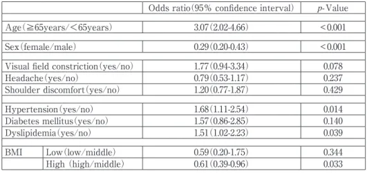
Table 2. result of the multiple logistic regression model on the associations of ptosis Odds ratio(95% confidence interval) p- Value
Age(≧65years/<65years) 3.07(2.02-4.66) < 0.001
Sex(female/male) 0.29(0.20-0.43) < 0.001
Visual field constriction(yes/no) 1.77(0.94-3.34) 0.078
Headache(yes/no) 0.79(0.53-1.17) 0.237 Shoulder discomfort(yes/no) 1.20(0.77-1.87) 0.429 Hypertension(yes/no) 1.68(1.11-2.54) 0.014 Diabetes mellitus(yes/no) 1.57(0.86-2.85) 0.140 Dyslipidemia(yes/no) 1.51(1.02-2.23) 0.039 BMI Low(low/middle) 0.59(0.20-1.75) 0.344 High (high/middle) 0.61(0.39-0.96) 0.033
interval [CI], 2.02–4.66; p < 0.001). Moreover, the proportion of patients with hypertension, dyslipidemia, and standard BMI (vs. high BMI) were significantly more prevalent in the ptosis group (ORs: 1.68, 1.51, and 0.61; 95%Cis: 1.11– 2.54, 1.02–2.23, and 0.39–0.96; p = 0.014, 0.039, and 0.033, respectively). The ptosis group also contained significantly more men than women (OR, 0.29; 95% CI, 0.20–0.43; p < 0.001). We found no significant difference between the two groups with regard to prevalence of diabetes, low BMI, difficulty in raising the eyelid, preva-lence of headache, or prevapreva-lence of stiff shoulder.
Discussion
Few studies have focused on the prevalence of aponeurotic ptosis in healthy individuals, and no studies have been conducted on Japanese subjects4,10). According to Kim et al, the
estimat-ed prevalence of degenerative ptosis in Koreans was 13.5% (MRD-1 of less than 2 mm was defined as ptosis) 4). In a British study of 400
randomly selected, age-stratified subjects older than 50 years of age, 11.5% had ptosis10). The
prevalence of ptosis in this study was 15.4%, and was slightly high level more than the above-mentioned previous researches.
In this study, rates of hypertension and dyslipidemia were significantly different between participants with and without ptosis4,11,12). It has
been said that steatosis of the aponeurosis as a result of aging, microcirculation disorders result-ing from hypertension, atherosclerotic microcir-culation disorders resulting from dyslipidemia, and tissue degeneration cause eyelid drooping4).
However, no actual research had been conducted previously in such a large number of healthy people in Japan.
Niimi described circulatory disturbances caused by hypertension13) : for example,
hypertro-phy of vessel walls, rarefaction of blood cells, and abnormalities in blood flow. These cause changes
in the microcirculation. In addition, they may play an important role in microcirculatory disorders caused by high blood pressure because they disrupt oxygen supply to organ tissue, gas exchange, and water flow13). The degeneration of
the aponeurosis as a result of microcirculation disorders may cause ptosis.
Shirado studied Japanese patients who had clinical signs of eyelid drooping, skin flaccidity, or eyebrow drooping or had a history of ptosis or blepharoplasty12). He statistically compared
plasma lipid data for patients with and without age-related ptosis and found that levels of total cholesterol and HDL-cholesterol were significant-ly and independentsignificant-ly associated with age-related ptosis.
Decreased HDL-cholesterol is known to be an independent risk factor for coronary artery disease and is one of the diagnostic criteria for dyslipidemia9). LDL-cholesterol, a component of
total cholesterol, is the first management target value in the guidelines of the Japanese Society of Atherosclerosis for preventing atherosclerotic disease9). Dyslipidemia is commonly associated
with decreased levels of HDL-cholesterol and elevated levels of LDL-cholesterol and may be one of the causes of age-related ptosis12).
There-fore, the results of this study were consistent with those of the studies mentioned previously. Although the reasons for the significant relation-ship between dyslipidemia and age-related ptosis have not been specified, a significant relationship between retinal microcirculatory disturbance and dyslipidemia has been described and may be the reason why circulatory disorders may be causing ptosis14,15).
Matsuda evaluated the length of the anterior layer of the levator aponeurosis that had no fatty deposits during transcutaneous aponeurotic repair3). If the eyelids contained more fatty
deposits during levator shortening operation, more levator aponeurosis shortening was needed, but surgical correction tended to be inadequate
at later.
As stated previously, this study also showed that dyslipidemia and hypertension were significantly related to ptosis and therefore might be among the risk factors for ptosis.
No significant difference was found in rates of diabetes in this study. Several reports have described the relationship between diabetes and ptosis4,11,14,16,17). In these researches, odds ratio of
diabetes for ptosis was reported as 1.32 (95%CI: 1.09-1.58)by Kim et al.4) and 1.53 (95%CI:
1.16-2.02) by Hashemi et al.11) According to these
reports, ptosis may occurs with microvascular dysfunction (caused by lipid deposition and oxidative stress on vascular walls) and neuropa-thy (caused by oculomotor paralysis and Horner syndrome) that result from diabetes11,17). But no
significant relationship between diabetes and ptosis was observed in this study. A further detailed investigation about relationship diabetes and ptosis is required.
In this study, high BMI was significantly less prevalent among people with ptosis than was standard BMI. According to some reports, BMI and ptosis are significantly related; according to others, they are not4,12,16-19). In this study, the
numbers of people with low and high BMI were lower than the number of people with standard BMI. In addition, the average BMI in this study was slightly lower than the average BMI in subjects in past studies in which BMI and ptosis were significantly related. It is possible that our results differ from those in reports in which BMI and ptosis were significantly related.
The prevalence of men with ptosis was significantly higher than that of women with ptosis. We found few reports in which gender differences in ptosis were described. Surveys in Tehran also revealed that men with ptosis were significantly more numerous than women with ptosis20). Although this study concerned
aponeu-rotic ptosis, there are reports that aging has more significant effects on cutaneous ptosis in
men than in women19,21,22). Surveys of Dutch and
British people also demonstrated that men with cutaneous ptosis were more numerous than women with cutaneous ptosis19). The skin in the
orbital region consists of the dermis, thin facial muscles, and a thin subcutaneous fat layer and is not attached to the bone. The orbital area is larger in men than in women, and men are more susceptible to age-related sagging21).
In addition, in a comparison of the deep muscle tendons involved in finger flexion in men and women, women reported less time-dependent elongation than did men; that is, women’s tendons were stiffer23). The sites of elongation
may have differed in men and women, but elongation may be common in tendons that undergo repeated loading. To determine whether male sex is a risk factor for ptosis, further research is needed.
This is the first study to investigate ptosis and subjective symptoms in the general population. According to some reports, headache is a functional indication for ptosis surgery24), and
headaches have been resolved with ptosis surgery. Although subjects with and without ptosis have exhibited generally significant differences in difficulty in raising the eyelids, headache, and stiff shoulders in other studies, we found no significant differences in this study. Eighty nine % of the ptosis group were unaware of visual field constriction despite the presence of ptosis. It was suggested that early ptosis may be asymptomatic due to compensation by Müller’s muscle contraction, occipitofrontalis muscle contraction and lateral flexion of the neck. Patients with ptosis are less likely to visit a hospital care if they do not have symptoms. Thus, a large number of ptosis patients are potentially present in the general population. Viewed from the opposite side, patients visiting a hospital were subjective and had significantly more advanced symptoms, suggesting that they may have symptoms of headache and stiffness.
Our study had several limitations. We used logistic regression analysis to analyze the data, and bias may have been present in our sample. The Iwaki project is based on statistics from volunteers from the Iwaki area, Hirosaki City, and data may differ from those of other Japanese populations.
This study was conducted on a large number of volunteers, and our findings may support a causal relationship between dyslipidemia/hyper-tension and ptosis. It has also been suggested that ptosis may contribute to sympathicotonia diseases such as hypertension. Early detection and treatment of potential ptosis patients by screening for ptosis may be helpful in the treatment of ptosis and associated symptoms.
Conclusions
In this study, we investigated the association between aponeurotic ptosis and its causes and the association between ptosis and symptoms. The present study provides the first representa-tive data on aponeurotic ptosis among a large-scale survey of the general population of a city in Japan. An association between ptosis and older age, male sex, hypertension, and dyslipid-emia was suggested. A significant relationship between dyslipidemia/hypertension and ptosis demonstrated in previous reports was found. In view of findings of previous studies and ours, we suggest that dyslipidemia, hypertension, and male sex may be included among the risk factors for aponeurotic ptosis in Japanese general population. It may be useful to educate people with the risk factors identified in this study about the risk of developing ptosis and to teach them to reduce eyelid irritation to prevent ptosis.
Financial Disclosure Statement
This work was supported by JST COI Grant Number JPMJCE1302.Conflict of Interests
All authors have no conflicts of interest di-rectly to the content of this article.
Acknowledgements
We thank Dr.Matsuzaka (Department of Medical Informatics, Hirosaki University Hospital), and Professor Ihara (Department of Social Medicine, Hirosaki University Graduate School of Medicine)for their valuable comments, and ENAGO (www.enago.jp)for the English language review.
References
1) Finsterer J. Ptosis: causes, presentation, and management. Aesthetic Plast Surg. 2003;27:193-204.
2) Watanabe A, Araki B, Noso K, Kakizaki H, Kinoshita S. Histopathology of blepharoptosis induced by prolonged hard contact lens wear. Am J Ophthalmol. 2006;141:1092-6.
3) Matsuda H, Sakai T, Takahashi Y, Nakamura M, Nakano T. Influence of fatty deposits in the levator aponeurosis/levator palpebrae superioris muscle on outcomes of aponeurotic repair in a Japanese population. Eye (Lond). 2018;32:1845-50. 4) Kim MH, Cho J, Zhao D, Woo KI, Kim YD, Kim S,
Yang SW. Prevalence and associated factors of blepharoptosis in Korean adult population: The Korea national health and nutrition examination survey 2008–2011. Eye (Lond). 2017;31:940-6. 5) Matsuo K, Osada Y, Ban R. Electrical stimulation
to the trigeminal proprioceptive fibres that innervate the mechanoreceptors in Müller's muscle induces involuntary reflex contraction of the frontalis muscles. J Plast Surg Hand Surg. 2013;47:14-20.
6) Japan Society of Plastic and Reconstructive Surgery, Japan Society for Surgical Wound Care, Japan Society of Cranio-Maxillo-Facial Surgery
eds. The guidelines of plastic surgery medical treatment. Vol.6. head, neck and facial disease. Tokyo. Kanehara & Co., Ltd. 2015. p.105-6. (in Japanese)
7) Japanese Society of Hypertension. Guidelines for the management of hypertension 2014. https:// www.jpnsh.jp/data/jsh2014/jsh2014v1_1.pdf (in Japanese) (accessed 2020.3.30)
8) The Japan Diabetes Association. The Diabetes Practice Guideline 2018-2019. Tokyo. Bunkodo. 2018. p.19-21. https://www.u-hyogo.ac.jp/shse/ sakaue/Lecture/file/2020_ref_DM.pdf (in Japanese) (accessed 2020.3.30)
9) Yamashita S. The atherosclerosis disease prevention guideline 2017 edition. Journal of the Japanese Society of Internal Medicine. 2018;107:73-80. (in Japanese)
10) Sridharan GV, Tallis RC, Leatherbarrow B, Forman WM. A community survey of ptosis of the eyelid and pupil size of elderly people. Age Ageing. 1995;24:21-4.
11) Hashemi H, Khabazkhoob M, Emamian MH, Yekta A, Jafari A, Nabovati P, Fotouhi A. The prevalence of ptosis in an Iranian adult popula-tion. J Curr Ophthalmol. 2016;28:142-5.
12) Shirado M. Dyslipidaemia and age-related involutional blepharoptosis. J Plast Reconstr Aesthet Surg. 2012;65:e146-50.
13) Niimi H, Yamakawa T. Hypertension and microvascular rheology. Nippon Rinsho. 1992;50: 444-53. (in Japanese)
14) Wong TY, Duncan BB, Golden SH, Klein R, Couper DJ, Klein BEK, Habbard LD, et al. Associations between the metabolic syndrome and retinal microvascular signs: the atherosclero-sis risk in communities study. Invest Ophthalmol Vis Sci. 2004;45:2949-54.
15) van Leiden HA, Dekker JM, Moll AC, Nijpels G, Heine RJ, Bouter LM, Stehouwer CDA, et al. Blood pressure, lipids, and obesity are associated with retinopathy: the hoorn study. Diabetes Care. 2002;25:1320-5.
16) Moon SS, Lee YS. Prevalence and clinical characteristics of blepharoptosis in patients with diabetes in the Korea national health and nutri-tion examinanutri-tion survey (KNHANES) 2009-2010. Endocrine. 2015;48:504-10.
17) Bosco D, Costa R, Plastino M, Branca D, Cotronei P, Sperli T, Santacroce N, et al. Glucose metabo-lism in the idiopathic blepharoptosis: utility of the oral glucose tolerance test (OGTT) and of the insulin resistance index. J Neurol Sci. 2009;284:24-8.
18) Paik JS, Jung SK, Han KD, Kim SD, Park YM, Yang SW. Obesity as a potential risk factor for blepharoptosis: the Korea national health and nutrition examination survey 2008-2010. PLoS One. 2015;10:e0131427.
19) Jacobs LC, Liu F, Bleyen I, Gunn DA, Hofman A, Klaver CCW, Uitterlinden AG, et al. Intrinsic and extrinsic risk factors for sagging eyelids. JAMA Dermatol. 2014;150:836-43.
20) Hashemi H, Khabazkhoob M, Yekta A, Mohammad K, Fotouhi A. The prevalence of eyelid ptosis in Tehran population: the Tehran eye study. Iran J Ophthalmol. 2010;22:3-6.
21) Ezure T, Yagi E, Kunizawa N, Hirao T, Amano S. Comparison of sagging at the cheek and lower eyelid between male and female faces. Skin Res Technol. 2011;17:510-5.
22) van den Bosch WA, Leenders I, Mulder P. Topographic anatomy of the eyelids, and the effects of sex and age. Br J Ophthalmol. 1999;83: 347-52.
23) Goldstein SA, Armstrong TJ, Chaffin DB, Matthews LS. Analysis of cumulative strain in tendons and tendon sheaths. J Biomech. 1987;20:1-6.
24) Matsuo K, Ban R. Surgical desensitisation of the mechanoreceptors in Müller's muscle relieves chronic tension-type headache caused by tonic reflexive contraction of the occipitofrontalis mus-cle in patients with aponeurotic blepharoptosis. J Plast Surg Hand Surg. 2013;47:21-9.