INTRODUCTION
Diabetic retinopathy (DR) is a major cause of visual loss in patients with diabetes mellitus. Dia-betic macular edema (DME), which can occur at any stage of DR, is characterised by increased vas-cular permeability and the deposition of hard exu-dates at the central retina. Diabetic macular edema is now the principal cause of vision loss in people
with diabetes (1).
Bevacizumab (AvastinTM, Genentech Inc. San
Francisco, California, USA) is a full length human-ized antibody that binds to all subtypes of vascular endothelial growth factor (VEGF) and has been ap-proved by the US Food and Drug Administration for the treatment of metastatic colorectal cancer (2). Re-cent reports have suggested that bevacizumab may be useful for the treatment of choroidal neovascu-larization (CNV), diabetic macular edema, DR and macular edema associated with retinal venous occlu-sive diseases (3-5).
The purpose of this study was to report the effi-cacy of intravitreal injections of bevacizumab by measuring visual acuity (VA) and foveal retinal
ORIGINAL
Efficacy of intravitreal bevacizumab (Avastin
TM) for
short-term treatment of diabetic macular edema
Toshihiko Nagasawa, Takeshi Naito, Shingo Matsushita, Hiroyuki Sato,
Takashi Katome, and Hiroshi Shiota
Department of Ophthalmology, Institute of Health Biosciences, the University of Tokushima Graduate School, Tokushima, Japan
Abstract : Purpose : To report the efficacy of intravitreal injections of bevacizumab for dia-betic macular edema (DME) in the short-term. Design : Retrospective, noncomparative, in-terventional case series. Methods : Medical records of 20 eyes of 19 patients who under-went intravitreal injections of bevacizumab for persistent diabetic macular edema were reviewed retrospectively. All eyes received intravitreal injections of bevacizumab (1.25 mg/ 0.05 ml). The clinical course of best-corrected visual acuity (BCVA) using a logarithm of the minimum angle of resolution chart, and averaged foveal retinal thickness using an op-tical coherence tomography (OCT) were monitored for up to four weeks after the injection. Results : BCVA at one week improved by two lines or more in six eyes (30%%) and in nine eyes (45%%) at four weeks. However, no significant improvement in the mean BCVA from baseline was observed at one week (P 0.05) and four weeks (P 0.05). Mean retinal thick-nesses (RT) were 411 170
!
m at baseline, 349 102!
m at one week after the injection (P 0.05), and 380 159!
m at four weeks (P 0.05). One week after the injection, significant re-gression of macular edema was seen. However, recurrence occurred at four weeks. No com-plications such as severe vision loss, endophthalmitis, or systemic events developed. Con-clusion : No changes in BCVA and RT were observed in the short-term observation after the intravitreal injection of bevacizumab for DME. J. Med. Invest. 56 : 111-115, August, 2009Keywords : bevacizumab, Avastin, diabetic macular edema, diabetic retinopathy
Received for publication December 25, 2008 ; accepted February 4, 2009.
Address correspondence and reprint requests to Takeshi Naito, MD, Ph.D., Department of Ophthalmology, Institute of Health Biosciences, the University of Tokushima Graduate School, Kuramoto cho, Tokushima 770 8503, Japan and Fax : +81 88 -631 - 4848.
thickness (RT) after intravitreal injection of bevaci-zumab for DME.
METHODS
We retrospectively reviewed 20 eyes of 19 con-secutive Japanese patients (12 males, 7 females) with DME treated with intravitreally administered bevacizumab. The patients were followed for four weeks at Tokushima University Hospital. The Ethi-cal Committee of the University of Tokushima ap-proved the off-label use of bevacizumab. The deci-sion to treat with intravitreally administered bevaci-zumab was made by the patient after a complete dis-cussion on its risks, benefits and alternative treat-ments. If the patient decided to proceed with bevaci-zumab therapy, they signed a consent form before administration.
The eye was prepared with a topical anesthetic and a drop of antibiotic before the injection. The eye-lid margin was prepared with a povidone/iodine so-lution. A wire speculum was placed followed by sev-eral drops of diluted povidone/iodine solution to the conjunctiva at the injection site. An injection of 1.25 mg of bevacizumab (0.05 ml of bevacizumab at a concentration of 25 mg/ml) was administered using a 30 gauge needle from the pars plana.
Patients were examined at baseline, at one week, two weeks, and four weeks. We recorded the BCVA measured with a Japanese standard decimal VA chart and calculated the mean BCVA using the loga-rithm of the minimum angle of resolution (logMAR) scale, intraocular pressure (IOP), and fluorescein an-giography (FA). Analysis of retinal anatomic features was performed using an optical coherence tomogra-phy (OCT, Stratus III OCT ; Carl Zeiss, Dublin, California, USA). The RT at the fovea of the 1-mm central retina was determined using six low-reso-lution diagonally oriented fast macula scans.
Statistical analysis was performed using the Stu-dent’s t-test to compare the VA, IOP and the cen-tral RT at one, two, and four weeks from baseline.
RESULTS
Twenty eyes of 19 patients (12 males, 7 females) with DME were studied. The ages of the patients ranged from 44 to 76 years with a mean of 59.7! 8.0 years. All patients had type II diabetes. There was no history of any other ocular disease except
for refractive errors or cataracts. All patients had panretinal photocoagulation. Three patients had sub-Tenon’s capsule injection of 20 mg triamcinolone acetonide (TA). Nine patients had cataract surgery with intraocular lens implantation, and nine pa-tients had vitrectomy. The glycosylated hemoglobin (HbA1c) averaged 7.1!1.1 before starting the study
(Table 1).
The BCVA at one week improved by two lines or more in six eyes (30%) (Figure 1) and the BCVA at
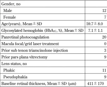
Table 1 Clinical characteristics of 19 patients (20 eyes) with diabetic macula edema at baseline.
Gender, no
Male 12
Female 7
Age(years), Mean!SD 59.7!8.0
Glycosylated hemoglobin (HbA1C, %), Mean!SD 7.1!1.1
Panretinal photocoagulation 20
Macula focal/grid laser treatment 0 Prior sub tenon triamcinolone injection 3
Prior pars plana vitrectomy 9
Lens status, no
Phakic 11
Pseudophakia 9
Baseline retinal thickness, Mean!SD (μm) 411!170
Figure 1 Scattergram of BCVA (1 week) and baseline. Changes in the best- corrected visual acuity (BCVA) at one week after treatment. Six eyes (30%) improved by two lines or more, one eye (5%) decreased by two lines or more.
There is no significant improvement between the mean BCVA and baseline (P!0.05, paired t-test).
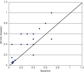
four weeks improved in nine eyes (45%) (Figure 2). No significant improvement in the mean BCVA from baseline was observed at one week (P"0.05), or four weeks (P"0.05).
Mean RT was 411!170 μm at baseline, 349!102 μm at one week, 365!149 μm at two weeks, and 380!159 μm at four weeks (Figure 3). The central RT significantly decreased from baseline at one week (P!0.05). However, no significant decrease was recognized at two weeks (P"0.05), and four weeks (P"0.05). At the four weeks follow-up visit,
FA showed resolution of leakage in some eyes. During the follow-up of the patients in this study, no complications such as inflammation, increased IOP (Figure 4), severe vision loss, endophthalmitis, or systemic events were observed.
DISCUSSION
DME has been characterized by inflammation, in-cluding intravitreous induction of proinflammatory cytokines (6), intraretinal expression of proinflam-matory caspases (7) and mediators. Many clinical investigators have found that an intravitreal injection of TA may reduce macular edema. However, the use of intravitreal TA may lead to complications such as increased IOP, progression of cataract and en-dophthalmitis (8).
Bevacizumab is an anti-VEGF agent that is ap-proved for the treatment of disseminated colorectal cancer but is not licensed for intraocular use. How-ever, bevacizumab appears to show a similar efficacy for the treatment of DME and proliferative DR (9). VEGF has been shown to be an endothelial cell-specific mitogen and an angiogenic inducer. It is also a vascular permeability factor ; it has been demon-strated to increase the permeability of retinal ves-sels by increasing the phosphorylation of tight junc-tion proteins (10). It was found that the concentra-tion of VEGF in the vitreous increased and corre-lated with the severity of macular edema in patients with DME (11), therefore anti-VEGF therapy is expected to show a dramatic reduction of DME. Bevacizumab has attracted interest because of its low cost ; however systemic safety is not approved
Figure 2 Scattergram of BCVA (4 weeks) and baseline. Changes in the best. - corrected visual acuity (BCVA) at four weeks after treatment. Nine eyes (45%) improved by two lines or more, 3 eyes (15%) decreased by two lines or more.
There is no significant improvement between the mean BCVA and baseline (P"0.05, paired t-test).
Figure 3 Changes in central retinal thickness throughout the study.
The central RT significantly decreased from baseline at one weeks (P!0.05). However, it did not significantly decrease at two weeks (P"0.05), and four weeks (P"0.05).
Figure 4 Changes of intra ocular pressure throughout the study.
(12, 13).
We conformed to the widely used concentration of the drug (1.25 mg bevacizumab) in this study. Anti-VEGF therapy of the intravitreally administered bevacizumab showed a marked reduction of macu-lar edema soon after the injection at one week. How-ever, recurrence of macular edema occurred within four weeks. A retinal penetration study revealed the absence of bevacizumab four weeks after the injec-tion (14), which may suggest the limited effect of bevacizumab on suppression of VEGF activity.
Mean retinal thickness was reduced at one week after the injection (P!0.05). However, no significant improvement in the mean BCVA. It is possible that visual acuity may not improve with the same time course as thickness of the macula. It depends on macular function improvement and it may have timelag (15).
Maia, et al. reported that a combination therapy of intravitreal triamcinolone and laser photocoagu-lation decreased DME (16). Recently, a combination therapy with intravitreal bevacizumab and photody-namic therapy (PDT) for CNV in patients with AMD has been reported to improve VA and anatomic changes and reduce retreatment rates (17). There-fore, we think that the combination of laser photo-coagulation with intravitreal bevacizumab may im-prove BCVA and decrease RT more than laser pho-tocoagulation alone or intravitreal bevacizumab alone for the treatment of moderate DME.
In summary, the therapy of intravitreal bevacizu-mab alone for DME is effective in the short-term, however it is not effective in the long-term.
REFERENCES
1. Klein R, Klein BE, Moss SE, Moss SE,
Cruickshanks KJ : The Wisconsin Epidemi-ologic Study of Diabetic, XVII : the 14-year in-cidence and progression of diabetic retinopathy and associated risk factors in type I diabetes. Ophthalmology 105 : 1801-1815, 1998
2. Ferrara N, Hillan KJ, Gerber HP, Novotny W : Discovery and development of bevacizumab, an anti-VEGF antibody for treating cancer. Nat Rev Drug Discov 3 : 391-400, 2004
3. Spaide RF, Fisher YL : lntravitreal bevacizumab (Avastin) treatment of proliferative diabetic ret-inopathy complicated by vitreous hemorrhage. Retina 26 : 275-278, 2006
4. Avery RL, Pieramici DJ, Rabena MD, Castellarin
AA, Nasir MA, Giust MJ : lntravitreal bevaci-zumab (Avastin) for neovascular age-related macular degeneration. Ophthalmology 113 : 363-372, 2006
5. Iturralde D, Spaide RF, Meyerle CB, Klancnik JM, Yannuzzi LA, Fisher YL, Sorenson J, Slakter JS, Freund KB, Cooney M, Fine HF : Intravitreal bevacizumab (Avastin) treatment of macular edema in central retinal vein occlu-sion : a short-term study. Retina 26 : 279-284, 2006
6. Funatsu H, Yamashita H, Ikeda T, Mimura T, Eguchi S, Hori S : Vitreous levels of interleukin-6 and vascular endothelial growth factor are re-lated to diabetic macular edema. Ophthalmol-ogy 110 : 1690-1696, 2003
7. Mohr S, Tang J, Kem TS : Caspase activation in retinas of diabetic and galactosemic mice and diabetic patients. Diabetes 51 : 1172-1179, 2002 8. Bhavsar AR, Glassman AR, DRCRnet Group, SCORE Study Group : The risk of endophthalmi-tis fbllowing intravitreal triamcinolone injection in the DRCRnet and SCORE clinical trials. Am J Ophthalmol 144 : 454-456, 2007
9. Scott lU, Edwards AR, Back RW, Diabetic ret-inopathy clinical research network : .A phase ll randomized clinical trial of intravitreal bevaci-zumab for diabetic macular edema. Ophthal-mology 114 : 1860-1867, 2007
10. Ferrara N : Vascular endothelial growth fctor : basic science and clinical progress. Endocr Rev 25 : 581-611, 2004
11. Funatsu N, Yamashita H, Sakata K, Noma H, Mimura T, Suzuki M, Eguchi S, Hori S : Vitre-ous levels of vascular endothelial growth factor and intracellular adhesion molecule 1 are re-lated to diabetic macular edema. Ophthalmol-ogy 112 : 806-816, 2005
12. Rosenfeld PJ : Intravitreal Avastin : the low cost alternative to Lucentis? Am J Ophthalmol 142 : 141-143, 2006
13. Gillies MC : What we don’t know about Avastin might hurt us. Arch Ophthalmol 124 : 1478-1479, 2006
14. Shahar J, Avery RL, Heilweil G, Barak A, Zemel E, Lewis GP, Johnson PT, Fisher SK, Perlman I, Loewenstein A : Electrophysiologic and reti-nal penetration studies following intravitreal injection of bevacizumab (Avastin). Retina 26 : 262-269, 2006
15. Diabetic Retinopathy Clinical Research Net-work : Relationship between optical coherence
tomography-measured central retinal thickness and visual acuity in diabetic macular edema. Ophthalmology 114 : 525-536, 2007
16. Maia Jr OO, Takahashi BS, Costa RA, Scott IU, Tkahashi WY : Combined laser and lntravitreal triamcinolone for proliferative diabetic retino-pathy and macular edema : one-year results of a randomized clinical trial. Am J Ophthalmol
146 : 930-941, 2008
17. Ladas ID, Kotsolis AI, Papakostas TD, Rouvas AA, Karagiannis DA, Vergados I : Intravitreal bevacizumab combined with photodynamic therapy for the treatment of occult choroidal ne-ovascularization associated with serous pigment epithelium detachment in age-related macular degeneration. Retina 27 : 891-896, 2007