previous sporting activities at 6 months.
At the 2 - year follow - up, the patient had no complaints of knee problems, and physical examination revealed no loss of function. Four follow - up MRI studies performed postoperatively confirmed survival of the fixed chondral fragment and a smooth articular surface without any sign of delamination, suggesting successful healing (Fig. 5).
DISCUSSION
Chondral and osteochondral fractures of the lateral femoral condyle are rare injuries. They were first reported by Makin in 1951 (24) and then by Rosenberg in 1964 (25) and Kennedyet al. in 1966
(4). In a later epidemiological study, Matthewsonet al. (26) identi-fied 20 patients with a diagnosis of osteochondral fracture of the femoral condyle overa 10 - year period. More recently, Uchidaet al. (9) reported the 10 - year incidence of these injuries to be 3 in 6000 cases (0.05%). Adolescents are predisposed to osteochondral injuries of the knee (27) and those with ligamentous laxity of the knee tend to have osteochondral fractures of the lateral femoral condyle (10). Our patient had a fracture of the lateral femoral condyle but no ligamentous laxity. The diagnosis, which is often impossible to make radiographically, is best obtained by performing arthroscopy or MRI (5). In our case, we used MRI to secure a definitive diagnosis.
There have been several reports on the mechanism of injury (4 - 8, 26, 28). Kennedyet al. (4) classified the injury into two main clinical groups according to the following mechanism : exogenous fractures resulting from direct injury and endogenous fractures resulting from a combination of rotation and compression forces. Huegliet al. (5) reported that most traumatic cartilage defects at the trochlear groove included both a flexion and a rotational Fig. 1 Imaging findings in a 13 - year - old boy with osteochondral
injury of the left lateral femoral condyle. a Lateral and axial plain radio-graphs of the knee. Lateral view showing irregularity of the subchondral bone at the lateral femoral condyle (arrow). b Volume rendering recon-struction from three-dimensional computed tomography of the knee showing a subchondral bone defect at the lateral femoral trochlea (arrow). c Fast spin-echo T2-weighted magnetic resonance image showing a cartilage defect on the lateral femoral condyle with an intra-articular fragment (arrows).
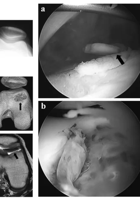
Fig. 2 Findings on arthroscopy. a Chondral fragment in the suprapa-tellar pouch (arrow). b Full-thickness chondral defect on the lateral femoral condyle.
component, and that the majority of patients with this condition sustained an indirect twisting injury. Oohashiet al. (8) reported a certain mechanism in a case that differed from the usual osteochon-dral fracture, whereby shear force was transmitted by the patella to the convex surface of the trochlea during rapid extension of the weight- bearing knee from a flexed position. Consequently, the cartilage of the trochlea was avulsed proximally. In our patient, the injury occurred while the knee was in the flexed position. Furthermore, the osteochondral fragment of the lateral femoral condyle was seen to be displaced distally on MRI. Therefore, in our patient, the fracture was caused by simultaneous twisting and compression forces. Chondral fracture of the trochlea has been reported to involve the lateral trochlea (6), potentially because of certain anatomical features. The lateral facet of the trochlea is larger and more prominent than the medial facet, so may be more easily impinged on by the patella.
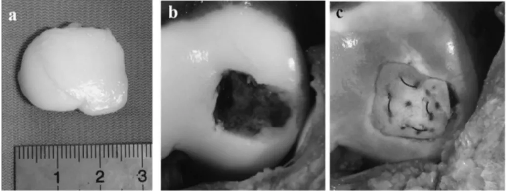
Most cases of chondral or osteochondral fracture are treated surgically. Depending on the size, condition, and location of the lesion, the appropriate method should be selected and should include reduction of the osteochondral fragment with internal fixation or excision and cartilage resurfacing (27). Kayaoglu and Binnet reported that internal fixation was the most effective method Fig. 3 Chondral defect and fragment. a Chondral fragment measuring 2.5 cm × 2 cm. b Macroscopic view shows the full - thickness chondral defect of the lateral femoral condyle and the surface of the bed covered with scar tissue. c The chondral fragment was trimmed to match the defect and fixed using the pull - out suture technique after curettage of the bed.
Fig. 4 Operative procedure for fixation using the pull - out suture technique.
Fig. 5 Follow - up fast spin - echo T2 - weighted magnetic resonance images of the lateral femoral condyle (sagittal view). Imaging studies at 2 years postoperatively showing that the articular surface remains smooth with no sign of delamination, suggesting successful healing.
for managing these fractures (29). ORIF is required in order to restore the congruity of the articular surface and to rigidly fix the fragment. Several reports have described use of a bioabsorbable implant to fix the osteochondral fragment with good clinical out-comes (9 - 14). There have also been several reports of articular cartilage damage with joint effusion and pain caused by pins that protruded from the bone or by catching of osteochondral frag-ments by the pins after fixation (30 , 31). Other methods used for fixation of osteochondral fractures have included drilling into the subchondral bone (1, 8, 15), bone peg fixation (21), fixation using compression screws (22), suture bridge fixation (16, 23), and use of fibrin adhesive (17). Recently, Song et al. (20) reported on three cases of osteochondral fractures of the lateral femoral condyle that were treated using three different methods : autologous bone pegs, headless screws, and transplantation of cultured chondro-cytes. The most favorable outcome was obtained using autologous bone pegs. In our case, we attempted pull - out fixation of the osteochondral fragment. This technique can anatomically reduce the fragment, compress the fragment into the subchondral bed, and provide enough fixation to allow immediate passive motion with comparatively less invasion of the articular cartilage. This method has the further advantages of being inexpensive, not needing removal of implants, and allowing for postoperative MRI to be performed without the presence of metal artifact. Several reports have described the use of suture fixation for acute osteo-chondral fractures (16, 23, 32 - 34).
Our patient was able to resume sports activity at his pre - injury level without knee pain, and follow - up imaging 2 years after sur-gery showed restoration of the congruity of the articular cartilage. This favorable outcome indicates that our method is a viable alternative to other accepted means of fixation for the treatment of osteochondral fragments in the knee. We will continue to follow up the patient to monitor the clinical outcomes in the long term.
CONFLICTS OF INTEREST
The authors declare that there is no conflict of interest regarding the publication of this article.
ACKNOWLEDGEMENTS
FundingThis report received no specific grant from any funding agency in the public, private, or not- for - profit sectors.
Ethical approval
All procedures performed were in accordance with the ethical standards of the institutional and/or national research committee and with the 1964 Helsinki Declaration and its later amendments or comparable ethical standards.
Informed consent
Informed consent was obtained from the parents of the patient presented in this report.
REFERENCES
1! Gilley JS, Gelman MI, Edson DM : Chondral fractures of the knee. Arthrographic, arthroscopic and clinical manifestations. Radiology 138 : 51 - 54, 1981
2! Hopkinson WJ, Mitchell WA, Curl WW : Chondral fractures of the knee : cause for confusion. Am J Sports Med 13 : 309
-312, 1985
3! Terry GC, Flandy F, Van Manen JW, Norwood LA : Isolated chondral fractures of the knee. Clin Orthop Relat Res 234 : 170 -177, 1988
4! Kennedy JC, Grainger RW, McGraw RW : Osteochondral fractures of the femoral condyles. J Bone Joint Surg Br 48 : 436 - 440, 1966
5! Huegli RW, Moelleken SM, Stoork A, Bonel HM, Bredella MA, Meckel S, Genant HK, Tirman PF : MR imaging of post-traumatic articular cartilage injuries confined to the femoral trochlea. Arthroscopic correlation and clinical significance. Eur J Radiol 53 : 90 - 95, 2005
6! Dory MA : Chondral fracture of the anterior intercondylar groove of the femur. Clin Rheumatol 2 : 175 - 177, 1983 7! Nakamura N, Horibe S, Iwahashi T, Kawano K, Shino K,
Yoshikawa H : Healing of a chondral fragment of the knee in an adolescent after internal fixation. A case report. J Bone Joint Surg Am 86 : 2741 - 2746, 2004
8! Oohashi Y, Oohashi Y : Chondral fracture of the lateral trochlea of the femur occurring in an adolescent : mechanism of injury. Arch Orthop Trauma Surg 127 : 791 - 794, 2007
9! Uchida R, Toritsuka Y, Yoneda K, Hamada M, Ohzono K, Horibe S : Chondral fragment of the lateral femoral trochlea of the knee in adolescents. Knee 19 : 719 - 723, 2012
10!Walsh SJ, Boyle MJ, Morganti V : Large osteochondral frac-tures of the lateral femoral condyle in the adolescent : outcome of bioabsorbable pin fixation. J Bone Joint Surg Am 90 : 1473 -1478, 2008
11!Matsusue Y, Nakamura T, Suzuki S, Iwasaki R : Biodegradable pin fixation of osteochondral fragments of the knee. Clin Orthop Relat Res 322 : 166 - 173, 1996
12!Braune C, Rehart S, Kerchbaumer F, Jäger A : Resorbable pin refixation of an osteochondral fracture of the lateral femoral condyle due to traumatic patellar dislocation : case manage-ment, follow - up and strategy in adolescent. Z Orthop Ihre Grenzgeb 142 : 103 - 108, 2004
13!Jehan S, Loeffler MD, Pervez H : Osteochondral fracture of the lateral femoral condyle involving the entire weight bearing articular surface fixed with biodegradable screws. J Pak Med Assoc 60 : 400 - 401, 2010
14!Mashoof AA, Scholl MD, Lahav A : Osteochondral injury to the mid - lateral weight- bearing portion of the lateral femoral condyle associated with patella dislocation. Arthroscopy 21 : 228 - 232, 2005
15!Jonson-Nurse C, Dandy DJ : Fracture-separation of articular cartilage in the adult knee. J Bone Joint Surg 67B : 42 - 43, 1985 16!Bowers AL, Huffman GR : Suture bridge fixation of a femoral condyle traumatic osteochondral defect. Clin Orthop Relat Res 466 : 2276 - 2281, 2008
17!Kaplonyi G, Zimmerman I, Frenyo AD, Farkas T, Nemes G : The use of fibrin adhesive in the repair of chondral and osteo-chondral injuries. Injury 19 : 267 - 272, 1988
18!Morelli M, Nagamori J, Miniaci A : Management of chondral injuries of the knee by osteochondral autogenous transfer (mosaicplasty). J Knee Surg 15 : 185 - 190, 2002
19!King PJ, Bryant T, Minas T : Autologous chondrocyte im-plantation for chondral defects of the knee : indications and technique. J Knee Surg 15 : 177 - 184, 2002
20!Song KS, Min BW, Bae KC, Cho CH, Lee SW : Chondral fracture of the lateral femoral condyle in children with different treatment methods. J Pediatr Orthop B 25 : 43 - 47, 2016 21!Nakayama H, Yoshiya S : Bone peg fixation of a large chondral
fragment in the weight- bearing portion of the lateral femoral condyle in an adolescent : a case report. J Med Case Rep 8 : 316, 2014
Rajaratnam S : Osteochondral fractures of the knee in skeletally immature patients : short term results of operative fixation using Omnitech screws. Acta Orthop Belg 82 : 762 - 767, 2016 23!Ng WM, Al-Fayyadh MZM, Kho J, Seow Hui T, Mohamed MRB : Crossing suture technique for the osteochondral frac-tures repair of patella. Arthrosc Tech 6 : 1035 - 1039, 2017 24!Makin M : Osteochondral fracture of the lateral femoral
con-dyle. J Bone Joint Surg Am 33 : 262 - 264, 1951
25!Rosenberg NJ : Osteochondral fractures of the lateral femoral condyle. J Bone Joint Surg Am 46 : 1013 - 1026, 1964 26!Matthewson MH, Dandy DJ : Osteochondral fractures of the
lateral femoral condyle : a result of indirect violence to the knee. J Bone Joint Surg Br 60 : 199 - 202, 1978
27!Kramer DE, Pace JL : Acute traumatic and sports-related osteochondral injury of the pediatric knee. Orthop Clin North Am 43 : 227 - 236, 2012
28!Milgram JW, Rogers LF, Miller JW : Osteochondral fractures : mechanism of injury and fate of fragments. Am J Roentgenol
130 : 651 - 658, 1978
29!Kayaoglu EE, Binnet MS : Chondral and osteochondral frac-tures. Acta Orthop Traumatol Turc 41 : 105 - 112, 2007 30!Friederichs MG, Greis PE, Burks RT : Pitfalls associated with
fixation of osteochondritis dissecans fragments using bioab-sorbable screws. Arthroscopy 17 : 542 - 545, 2001
31!Scioscia TN, Giffin JR, Allen CR : Potential complication of bioabsorbable screw fixation for osteochondritis dissecans of the knee. Arthroscopy 17 : E7, 2001
32!Dhawan A, Hospodar PP : Suture fixation as a treatment for acute traumatic osteochondral lesions. Arthroscopy 15 : 307 -311, 1999
33!Pritsch M, Velkes S, Levy O, Greental A : Suture fixation of osteochondral fractures of the patella. J Bone Joint Surg Br 77 : 154 - 155, 1995
34!Sodl JF, Ricchetti ET, Huffman GR : Acute osteochondral shear fracture of the capitellum in a 12-year-old patient : a case report. J Bone Joint Surg Am 90 : 629 - 633, 2008