IRUCAA@TDC : A 5-year activity report from the Oral Cancer Center, Tokyo Dental College
10
0
0
全文
(2) Bull Tokyo Dent Coll (2013) 54(4): 265–273. Clinical Report. A 5-year Activity Report from the Oral Cancer Center, Tokyo Dental College Nobuharu Yamamoto, Kazumichi Sato, Tomohiro Yamauchi, Taiki Suzuki, Ryuta Osaka, Mira Kin, Yoshifumi Yoshida, Sunaki Noguchi, Ken Ishizaki*, Masayuki Takano, Akira Katakura, Yoichi Tanaka**, Takahiko Shibahara and Nobuo Takano Oral Cancer Center, Tokyo Dental College, 5-11-13 Sugano, Ichikawa, Chiba 272-8513, Japan * Department of Removable Prosthodontics and Gerodontology, Tokyo Dental College, 1-2-2 Masago, Mihama-ku, Chiba 261-8502, Japan ** Division of Surgical Pathology, Clinical Laboratory, Ichikawa General Hospital, Tokyo Dental College, 5-11-13 Sugano, Ichikawa, Chiba 272-8513, Japan Received 24 May, 2013/Accepted for publication 3 July, 2013. Abstract The Tokyo Dental College Oral Cancer Center was established on April 1st, 2006 at our Ichikawa General Hospital for the purpose of providing multimodal treatment for oral cancer. This report summarizes the Center’s activities over the last 5 years. The total number of oral cancer patients treated was 360 (April 2006 to March 2011), with 205 primary cases. We investigated the following treatment-related items: 1) site, 2) age, 3) sex, 4) pathological examination, 5) staging, 6) systemic disorder, 7) double cancer, 8) treatment, and 9) prognosis. Out of 205 patients, 60% were men and 40% were women. Men in their 60s and women in their 80s were seen the most. The most common site was the tongue, at 42%, followed by the mandibular gingiva, maxillary gingiva, oral floor, and buccal mucosa. Squamous cell carcinomas were seen most frequently, at 94% (15% were stage I, 33% stage II, 15% stage III, and 34% stage IV). The most common treatment method was surgical treatment, at 83%. The 5-year survival rate at all stages was 85.4%. At the Oral Cancer Center, oral surgeons take the initiative in establishing treatment in cooperation with other departments and branches. Since the establishment of the Ambulatory Center for Maxillary Prosthetics in October 2011, 26 patients have undergone treatment. Related departments and branches work in teams, enabling comprehensive treatment, from the preoperative state to postoperative functional recovery. We wish to use these strengths to improve oral cancer treatment in Japan and will continue to work toward providing the best possible care for our patients. Key words:. Oral cancer — Clinical statistics — Oral Cancer Center. 265.
(3) 266. Yamamoto N et al.. Fig. 1 Therapy algorithm of our center. Introduction The Tokyo Dental College Oral Cancer Center was established on April 1st, 2006, at our Ichikawa General Hospital, and was the first center of its kind at a general hospital in Japan. The Ichikawa General Hospital was newly-built and relocated in July 1992, 66 years after its establishment in 1946. Today, it comprises 21 Departments, 6 Centers, including the Oral Cancer Center, and 570 beds, and serves as a mid-scale regional foundation hospital. Here, we report the results of treatments carried out at the Oral Cancer Center over the 5 years since its establishment, and describe its current activities and future goals.. Patients and Methods A total of 360 patients visited the Oral. ancer Center in the 5 years between April, C 2006 and March, 2011. We reviewed these cases, focusing on the following items: 1) site, 2) age, 3) sex, 4) pathological examination, 5) staging, 6) systemic disorder, 7) double cancer, 8) treatment, and 9) prognosis. Maxillary prosthetic cases seen at the Ambulatory Center for Maxillary Prosthetics, established in October 2011, were also included. The treatments available at the Center are similar to the standard treatments offered at other multiple facilities. Where radical surgery is selected, postoperative adjuvant chemo radiation therapy (3-weekly CDDP+RT) is performed when any of the following highrisk recurrence factors is found: 1) positive microscopic margin, 2) extra-nodal invasion, or 3) multiple cervical lymph node metastases (three or more). In addition, preoperative chemotherapy is performed for tumors rated T2 or above. S-1 or TPF (TXT+CDDP+5FU) is used in cases with excessive peplomycin..
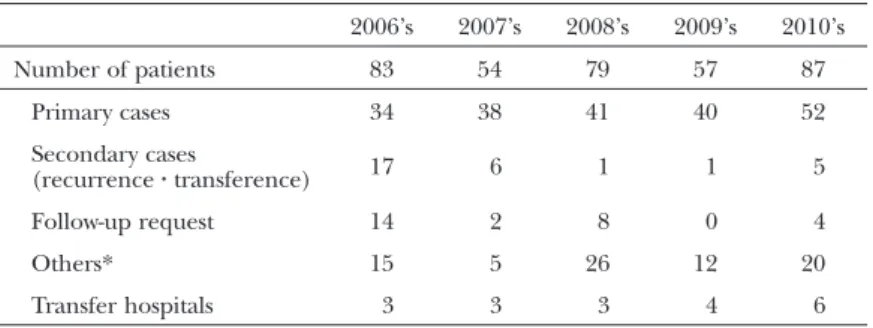
(4) 267. Activity Report from the Oral Cancer Center. Table 1 Number of patients (April 2006–March 2011: 360 cases). Number of patients. 2006’s. 2007’s. 2008’s. 2009’s. 2010’s. 83. 54. 79. 57. 87. Primary cases. 34. 38. 41. 40. 52. Secondary cases (recurrence z transference). 17. 6. 1. 1. 5. Follow-up request. 14. 2. 8. 0. 4. Others*. 15. 5. 26. 12. 20. Transfer hospitals. 3. 3. 3. 4. 6. *: Radiation osteomyelitis, HPV examination request, oral care, etc.. As postoperative chemotherapy, only S-1 or TPF (TXT+CDDP+5-FU) is administered, depending on the general condition of the patient. Figure 1 shows a schematic of the treatment algorithm applied. The TNM classification follows the 2009 UICC Classification11). The classification of cervical lymph nodes follows the General Rules for Clinical and Pathological Studies on Oral Cancer issued by the Japan Society for Oral Tumors4). The cumulative survival rate was calculated from data collected at the end of August, 2012.. Results. Fig. 2 Percentage of site of occurrence (April 2006– March 2011: Primary 205 cases). 1. Patients Of a total of 360 patients, 205 (57%) were primary cases, 30 (8%) were secondary cases (recurrence and metastasis), 28 (8%) were follow-up requests, 19 (5%) transferred to another hospital, and 78 (22%) were other cases (e.g., radiation osteomyelitis, HPV examination request, oral care, etc.) (Table 1).. 3. Age and sex A total of 123 of the patients (60%) were men and 82 (40%) were women; the male-tofemale ratio was 3:2, with more male cases. The age range was from 24 to 95 years, with an average age of 68 years. Men in their 60s and women in their 80s were seen the most (Fig. 3).. 2. Site The tongue was the most frequent site with 86 cases (42%), followed by 52 (25.4%) in the mandibular gingiva, 21 (10.2%) in the maxillary gingiva, 21 (10.2%) in the oral floor, 12 (5.9%) in the buccal mucosa, 5 (2.4%) in the lip, 5 (2.4%) in the mandible, and 3 (1.5%) in the palate (Fig. 2).. 4. Pathological diagnosis Histopathologic type was as follows: 192 cases (94%) of squamous cell carcinoma; 6 of carcinoma in situ; 5 of papillary squamous cell carcinoma; 2 each of verrucous carcinoma, adenoid cystic carcinoma, and malignant fibrous histiocytoma; and 1 each of spindle cell carcinoma, polymorphous low malignant.
(5) 268. Yamamoto N et al.. Fig. 3 Age, Gender (April 2006–March 2011: Primary 205 cases). Table 2 Classification of histopathology (April 2006–March 2011: Primary 205 cases) Squamous cell carcinoma. 192 cases (94%). Adenoid cystic carcinoma. Well-differentiated type. 140 cases. Polymorphous low grade adenocarcinoma 1 case. Moderately differentiated type. 20 cases. Basal cell carcinoma. 1 case. Poorly differentiated type. 6 cases. Malignant melanoma. 1 case. Differentiated type unknown. 12 cases. Carcinoma in situ. 6 cases. Papillary squamous cell carcinoma 5 cases Verrucous carcinoma. 2 cases. Spindle cell carcinoma. 1 case. 2 cases. Osteosarcoma. 1 case. Malignant fibrous histiocytoma. 2 cases. Metastatic tumor (Breast cancer). 1 case. Unidentified. 4 cases. adenocarcinoma, basal cell carcinoma, malignant melanoma, osteosarcoma, and metastatic tumor (breast cancer); four were unidentified (Table 2). 5. Cancer stage Six cases (3.1%) were stage 0, 29 (15.1%) were stage I, 64 (33.3%) were stage II, 28 (14.6%) were stage III, and 65 (33.9%) were stage IV, showing a similar ratio of early-stage and advanced-stage cancer. The staging percentage of each site is shown in Fig. 4. 6. Systemic disorder Regarding systemic disorders, 110 cases (54%) were cardiovascular, taking up over. Fig. 4 Percentage of different stages of squamous cells carcinoma (April 2006–March 2011: Primary 192 cases).
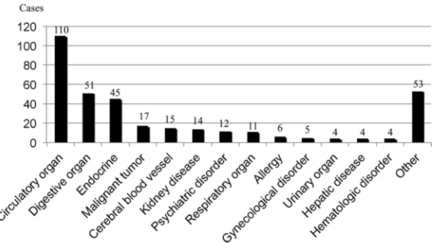
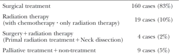
(6) Activity Report from the Oral Cancer Center. 269. Fig. 5 Systemic disease (April 2006–March 2011: Primary 205 cases). Table 3 Cases of double cancers (April 2006–March 2011: Primary 205 cases) Double cancer. 22/205 Cases (10.7%). Gastric cancer. 7. Esophagus cancer. 5. Lung cancer. 3. Breast cancer. 3. Colon cancer. 1. Bladder cancer. 1. Kidney cancer. 1. Non-Hodgkin’s lymphoma. 1. half of all cases. Fifty-one (25%) were digestive system disorders, 45 (22%) were endocrine system disorders, 17 (8%) were malignant tumors, 15 (7%) were cerebrovascular disorders, 14 (7%) were kidney disorders, 12 (6%) were psychiatric disorders, 11 (5%) were respiratory disorders, 6 (3%) were allergic disorders, 5 (2%) were gynecological disorders, 4 (2%) each were urologic disorders, liver disorder, or hematologic disorders, and 53 (26%) were other disorders (e.g., osteoporosis, rheumatism, etc.) (Fig. 5). 7. Double cancers At this center, PET-CT and upper/lower gastrointestinal endoscopy are used to detect. double cancers. Double cancers were seen in 22 (10.7%) out of 205 primary cases. Gastric cancer was observed most frequently with 7 cases, followed by 5 of esophageal carcinoma, 3 of lung cancer, 3 of breast cancer, and 1 each of colorectal cancer, bladder cancer, kidney cancer, and non-Hodgkin lymphoma. Triple cancer (gastric cancer and lung cancer) was seen in 1 case (Table 3). 8. Treatment The treatment methods used for the 192 primary squamous cell carcinoma cases were as follows: 160 patients (83%) underwent surgical treatment, 19 (10%) underwent radiation treatment (chemotherapy in combination with radiation), 4 (2%) underwent surgery plus radiation therapy (primal radiation treatment plus neck dissection), and 9 (5%) underwent palliative treatment plus nontreatment (Table 4). 9. Prognosis The prognosis in the 160 primary squamous cell carcinoma patients who underwent sur gical treatment was as follows: 138 patients (86%) survived and 22 (14%) died. The 5-year survival rate in the 160 patients was 85.4%. The 5-year survival rate by stage was as follows: 100% in stage 0, 96.2% in stage I, 87.3% in stage II, 86.5% in stage III, and 72.7% in stage IV (Fig. 6)..
(7) 270. Yamamoto N et al.. Table 4 Treatments (April 2006–March 2011: 192 primary squamous cells carcinoma cases) Surgical treatment. 160 cases (83%). Radiation therapy (with chemotherapy z only radiation therapy). 19 cases (10%). Surgery+radiation therapy (Primal radiation treatment+Neck dissection). 4 cases (2%). Palliative treatment+non-treatment. 9 cases (5%). Fig. 6 Survival rates of 5 stages (April 2006–March 2011: 160 cases of primary operation in squamous cell carcinoma). 10. Maxillary prosthetics cases Of the 26 patients that underwent maxillary prosthetic treatment, 14 were men and 12 were women. The ages ranged between 57 and 89 years, with an average age of 73 years. Site was as follows: the mandibular gingiva in 9 cases (34.6%); the maxillary gingiva in 9 (34.6%); the tongue in 3 (11.5%); the oral floor in 3 (11.5%); and the buccal mucosa and mandible in 1 each (3.9% and 3.9%, respectively).. Discussion The mission of the Oral Cancer Center is to provide a safe, high-quality medical service. (Fig. 7) for oral cancer patients. According to the Ministry of Health, Labour and Welfare, the Grants-in-Aid for Cancer Research “Regional Cancer Registration” Research Group (1975– 1999), and Health and Labour Sciences Research Grant for Third-Term Comprehensive Control Research for Cancer “Cancer Morbidity and Death Trend Survey” (from 2000–), for every 100,000 people, the morbidity rate of oral and pharyngeal cancers was 2.7– 5.3 people (average of 4 people), which is 1/17 the rate of gastric cancer and 1/9 that of colorectal cancers12). The number of oral cancer patients in Japan was only 2,100 in 1975, but increased to 6,900 in 2005, and is expected to grow to 7,800 by 20155). Although this represents only 1–2% of all cancers, it.
(8) Activity Report from the Oral Cancer Center. 271. Fig. 7 From early diagnosis in our center to rehabilitation after the treatment. represents 40% of head and neck cancers, indicating its great importance in head and neck cancer treatment8,9). The tongue was the most common site, accounting for 40–60% of all oral cancers1,3,6,14), followed by the gingiva, accounting for 20–30%1,3,6). The morbidity age ranged from younger to older age groups, but was seen most frequently in 50- to 60-year-olds2,7,8,14), with an average age of 63.78) and median age of 59 years7). The number of elderly patients is increasing due to today’s aging society. There were twice the number of male to female patients2,8), with men in the lower 60% and women in the higher 30%7,14). Pathological examination revealed that the majority had squamous cell carcinomas, ranging from 80–90% in previous reports3,6,10,14). The distribution of site of oral cancer was as follows: the tongue was the most frequent site, with 42%, followed by 25% in the man-. dibular gingiva, 10% in the maxillary gingiva, and 10% in the oral floor, which is similar to patterns observed in earlier reports1,3,6,14). Sixty per cent of the patients were men and 40% women, with a male-to-female ratio of 3:2, showing an increase in the number of female patients. The average age was 68 years, which was 5 years older than in the past. Moreover, as the age of the patients increased, more concomitant systemic disorders were observed, such as those of the cardiovascular (hypertension), digestive, and endocrine (diabetes) systems. This is due to improvements in the diagnostic accuracy of clinical exami nation, such as diagnostic imaging, which increases the possibility of detecting under lying disorder(s) not found at preoperative examination. In most epidemiological studies on oral cancer, stage II is observed most frequently1,3,7,14) with stage I and II together accounting for.
(9) 272. Yamamoto N et al.. 50–70%1,3,7,14). On the other hand, some facilities deal mostly with stage IV 6,10). A unique aspect of the stage distribution observed at our center was that stage II and IV showed the same percentage. This was because the majority of patients treated at our center had developed a concomitant systemic disorder, with such patients being referred to our center, regardless of cancer stage, including those with early- or progressive stage disease. The high frequency of 34% in stage IV was problematic. Although Tokyo Dental College has been actively holding oral cancer examinations with regional dental associations for over 20 years, the number of higher stage cases has not decreased. We believe that informing the general public of the availability and importance of undergoing such examinations remains as an important challenge for the future. Recent years have seen an increase in the frequency of multiple primary cancers in oral cancer patients13). The present results showed that 10.7% of the patients developed double cancer, and 1 case of triple cancer was also observed. Most of the multiple cancers at our center are found at an early stage, during preoperative examination or followup. Because the majority of those cancers are resectable, the importance of close exami nation of pre- and post-operative local and systemic images is clear. A pathological classification of squamous cell carcinoma was observed in over 90% of the cases reviewed, indicating the importance of radiation therapy. However, because oral cancer is prone to develop into cervical lymph node metastases, surgery is the primary method of treatment13). The prognosis of each stage was as follows: the 5-year survival rate was 70–90% in stage I1,6,7,10,14), 60–80% in stage II1,6,7,10,14), 50–90% in stage III6,7,10,14), and 0–40% in stage IV1,6,7,10,14). The total percentage of the 5-year survival rate at our center showed a good result, with 85.4%. The 5-year survival rate in each stage tended to decrease as the staging became higher. This suggests the importance of early-stage detection. The survival rate in progressive-stage cancers showed. a positive result, at 86.5% in stage III and 72.7% in stage IV, compared to that seen in previous reports1,6,7,10,14). This result supports the validity of our policy of treating such cancers aggressively by surgical means and applying post-operative radiation therapy whenever appropriate. One special feature of our center is that both pathological and radio-diagnoses are obtained. Prior to treatment, we always consult closely with specialists in oral pathology and radiology. Evaluation of oral function, focusing on eating, swallowing, and lingual function, is also conducted. Endoscopy at our center is used to evaluate eating and swallowing function in the pre- and postoperative state. To recover chewing function, implants are used with the cooperation of the Ambulatory Center for Maxillary Prosthetics. Furthermore, pre-operative palliative care along with pain control and nutritional guidance by the Nutrition Support Team is also provided, implementing perioperative patient care by making the best use of the kind of facilities that such a general hospital is able to offer. In 2008, the Oral Cancer Center was designated a central hospital for local cancer treatment by the Ministry of Health, Labour and Welfare after receiving a high evaluation. The majority of oral cancer cases are treated by oral surgeons. This is apparent from the statistics, as well as from surveys by the mass media. Post-operative reconstruction is not limited to morphologic appearance; the recovery of oral function is also considered important, something in which dentists play an important role. However, oral cancers cause not only cervical lymph node metastases, but also distant metastases such as those to the lung, sometimes causing systemic symptoms during follow-up. This means that the diagnosis and treatment of oral cancer cannot be dealt with solely by an oral surgeon. With these issues in mind, working in conjunction with other departments and branches is indispensable for oral surgeons in providing proper treatment. The Oral Cancer Center is a wellequipped, far-sighted facility that has become.
(10) Activity Report from the Oral Cancer Center. a front-line Oral Cancer Treatment Center. We wish to further improve our performance with the aim of becoming an exemplary facility for oral cancer treatment.. Conclusion We have reported a 5-year summary of our activities (April 2006 to March 2011) at the Oral Cancer Center, Tokyo Dental College.. Acknowledgements The authors would like to thank Dr. Elizabeth Hearing, DASS Manuscript, for her assistance with the English of this manuscript. A summary of this report was presented at the 57th Japanese Society of Oral and Maxillofacial Surgeons Meeting (October 20, 2012, in Yokohama), the 294th Tokyo Dental College Association Meeting (October 20, 2012, in Chiba), the 22nd Japanese Association for Dental Science Meeting (November 10, 2012, in Osaka), and the 31st Japan Society for Oral Tumors Meeting ( January 25, 2013, in Tokyo).. References 1) Aoki S, Kawabe R, Chikumaru H, Hirota M, Miyake T, Omura S, Fujita K (2003) Ten-year clinico-statistical study of oral squamous cell carcinoma. Yokohama Igaku 54:539–546. (in Japanese) 2) Hasegawa Y, Takezaki T (2000) Epidemiology of tongue cancer. JOHNS 16:549–553. (in Japanese) 3) Japan Society for Head and Neck Cancer Registry Committee (2006) Report of head and neck cancer registry of Japan clinical statistics of registered patients, 2002. Jpn J Head Neck Cancer 32 (Supple):1–98. 4) Japan Society for Oral Tumors (Eds) (2010) General Rules for Clinical and Pathological Studies on Oral Cancer, 1st ed., pp.34, Kanehara Shuppan, Tokyo. (in Japanese) 5) Japan Society for Oral Tumors, Japanese. 273. Society of Oral and Maxillofacial Surgeons (Eds) (2009) Evidence-Based Oral Cancer Treatment Guideline, 2009 ed., pp.11–13, Kanehara Shuppan, Tokyo. (in Japanese) 6) Koike T, Kurita H, Ohtsuka A, Narikawa J, Nakatsuka A, Kojima Y, Fujimori S, Kobayashi H, Kurashina K (2003) Clinico-statistical studies of primary malignant tumors in the oral and maxillofacial regions. Shinshu Med J 51:15– 23. (in Japanese) 7) Matsuura K, Hayashi R, Ebihara S, Saikawa M, Yamazaki M, Monden N, Seino Y, Kimata Y, Sakuraba M, Hishinuma S (2004) Result of surgical treatment for squamous cell carcinoma of tongue. Head and Neck Cancer 30:550– 557. (in Japanese) 8) Miyahara H (2004) Primer of Diagnosis and Treatment for Malignant Disease in Otolaryngology, Head and Neck Surgery, pp.175–197, Tokyo Igakusha, Tokyo. (in Japanese) 9) Sato K, Tomita M, Watanabe J, Matsuyama H, Takahashi S (2007) Clinical analysis of oral carcinoma treated in the Department of Otolaryngology, Niigata University Hospital. Head and Neck Cancer 33:439–443. (in Japanese) 10) Shibayama M, Hanamitsu M, Sonoda S, Shimizu T (2006) Treatment of tongue cancer. Pract Otol 99:743–749. (in Japanese) 11) Sobin LH, Gospodarowicz MK, Wittekind Ch, International Union against Cancer (2009) TNM Classification of Malignant Tumours, 7th ed., pp.25–29, UICC, Geneva. 12) The Japan Cancer Surveillance Research Group (2006) Cancer incidence and incidence rates in Japan in 2000: estimates based on data from 11 population-based cancer registries. Jpn J Clin Oncol 36:668–675. 13) Yamamoto N, Okubo T, Sakuma T, Sugahara K, Yamamoto M, Muramatsu K, Watanabe A, Yakushiji T, Nomura T, Takano N, Shibahara T (2012) Clinical study of multiple primary and double cancers including oral squamous cell carcinoma. J Oral Maxillofac Sur Med Path 24:189–194. 14) Yuta A, Kishioka C, Tatematsu M, Majima Y (2002) Clinical review of oral cancers. Pract Otol 95:933–937. (in Japanese) Reprint requests to: Dr. Nobuharu Yamamoto Department of Oral and Maxillofacial Surgery, Tokyo Dental College, 1-2-2 Masago, Mihama-ku, Chiba 261-8502, Japan E-mail: nyamamot@tdc.ac.jp.
(11)
図


+3

関連したドキュメント
Treatment with CH11 caused a relocalization of the 681 antigen: signals were no longer detectable in the cell nucleus, and instead cell bodies, in particular the region near
The objectives of this study were to evaluate the formation of lymphvascular niches in lymph nodes of patients with oral squamous cell carcinoma (OSCC), and investigate the roles
In conclusion, IFN-α alternation therapy is one treatment option for mRCC patients in whom first- line IFN-α treatment failed if the patient has only lung or
We have studied the effects of different treatment regimens on both the tumour growth and the immune response within the simple ODE model that describes tumour-immune dynamics
The analysis presented in this article has been motivated by numerical studies obtained by the model both for the case of curve dynamics in the plane (see [8], and [10]), and for
Then the center-valued Atiyah conjecture is true for all elementary amenable extensions of pure braid groups, of right-angled Artin groups, of prim- itive link groups, of
The explicit treatment of the metaplectic representa- tion requires various methods from analysis and geometry, in addition to the algebraic methods; and it is our aim in a series
・Mozaffari E, et al. Remdesivir treatment in hospitalized patients with COVID-19: a comparative analysis of in- hospital all-cause mortality in a large multi-center
