〈
Brief Report
〉
Microfloral analysis of pediatric respiratory syncytial
virus infection, with and without the use of antibiotics
Koo Nagasawa1,2, Kenichi Takeshita3, Hatsumi Taniguchi4, Hiroshi Suzuki3, Sachiko Naito1, Haruka Hishiki1, Tadashi Hoshino5, Hirokazu Kimura2,6, Kazuo Suzuki7,
Naoki Shimojo1 and Naruhiko Ishiwada8
1 Department of Pediatrics, Graduate School of Medicine, Chiba University 2 Infectious Disease Surveillance Center, National Institute of Infectious Diseases
3 Department of Pediatrics, Chiba Rosai Hospital
4 Department of Microbiology, University of Occupational and Environmental Health 5 Division of Infectious Diseases, Chiba Children s Hospital
6 School of Medical Technology, Faculty of Health Science, Gunma Paz University 7 Asia International Institute of Infectious Diseases Control, Teikyo University 8 Department of Infectious Diseases, Medical Mycology Research Center, Chiba University
(Received for publication October 29, 2017)
Objectives To describe, in detail, the influence of antibiotics on the microflora of children with respiratory syncytial virus (RSV) infection.
Methods Eight hospitalized infants with RSV infection were included in the study. To examine the change in the microflora before and after the use of antibiotics, we simultaneously collected nasopharyngeal and stool swabs from each patient at 3 points; before antibiotics use, during antibiotics use, after use. The use of antibiotics was determined by clinicians. In 6 patients, antibiotics were used, and in 2, they were not. We analyzed the nasopharyngeal and fecal microflora through the clone library method, using amplified fragments of the 16S ribosomal RNA gene with universal primers.
Results With regards to nasopharyngeal microflora, of the 6 patients who were prescribed antibiotics, 4 had pathogenic bacteria including Haemophilus influenzae and Moraxella catarrhalis, on admission, and only 1 patient had M. catarrhalis 3–5 days after admission. However, in 3 patients, M. catarrhalis was the dominant bacterium, 1–2 weeks after hospital discharge. Among the patients who were not
*Corresponding author: Naruhiko Ishiwada, Department of Infectious Diseases, Medical Mycology Research Center, Chiba
University, 1–8–1 Inohana, Chuo-ku, Chiba-shi, Chiba 260–8677, Japan. Phone: +81–43–226–2799; Fax: +81–43–226–2786; E-mail: ishiwada@faculty.chiba-u.jp
prescribed antibiotics, the representative pathogens of childhood pneumonia were not observed in any of the patients 1–2 weeks after hospital discharge, despite them having those pathogens on admission or 3–5 days after. With regards to fecal microflora, the number of bacterial species decreased after antibiotics use, except in 1 patient. One to 2 weeks after hospital discharge, the number of bacterial species was lower than that observed on admission, in all the cases in which antibiotics were used.
Conclusions The number of bacterial species both in the nasopharyngeal and fecal samples decreased after antibiotic use. The change in the dominant bacteria of the nasopharynx may be different between the cases in which antibiotics were administered and those in which they were not. These results suggest that clinicians should administer antibiotics, taking into consideration their influence on patients microflora.
1. Introduction
Respiratory tract infections (RTIs) occur commonly in children. RTIs are induced by many microorganisms, and, in children, viruses are the major cause1). The respiratory syncytial virus (RSV) is one of the major causative viruses of RTI in children1). Generally, patients with RSV in-fection receive symptomatic treatment. If the patients are considered as having a bacterial co-in-fection, they are treated with antibiotics. However, it is not always easy to assess if patients have a bacterial co-infection, at the first medical examination. Moreover, the bacterial co-infection rates of RTIs, caused by RSV, vary, by study design1,2). Therefore, in the clinical setting, antibiot-ics are sometimes prescribed to children with RSV infection, even when they are not required.
The use of antibiotics has its own demerits. Diarrhea is one of the common side effects of antibiotics use. Although several previously conducted studies have suggested that the use of an-tibiotics influences microflora, those reports used bacterial cultures3–5). The technique involved in bacterial culture is relatively easy and can be conducted easily in hospitals. However, in bacterial cultures, non-culturable bacteria cannot be detected, and the percentage of each bacteria in a mi-croflora cannot be estimated. To overcome these drawbacks, a method using a sequencer and polymerase chain reaction (PCR) for 16S ribosomal RNA was developed6). To examine, in detail, the influence of antibiotics on the microflora of children with RSV infection, we analyzed naso-pharyngeal and fecal microflora simultaneously through this method, before and after antibiotics were used. In this study, we focused on the relationships between viral infection, use of antibiot-ics and microflora. To the best of our knowledge, there are no reports, till date, on how antibiotantibiot-ics change both nasopharyngeal and stool microflora, using this new molecular biological method. We aimed to estimate the change in the microflora of children with viral infection, and, thereby, guide the proper use of antibiotics in these patients.
2. Materials and Methods
2.1. ParticipantsThe patients in the present study were hospitalized infants with RSV infection, as diagnosed by rapid test kits (Chiba University: RSV examen™, Becton Dickinson and Company; Franklin Lakes, NJ. Chiba Rosai hospital and Chiba Children s hospital: QuickNavi™ RSV, DENKA SEIKEN Co., Ltd.: Tokyo, Japan), between September 2014 and March 2015, in Chiba Univer-sity Hospital, Chiba Rosai Hospital and Chiba Children s Hospital. We excluded patients in whom antibiotics were administered within 2 weeks from the date of hospital admission. We also excluded patients in whom the injection of palivizumab, an anti-RSV monoclonal antibody, was recommended. Written informed consent was obtained from the participants guardians.
2.2. Study protocol
To examine the change in the microflora, before and after the use of antibiotics, we collected nasopharyngeal and stool swabs, simultaneously, from each patient at 3 points; on admission (be-fore antibiotic use), 3–5 days after admission (during antibiotic use), and 1–2 weeks after dis-charge from the hospital (after antibiotic use). The use of antibiotics was determined by clini-cians. The severity of the condition, in each patient, was scored, as previously described7,8). The study protocols were approved by the Ethics Committee of the Chiba University (approval no. 1857), the Chiba Rosai hospital (approval no. 26–22), and the Chiba Children s hospital (ap-proval no. 2014-01-02). This study was carried out in accordance with the protocol.
2.3. Analysis of the nasopharyngeal and fecal microflora
We analyzed the nasopharyngeal and fecal microflora through the clone library method, using amplified fragments of the 16S ribosomal RNA gene with universal primers9–12). After bac-terial cell staining with ethidium bromide, we counted the total number of bacbac-terial cells in the sample13). DNA was extracted from the samples by adding 2 ml of sterilized water and vigorously shaking the samples with sodium dodecyl sulfate (final concentration: 3.0%) and glass beads9,11). Then, the total number of bacterial cells in the sample was counted again, and we evaluated the efficacy of cell lysis, as previously described9–12). PCR of 16S rRNA gene was subsequently per-formed, using universal primers, under the following conditions; 95°C for 5 minutes, 30 cycles at 94°C for 1 minute, 50°C for 1 minute, and 72°C for 2 minutes, followed by a final extension at 72°C for 7 minutes11,13). The PCR amplicons were cloned using a TOPO TA cloning kit (Invitro-gen; Carlsbad, CA). Of them, a total of 96 colonies were randomly selected for the sequencing analysis. The analysis was performed using a 3130xl Genetic Analyzer (Applied Biosystems), ac-cording to the instrument manual. A Blast search was used to determine the species of each clone library. These analyses were performed as soon as possible, after the samples were collected.
3. Results
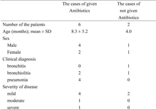
3.1. Patients characteristicsA total of 8 patients were assessed in this study; of them, antibiotics were used in 6 (ampicil-lin; 1 patient, ampicillin/sulbactam; 5 patients). As shown in Table 1, the age of the patients in whom antibiotics were used was 8.3±5.2 months (mean±standard deviation, SD), while that of the patients in whom antibiotics were not used was 4.0 months. The SD of the latter could not be calculated because of the small number. All the patients had a lower respiratory infection, such as bronchitis, bronchiolitis, and pneumonia. With regards to the severity of the RTIs, in this study, 6 out of the 8 patients were categorized as having a mild form of the infection, while 1 each were considered as having moderate and severe infection, respectively.
3.2. Total bacterial cell numbers and cell lysis
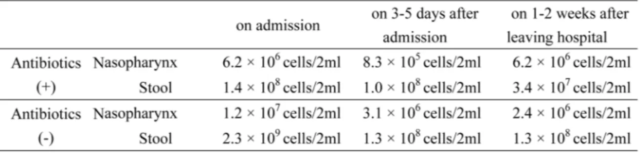
All the samples collected in the present study had the sufficient number of bacterial cells for analysis. The medians of the total bacterial cell number are shown in Table 2. A larger number of bacterial cells was found in the stool than the nasopharynx. Furthermore, the total number of bac-terial cells tended to decrease after admission, regardless of the use of antibiotics. The efficiency of cell lysis ranged from 74.5% to 99.9% (median 91.0%).
3.3. Nasopharyngeal microflora
We analyzed 22 nasopharyngeal swabs, including 8 samples on admission, 8 samples 3–5 days after admission, and 6 samples 1–2 weeks after hospital discharge. In the case of 2 patients, samples could not be collected at the time of hospital discharge, as 1 patient was transferred to another hospital and the other did not allow the collection of nasopharyngeal samples 1–2 weeks after discharge.
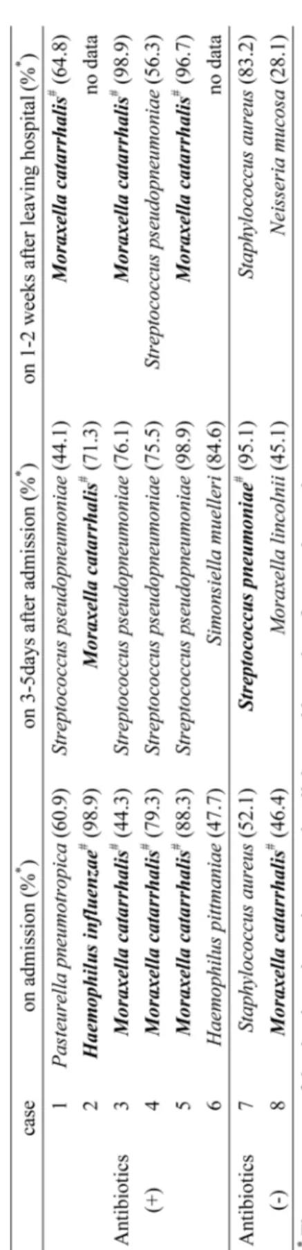
The number of nasopharyngeal bacterial species is shown in Fig. 1. As shown in Fig. 1, the number tended to decrease after the use of antibiotics, while the number of patients in whom anti-biotics were not used was either constant or tended to increase. Furthermore, we examined the change in the dominant bacteria, in each sample (Table 3). Of the 6 patients in whom antibiotics were prescribed, the representative pathogens of childhood pneumonia, including Haemophilus influenzae and Moraxella catarrhalis, were noted in 4 patients, on admission, while M. catarrha-lis was observed in 1 patient, 3–5 days after admission. However, in 3 patients, M. catarrhacatarrha-lis was the dominant bacterium 1–2 weeks after hospital discharge. However, among patients who were not prescribed antibiotics, none of the patients had the representative pathogens of
child-Table 2. The median of total bacterial cell numbers
Fig. 1. The number of nasopharyngeal bacterial species in a) the patients in whom antibiotics were used and b) patients in whom antibiotics were not used
Table 3.
hood pneumonia 1–2 weeks after hospital discharge, although they had those pathogens on ad-mission or 3–5 days after adad-mission.
3.4. Fecal microflora
Twenty-three stool samples, including 8 samples collected on admission, 8 samples 3–5 days after admission, and 7 samples 1–2 weeks after hospital discharge were analyzed. One sample could not be obtained due to hospital transfer. The number of bacterial species in each stool sam-ple is shown in Fig. 2. The number decreased after the use of antibiotics, except in the case of one patient (case 3). However, the number decreased in case 3 too, after hospital discharge. One to 2 weeks after hospital discharge, the number of bacterial cells was lower than that observed on ad-mission, in all the cases in which antibiotics were used. We could not find a tendency for the re-duction in the number of bacterial cells and species in the no antibiotics admission cases. Further-more, in the present study, we could not detect the dominant bacterium in the stool.
3.5. Correlations between nasopharyngeal and fecal microflora during the clinical course
Table 4 shows the number of bacterial species in the nasopharyngeal and fecal microflora, in the patients, during the clinical course. The number of bacterial species in both the nasopharynx and fecal microflora decreased in 4 of 6 patients treated with antibiotics. One to 2 weeks after an-tibiotic treatment, in 2 specimens—the nasopharynx specimen of case 4 and fecal specimen of case 6—were recovered number of bacterial species before antibiotic treatment.
Fig. 2. The number of fecal bacterial species in a) the patients in whom antibiotics were used and b) those in whom antibiotics were not used
Table 4.
Number
of bacterial species of nasopharyngeal and fecal micr
4. Discussion
In this study, we described the change in the microflora, in children with a viral infection. Our main findings are as follows: First, the number of bacterial species in both the nasopharyn-geal and fecal samples decreased after the use of antibiotics. Second, the change in the dominant bacteria of the nasopharynx may be different between cases in which antibiotics were adminis-tered and those in which they were not. Reports suggest that the divergence of the microflora in the stool decreased through antibiotic administration14,15). Furthermore, Brook et al and Konno et
al showed, through bacterial cultures, that antibiotics affected the nasopharyngeal microflora4,5). Those studies, however, did not examine the microflora of the upper respiratory tract, using mo-lecular biological methods. In contrast, we examined both nasopharyngeal and fecal microflora, simultaneously, using the clone library method. As a result, we could accurately evaluate fluctua-tions in the microflora, in the nasopharynx and stool, including bacteria that are hard to culture.
In the present study, we focused on patients with a viral infection. Hamano-Hasegawa et al. reported that 15.2% of pediatric patients with community-acquired pneumonia have bacterial co-infection, using the broad-range PCR method1). Hishiki et al. showed that 45% of patients with lower respiratory tract infection, caused by RSV—which is one of the most common causes of infection in children—have bacterial co-infection, through the washed sputum culture method2). In our study too, 5 of 8 patients had the pathogenic bacteria of pediatric pneumonia on admission, and another patient had Streptococcus pneumoniae 3–5 days after admission. However, in the pa-tients in whom antibiotics were not prescribed, those pathogenic bacteria were not detected 1–2 weeks after hospital discharge. Based on this result, it is suggested that not all detected patho-genic bacteria may be causative pathogens in patients with a viral infection; in fact, bacterial in-fections may be more over-diagnosed than expected.
In this study, the number of bacterial species decreased after the use of antibiotics, in the na-sopharyngeal swab samples and stool samples, while the number of bacterial species did not de-crease, in cases in which antibiotics were not administered. Many studies reported that the diver-gence of fecal microbiota decreased through the use of antibiotics14–17). The divergence of nasopharynx microbiota may also decrease through antibiotics use, with regular dosage.
The dominant bacterium of the nasopharynx differed, depending on the phase at which each sample was collected. Three to 5 days after admission, in the cases in which antibiotics were ad-ministered, the changes in the number of bacterial species could be attributed to the antibacterial activity of the antibiotics. After the use of antibiotics, pathogenic bacteria, including H. influenzae and M. catarrhalis, disappeared. However, M. catarrhalis was recovered 1–2 weeks after hospital discharge, in cases in which antibiotics were used. Brook et al suggested that potential pathogens including S. pneumoniae, H. influenzae, and M. catarrhalis rebound faster in those treated with amoxicillin/clavulanate compared to those in whom cefdinir was administered, although the
study used the bacterial culture method5). We used the clone library method, and, therefore, were able to evaluate a large number of bacteria, including those species that are difficult to culture. Our results were compatible with those of the study by Brook et al.5).
To estimate the pathogens in patients with RTI, at the initial diagnosis, may be difficult as the symptoms of RTI do not vary from those of other microorganisms. In addition, the results of bacterial culture, which is the standard for bacterial infection, are obtained only after a few days. A substantial number of instances of bacterial and viral co-infections have been reported1,2). Therefore, it can be assumed that antibiotics are used in several cases even when they are not re-quired. Some reports suggest that the use of antibiotics in infants may raise the risk of asthma18,19). It was thought that these demerits were caused by the change in the microflora18). Our results showed that antibiotics may change the microflora of both the nasopharynx and bowel.
Our study had some limitations. First, the number of patients in the present study was slightly small. Second, samples from two patients that could not be collected at all 3 points were also included in the present study. To confirm these effects of antibiotics on microflora, further studies are needed. Based on the results of the present study, clinicians are urged to use antibiot-ics taking into consideration their influence on microflora.
Acknowledgements
This work was supported by JSPS KAKENHI Grant Number G17K16288, and Special Re-search fund of Nurture of Creative ReRe-search Leaders in Immune System Regulation and Innova-tive Therapeutics Leading Graduate School at Chiba University. We would like to thank Editage (www.editage.jp) for English language editing.
Competing interests
The authors declare that they have no competing interests.
References
1) Hamano-Hasegawa K, Morozumi M, Nakayama E, et al.: Comprehensive detection of causative pathogens using real-time PCR to diagnose pediatric community-acquired pneumonia. J Infect Chemother. 2008; 14: 424–32.
2) Hishiki H, Ishiwada N, Fukasawa C, et al.: Incidence of bacterial coinfection with respiratory syncytial virus bronchopulmonary infection in pediatric inpatients. J Infect Chemother. 2011; 17: 87–90.
3) Savino F, Roana J, Mandras N, et al.: Faecal microbiota in breast-fed infants after antibiotic ther-apy. Acta Paediatr. 2011; 100: 75–8.
4) Konno M, Baba S, Mikawa H, et al.: Study of nasopharyngeal bacterial flora. Variations in naso-pharyngeal bacterial flora in schoolchildren and adults when administered antimicrobial agents. J
Infect Chemother. 2007; 13: 235–54.
5) Brook I, Gober AE: Long-term effects on the nasopharyngeal flora of children following antimi-crobial therapy of acute otitis media with cefdinir or amoxycillin-clavulanate. J Med Microbiol. 2005; 54: 553–6.
6) Woo PCY, Lau SKP, Teng JLL, et al.: Then and now: use of 16S rDNA gene sequencing for bac-terial identification and discovery of novel bacteria in clinical microbiology laboratories. Clin Mi-crobiol Infect. 2008; 14: 908–34.
7) McCallum GB, Morris PS, Wilson CC, et al.: Severity scoring systems: Are they internally valid, reliable and predictive of oxygen use in children with acute bronchiolitis? Pediatr Pulmonol. 2013; 48: 797–803.
8) Miyaji Y, Kobayashi M, Sugai K, et al.: Severity of respiratory signs and symptoms and virus profiles in Japanese children with acute respiratory illness. Microbiol Immunol. 2013; 57: 811–21. 9) Yamasaki K, Kawanami T, Yatera K, et al.: Significance of anaerobes and oral bacteria in
commu-nity-acquired pneumonia. PLoS ONE. 2013; 8: e63103.
10) Fukuda K, Yatera K, Ogawa M, et al.: An unclassified microorganism: Novel pathogen candidate lurking in human airways. PLoS ONE. 2014; 9: e103646.
11) Noguchi S, Mukae H, Kawanami T, et al.: Bacteriological assessment of healthcare-associated pneumonia using a clone library analysis. PLoS ONE. 2015; 10: e0124697.
12) Kawanami T, Yatera K, Yamasaki K, et al.: Clinical impact of methicillin-resistant staphylococcus aureus on bacterial pneumonia: Cultivation and 16S ribosomal RNA gene analysis of bronchoal-veolar lavage fluid. BMC Infect Dis. 2014; 16: 155.
13) Akiyama T, Miyamoto H, Fukuda K, et al.: Development of a novel PCR method to comprehen-sively analyze salivary bacterial flora and its application to patients with odontogenic infections. Oral Surg Oral Med Oral Pathol Oral Radiol Endod. 2010; 109: 669–76.
14) Koning CJ, Jonkers D, Smidt H, et al.: The effect of a multispecies probiotic on the composition of the faecal microbiota and bowel habits in chronic obstructive pulmonary disease patients treated with antibiotics. Br J Nutr. 2010; 103: 1452–60.
15) Ferrer M, Méndez-García C, Rojo D, et al.: Antibiotic use and microbiome function. Biochem Pharmacol. 2017; 134: 114–26.
16) Dubourg G, Lagier JC, Robert C, et al.: Culturomics and pyroseauencing evidence of the reduc-tion in gut microbiota diversity in patients with broad-spectrum antibiotics. Int J Antimicrob Agents. 2014; 44: 117–24.
17) Bacattini S, Taur Y, Pamer EG, et al.: Antibiotic-induced changes in the intestinal microbiota and disease. Trends Mol Med. 2016; 22: 458–78.
18) Hoskin-Parr L, Teyhan A, Blocker A, et al.: Antibiotic exposure in the first two years of life and development of asthma and other allergic diseases by 7.5 yr: A dose-dependent relationship. Pedi-atr Allergy Immunol. 2013; 24: 762–71.
19) Penders J, Kummeling I, Thijs C: Infant antibiotic use and wheeze and asthma risk: A systematic review and meta-analysis. Eur Respir J. 2011; 38: 295–302.