Title Studies on the Clinical Usefulness of Serum and GeneBiomarkers for Diagnosis of Bovine Leukosis( 本文(Fulltext) )
Author(s) Mohammad Monir Tawfeeq
Report No.(Doctoral Degree) 博士(獣医学) 甲第420号 Issue Date 2014-03-13 Type 博士論文 Version ETD URL http://hdl.handle.net/20.500.12099/49043 ※この資料の著作権は、各資料の著者・学協会・出版社等に帰属します。
I
Studies on the Clinical Usefulness of Serum and Gene Biomarkers for Diagnosis of Bovine Leukosis
2013
The United Graduate School of Veterinary Sciences, Gifu University (Obihiro University of Agriculture and Veterinary Medicine)
II Table of Contents Title page……….………. II Table of Contents ……….…II Abbreviations………... II General introduction………...1
Chapter 1. Utility of serum thymidine kinase activity measurements for detection of cases of bovine leukosis with difficult clinical diagnoses……….…...10
Chapter 2. The preliminary evaluation of gene expression as biomarkers in clinical cases of bovine leukosis……….22
Chapter 3. Evaluation of gene expression in peripheral blood cells as a potential biomarker for enzootic bovine leukosis……….49
Summary and conclusion..………...66
References …… ………..……….70
III ABBREVIATION
BL: bovine leukosis
EBL: enzootic bovine leukosis SBL: sporadic bovine leukosis BLV: bovine leukemia virus PL: persistent lymphocytosis AGID: agar gel immunodiffusion ELISA: enzyme immune sorbent assay PCR: polymerase chain reaction FNA: fine needle aspiration LDH: lactate dehydrogenase
RT-PCR: reverse transcription polymerase chain reaction
CYP1B1: cytochrome P450, family1- subfamily B- polypeptide 1
CDKN2A: cyclin-dependent kinase inhibitor 2A
IL2R: interleukin-2 receptor
WT1: wilms’ tumor 1
BCL2: B-cell leukemia/lymphoma protein 2
PDE7B: phosphodiesterase isoform 7B
TK1: thymidine kinase 1
MB1: immunoglobulin associated alpha-1
IV YKL-40: chitinase-3 like 1
1
Studies on the Clinical Usefulness of Serum and Gene Biomarkers for Clinical Diagnosis of Bovine Leukosis
GENERAL INTRODUCTION
Bovine leukosis (BL), a disease afflicting cows that was first reported by Leisering in 1871, is characterized by yellowish nodules within an enlarged spleen (Leisering 1871; Rodriguez et al. 2011). The well-defined clinical entity of bovine leukemia was described by Bollinger several years later, and by 1876, the first case of bovine lymphocytic malignancy had been reported (Siedamgrotzky et al. 1876; Rodriguez et al. 2011). In 1969, bovine leukemia virus (BLV), the etiological agent of bovine leukemia, was isolated in culture (Miller et al. 1969). BL is now one of the most commonly reported neoplastic diseases in cattle, having been detected in cattle worldwide and causing significant economic losses. In general, BL is categorized according to its epizootiology, etiology, clinicopathology, and clinical signs, into two types called: (1) enzootic bovine leukosis (EBL) and (2) sporadic bovine leukosis (SBL) (Radostits et al. 2007; Angelos and Thurmond 2008).
EBL is an infectious and contagious disease of cattle; the causative agent is BLV, which belongs to the genus Deltaretrovirus within the subfamily Orthoretrovirinae and the family Retroviridae (Jacobs et al. 2002; Radostits et al. 2007). The main natural hosts for BLV are domestic cattle; however, the existence of a wild reservoir remains controversial, and there is some evidence indicating that BLV may also persist in water buffaloes (Meas et al. 2000). Sero-epidemiological surveys conducted in different countries have indicated that EBL infection is widespread throughout all continents except Europe, and that the prevalence rates vary dramatically between countries (Rodriguez et al. 2011). In Japan, EBL is one of the most prominent diseases among
2
cattle, and the prevalence rate is increasing every year. According to animal hygiene statistics from the Ministry of Agriculture, Forestry, and Fisheries of Japan (MAFF 2008), 838 outbreaks of EBL in 677 farms were reported in 2007, while only 159 outbreaks in 157 farms were reported in 2000 (Animal Hygiene Weekly 2008). In this regard, it can be speculated that the number of infected cattle are increasing (Figure. 1), which highlights the importance of controlling this disease.
Although cattle can be infected with BLV at any age, BLV infection is typically seen in animals over three years of age (Radostits et al. 2007; Fry and McGavin 2007). Once BLV infects a herd, most cattle will be seropositive for BLV, although the great majority of animals infected are asymptomatic carriers of the virus (Ferrer 1979). In animals seropositive for BLV, neither clinical symptoms nor alteration of total lymphocyte count become evident, and these cattle may remain in a clinically-silent, aleukemic state for their entire lives. These carrier animals can only be identified serologically by tests for BLV antibodies or proviral DNA (Ferrer 1979). Among all seropositive cattle, about 29% may develop a benign proliferation of B cells called persistent lymphocytosis (PL). Despite hematological alterations, PL cattle do not develop any other clinical signs and may be stable for several years without any other clinical manifestations of BL (Radostits et al. 2007; Ferrer 1979). Less than 5% of cattle infected with BLV will develop B-cell lymphosarcoma, the last stage of BL that affects various lymph nodes and organs (Radostits et al. 2007; Pollari et al. 1992).
BLV can be transmitted either horizontally or iatrogenically via biological materials containing B lymphocytes, such as blood, milk, colostrum, or saliva. Insect vectors may also play a role in BLV transmission, as the exchange of infected cells is important for disease transmission. Additionally, cattle with PL appear to transmit the
3
virus more readily than those that are only positive for BLV antibodies (Jacobs et al. 2002).
Tumor-harboring cattle generally show clinical signs of the disease, such as loss of condition, decrease in milk production, enlargement of superficial and internal lymph nodes, anorexia, cardiac lesions, constipation, fever, posterior paresis, and exophthalmos (Angelos and Thurmond 2008; Jacobs et al. 2002; Radostits et al. 2007). Tumors in the alimentary tract often cause symptoms of vagus indigestion, which interfere with rumen motility and cause abomasal dilution, melena, and diarrhea. In some clinical cases (peracute) sudden death will occur due to rupture of the abomasum or spleen, which can cause severe internal hemorrhage (Jacobs et al. 2002). In addition, atypical targets such as the upper and lower respiratory tract, udder, kidney, ureter, liver, spleen, or bone marrow may be affected (Simon et al. 2008). Figure 2 details clinical signs and involvement of body organs by percentage.
Of the two types of BL, SBL is usually found in young animals and has no known cause. BLV is not cultured from animals infected with SBL, nor are antibodies against BLV detected in these cases. As compared to EBL, SBL has much lower prevalence rates, affecting only 0.5 to 1.2 out of every 100,000 cattle (Angelos and Thurmond 2008; Radostits et al. 2007). Three different types of SBL have been described in the literature: (1) calf/juvenile, (2) thymic, and (3) cutaneous (Valli 2007; Radostits et al. 2007).
The juvenile form of SBL is a multi-systemic, neoplastic disease that generally occurs in calves aged three to six months, but can also occur in calves as young as one month or in cattle as old as three years (Pasquini and Pasquini 1996; Angelos and Thurmond 2008). Animals affected by the juvenile form of SBL often show signs of
4
weight loss, decreased appetite, depression, anemia, fever, and enlargement of superficial lymph nodes. However, the clinical signs vary due to organ infiltration and involvement (Dubreulil et al. 1998).
The thymic form of SBL has been reported more commonly in beef cattle than diary calves (Jacobs et al. 2002). It is usually observed in animals aged less than two years, and is characterized by a large, firm swelling of the ventral neck region that causes bloating (Braun et al. 2007; Divers and Peek 2008). Progressive thymic enlargement occurs in all cases and is clinically apparent when cervical enlargement develops. Cattle with thymic lymphosarcoma often display clinical signs of brisket and submandibular edema, as well as jugular vein distension (Dungworth et al. 1964; Grimshaw et al. 1979).
The cutaneous form of SBL is not as age-specific as other forms and primarily affects cattle aged between one and three years that are negative for BLV. In this type of SBL, many skin nodules appear in the neck, trunk, and rear quarters, and although it may regress, it often returns as generalized lymphosarcoma and carries a fatal prognosis (Pasquini and Pasquini 1996).
Several serological tests are currently in use for the diagnosis of BLV infection in cattle, including the agar gel immunodiffusion (AGID) test, enzyme-linked immunosorbent assay (ELISA), and radioimmunoassay (RIA). In addition, the polymerase chain reaction (PCR) assay, which is more sensitive, has recently been developed (Simon et al. 2008). Positive results on BLV-AGID, ELISA, RIA, or PCR tests do not ensure an accurate diagnosis of BL, because most cattle that test positive via these methods never develop tumors, and are stable for several years without any
5
clinical signs. Additionally, these tests cannot detect SBL forms that have no known cause.
Cattle that develop tumors, referred to as BL ‘clinical cases,’ can be identified by direct physical examination, and can be diagnosed by cytological evaluation of aspirates from primary neoplasms or enlarged neoplastic lymph nodes by fine needle aspiration (FNA). However, the sensitivity and specificity of FNA do not always allow for definitive diagnosis of BL (Washburn et al. 2007). Alternatively, a diagnosis of BL can be made based on the presence of atypical lymphocytes and/or lymphocytosis in peripheral blood,although this technique is not always reliable. While current diagnostic methods are imperfect, it would be much more difficult to suspect and diagnose an atypical BL case through clinical examination alone, particularly when more specific areas such as the spinal cord or abomasum are involved or when lymph node enlargement are not observed. In such cases, ultrasound-guided FNA or biopsy of vascular masses located in the body cavity, retrobulbar space, or heart is helpful (Garry 2008), but the aforementioned diagnostic tools are not always available for such cases. Illustrating problems with the clinical diagnosis of some BL cases, Tagawa et al. (2008) reported three different cases of BL that lacked any typical clinical manifestations of BL, and were suspected of having amyloidosis, pleurisy, and cardiac disease respectively. Necropsy findings, however, revealed all three cases to be atypical forms of BL. Diagnosing such cases is very difficult to do in routine clinical examination, and in this regard, biomarkers are anticipated to greatly improve detection of clinical BL cases, including EBL and SBL, in the field of veterinary medicine.
Several substances, collectively termed ‘tumor markers,’ are present in the blood and are correlated with the appearance of tumors (Cullen et al. 2002). In routine clinical
6
diagnosis of certain diseases, including tumors, different types of biomarkers are measured as diagnostic and therapeutic tools, especially in human medicine. Nowadays, much more interest has been focused on the identification and validation of different types of biomarkers for the early diagnosis of many kinds of tumors and other diseases (Hoffmann 2006). Meanwhile, this field has gained much more interest in recent decades among both clinicians and researchers.
Within the context of clinical trials, biomarkers may prove useful in the identification of patients who are suitable for enrollment in a clinical study. Biomarkers may also be used to define the nature of a disease or its severity, or may provide insights into a drug’s mechanism of action (Christ-Crain and Opal 2010). Biomarkers are produced either by affected organs or by the body’s response to disease, and are present throughout the entire course of the disease process. For example, plasma-soluble interleukin receptors have been shown to be potential biomarkers for the diagnosis of acute lymphoblastic leukemia and acute myeloid leukemia in humans (Moon et al. 2004). Genetic biomarkers are increasingly used to diagnose certain diseases in human medicine, such as Wilms’ tumor 1 (WT1) (Oji et al. 2002; Ueda et al. 2003) or B-cell lymphoma/leukemia protein 2 (BCL2) (Adachi et al. 1990). High levels of these proteins are expressed in hematological malignancies and various cancers, while normal tissues show only low levels of expression, allowing their use as biomarkers for clinical diagnosis (Handy 2009).
In bovine leukosis, increased serum lactate dehydrogenase (LDH) activity and LDH isozymes, such as LDH2 and LDH3, have been used as biomarkers for the diagnosis of lymphosarcoma (Ishihara et al. 1980). However, LDH does not have a higher specificity than other substances for confirming a diagnosis of BL, as LDH and
7
its isozymes are increased in other diseases as well (Garry 2008). Sakamoto, L et al. reported higher serum thymidine kinase (TK) activity in cattle as a potential biomarker for the diagnosis of clinically-confirmed BL (Sakamoto, L et al. 2009); however, the usefulness of serum TK activity has not been evaluated for the diagnosis of BL with difficult diagnosis cases. Additionally, the effectiveness of genetic biomarkers in the diagnosis of BL has yet to be evaluated.
According to the aforementioned references, there is still a great desire and need for biomarkers capable of detecting clinical cases of EBL and SBL. For this purpose, such markers, which are currently used in the field of human medicine, must necessarily be developed for the field of veterinary medicine. Thus, this dissertation aims to examine some serum and genetic biomarkers for the diagnosis of BL.
The following dissertation has been divided into three chapters. In the first chapter, the utility of serum TK activity was evaluated by using both typical and atypical BL cases, as well as other diseases. In the second chapter, the expression of some genes as biomarkers for BL was preliminary evaluated in three clinical cases with different types of BL. Finally, in the last chapter, gene expression in peripheral blood cells was evaluated as a potential biomarker for the clinical diagnosis of enzootic BL.
8
2002
2003
2004
2005
2006
2007
2008
2009
2010
2011
0
500
1000
1500
2000
N
um
ber
of
B
L
ca
se
s
Fig.1: Number of cattle affected by BL in Japan. The number of affected cattle is
9
Enzootic bovine leukosis (Bovine lymphosarcoma )
0 20 40 60 80 100
Cardiovascular (7)
Unilateral exophthalmos (7.4)
Constipation (8.7)
Diarrhea (12.7)
Bilateral exphthalmos (13.2)
Respiratory involvement (14.3)
Fever(23)
Posterior paresis (41)
Internal lymphoadenopathy (43)
Decreased appetite (52)
External lymphadenopathy (58)
Decreased milk(77)
Loss of weight(80)
Clinical signs and involvement of body organs (%)
Fig. 2: Clinical diagnosis: frequency of predominant signs of bovine leukemia-1100
10 Chapter 1
Utility of Serum Thymidine Kinase Activity Measurements for Detection of Cases of Bovine Leukosis with Difficult Clinical Diagnoses
INTRODUCTION
Bovine leukosis (BL) is one of the most common neoplastic diseases of cattle. Cattle with BL often present with loss of condition, an abrupt drop in milk production, enlarged superficial lymph nodes and exophthalmos, and are partial to complete anorexia, particularly with regard to grain or concentrates (Angelos and Thurmond 2008). Once clinical signs appear, there is no cure for the disease (Reed 1981). Clinical findings, including superficial lymph node swelling, lymphocytosis and detection of neoplastic lymphocytes in peripheral blood, are sufficient for suspicion of BL (Reed 1981). A definitive diagnosis is usually obtained by cytology of aspirates from tumors or tumorous nodes; however, the sensitivity and specificity of fine-needle aspiration (FNA) of enlarged peripheral lymph nodes are not always reliable (Washburn et al. 2007). Definitive diagnosis of BL is difficult in cattle without lymphadenopathy, even with evidence of lymphocytosis and atypical lymphocytes in the peripheral blood (Reed 1981).
Serum thymidine kinase (TK) activity has been evaluated as a serum marker for human and canine hematopoietic tumors (Filanovskaia et al. 1994; Gronowitz et al. 1983; Luoni et al. 1992; Musto et al. 1995; and von et al. 2004), and is potentially a marker for BL with higher sensitivity than FNA (Sakamoto, L et al. 2009). The cut-off point for TK (>5.4 IU/L) has recently been reported with 95 and 95.9 % sensitivity and specificity respectively (Sakamoto, L et al. 2009). In the previous study, 19 of 20 cows (95.0%) with BL showed serum TK activities above the cut-off point (>5.4 IU/L).
11
However, the clinical usefulness of measuring serum TK activity in cattle as a diagnostic marker of BL for which clinical diagnosis is difficult has yet to be evaluated. There are also few data available for the specificity of TK activity in BL diagnosis. Thus, the present study evaluated the clinical usefulness of measuring serum TK activity for BL cases for which clinical diagnosis is difficult.
MATERIALS AND METHODS
Samples
Sera from 87 cows, including 60 with BL and 27 with other diseases, were used in this study. Among these, definitive diagnoses were made for 47 of the cows with BL and all 27 cows with other diseases by post-mortem examination and histopathological findings at the Obihiro University of Agriculture and Veterinary Medicine from April 2007 to November 2012. Diagnoses in 13 other cows with BL were clinically confirmed by FNA cytology of enlarged superficial lymph nodes. In most cases, routine blood and blood chemical examinations were performed, and included complete blood counts and measurement of lactate dehydrogenase (LDH) activity. Peripheral lymphocyte numbers were evaluated by Bendixen’s key criteria (Bendixen 1965). Antibodies against bovine leukemia virus (BLV) were detected by agar-gel immunodiffusion (Kitasato Institute Research Center for Biologicals, Kitamoto, Japan).
The 60 cows with BL were divided into two groups: ‘BL with difficult diagnosis’ (N = 24) and ‘clinically confirmed BL’ (N = 36) (Tables 1 and 2). None of the cows in the ‘BL with difficult diagnosis’ group showed enlarged superficial lymph nodes. As such, a definitive diagnosis of BL was impossible when these cows were alive, despite the fact that some of the cows showed lymphocytosis and evidence of atypical
12
lymphocytes in the peripheral blood (Table 1-1). Definitive diagnoses for all 24 cows in the ‘BL with difficult diagnosis’ group were made by post-mortem examination and histopathological findings. Among these, we found enzootic type (N = 17), calf type (N = 1), thymus type (N = 1) and an unknown type (N = 5) of BL. In contrast, all ‘clinically confirmed BL’ cows showed superficial lymph node swelling and/or lymphocytosis. Neoplastic lymphocytes in peripheral blood or FNA samples were detected in all cows of this group. Among these 36 cows with BL, we found enzootic type (N = 27), calf type (N = 2), skin type (N = 2), thymus type (N = 3) and an unknown type (N = 2) of BL.
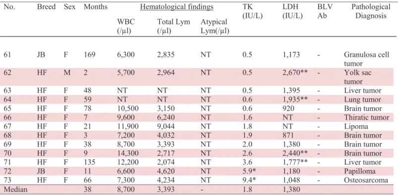
The 27 cows with other diseases were also divided into two groups: 13 with other tumors and 14 with inflammatory diseases (Table 1-3). The group with other tumors included 4 brain tumors, 2 liver tumors, 1 lung tumor, 1 osteosarcoma, 1 leiomyosarcoma, 1 lipoma, 1 granulose cell tumor, 1 papilloma and 1 yolk sac tumor. All cows with inflammatory diseases showed lymphadenopathy or palpable masses in the pelvic cavity, and BL was suspected as part of the differential diagnosis. Post-mortem examination and histopathological findings for cows in this group revealed 6 cases of abscess in the pelvic cavity, 3 of mastitis, 2 of pneumonia, 1 of pericarditis and 1 of polyarthritis with amyloidosis.
TK activity assay
TK activity assays were performed on serum samples using a commercial radioenzyme TK-assay kit and 125I-iododeoxyuridine tracer (Kishimoto Clinical Laboratory, Inc., Obihiro, Japan). TK activity was expressed as units per liter (U/L). The reportable range of the assay was 0.5 to 1,000 U/L. TK activities of 20 cattle among 60 with BL have been already reported in a previous report (Sakamoto, L et al. 2009).
13
Statistical analysis
Mann-Whitney U tests were used to compare TK activity levels of ‘BL with difficult diagnosis’ cases with that of other groups. Chi square analysis was also used to compare the positive rates of each group. A P-value of less than 0.05 was considered statistically significant.
RESULTS
Results of TK activity assays for each group are shown in Tables 1-1, 1-2 and 1- 3 and Fig.1-1. Median TK activity values for cows in the ‘BL with difficult diagnosis’ and ‘clinically confirmed BL’ groups were 36.8 and 39.4 IU/L, respectively (Fig. 1-1), with no significant difference between the two groups of BL. Although the percentage of cows with positive TK activity (>5.4 IU/L) was higher in those with ‘clinically confirmed BL’ (97.2%) than in those with ‘BL with difficult diagnosis’ (83.3%), this difference was not statistically significant (Table 1-4). Of the 24 cows with ‘BL with difficult diagnosis’, four (Nos. 1, 2, 3 and 18) showed TK activities lower than the cut-off point. This was also the case for one (No. 25) of the 36 cows with ‘clinically confirmed BL’. However, 4 of these 5 cows showed higher LDH activity than the reference range (Table 1-1 and 1-2).
TK activity was significantly higher in cows with ‘BL with difficult diagnosis’ compared to that measured in cows with other tumors and inflammatory diseases (Fig.1-1). Median TK activities in cows with other tumors and inflammatory diseases were 1.8 and 1.4 IU/L, respectively (Table 1-3). The maximum TK activities in cows with other tumors and inflammatory diseases were 9.4 and 6.9 IU/L, respectively. The percentage of cows with positive TK activity was significantly higher in cows with ‘BL with
14
difficult diagnosis’ (83.3%) compared to cows with other tumors (15.3 %) and inflammatory diseases (21.4 %). Of the cows with positive TK activity, two cows (Nos. 72 and 73) with other tumors included a cow with papilloma and another with osteosarcoma. Three cows with inflammatory diseases included two with pelvic cavity abscesses (Nos. 85 and 87) and one with pneumonia (No. 86) (Table 1-3). LDH activity was lower than the reference value of 1,445 IU/L for all five of these cows.
DISCUSSION
TK converts thymidine to thymidine monophosphate in rapidly proliferating cells and serves as part of a DNA synthesis salvage pathway. TK is activated during the G1/S phase of the cell cycle, and its activity correlates with tumor cell proliferation (Bello 1974). Serum TK concentrations increase in patients with several types of hematopoietic tumors (Filanovskaia et al. 1994; Gronowitz et al. 1983; Luoni et al. 1992 and Musto et al. 1995). Serum TK activity is useful for detecting, grading and monitoring tumors in lymphoma and leukemia patients, may also be helpful in diagnosing and monitoring canine lymphoma and leukemia (Tanaka et al. 1993) and is also a possible marker of bovine leukosis (Sakamoto, L et al. 2009). However, BLV infection with no onset of BL would not induce TK activities (Sakamoto, L et al. 2009). The present study evaluated the clinical utility of serum TK activity for diagnosis of ‘BL with difficult diagnosis’ by comparing TK activity in cows with ‘BL with difficult diagnosis’ to that in cows with clinically confirmed BL, those with lymphadenopathy, or those with other diseases.
First, TK activity in cows with ‘BL with difficult diagnosis’ was compared to that in cows with ‘clinically confirmed BL’ significant differences were not found in
15
activities or positive rates between the two groups. These results suggest that TK activity is a useful marker to detect BL, even when typical clinical signs of superficial lymph node enlargement as well as typical forms are not clear. TK activity can be used as a marker for BL in suspected BL cases without apparent clinical evidence, such as lymphadenopathy, lymphocytosis and/or increased neoplastic lymphocytes in peripheral blood.
Then, the specificity of TK activity was evaluated and found that TK values in cows with ‘BL with difficult diagnosis’ were significantly higher than in those with other tumors and inflammatory diseases. The percentage of cows that showed positive TK activity was also significantly higher in cows with ‘BL with difficult diagnosis’ compared to the other two groups. These results suggest that TK activity can be a useful BL marker when BL is suspected. However, 15.3% of cows with other tumors and 21.4% of those with inflammatory diseases also showed TK activity levels above the cut-off point (5.4IU/L). The maximum TK activities in cows with other tumors and inflammatory diseases were 9.4 and 6.9 IU/L, respectively. Higher cut-off point of TK may be more appropriate, however, it may have some benefits and weaknesses, as cases of other tumors and inflammatory diseases may not have higher TK, while, some cases of BL may have lower than cut-off point. TK activity can be induced by the herpes virus infection (Jamieson et al. 1974), and elevated serum TK activity was reported for human patients with acute viral hepatitis (Tanaka et al. 1993). With the exception of BLV, viral infections were not evaluated in the present study, but it is certainly possible that a herpes virus infection (e.g., infectious bovine rhinotracheitis) affected the serum TK activity in our cattle. Future studies should evaluate the effects of viral infection on serum TK activity in cattle.
16
In the present study, 4 of the 5 cows with BL who had lower TK values showed higher LDH activity. In contrast, all 5 non-BL cows with higher TK activity had LDH activity lower than the reference value (Kaneko et al. 1997). Higher activity of LDH and LDH isozymes like LDH2 and 3 have already been reported as biomarker for diagnosis of BL, however, it may not be enough specific for definitive diagnosis of BL, because it increase in other diseases as well (Keller 2001). Since in this study most of the cases showed higher activity of TK as well as LDH, double screening of serum LDH and TK for clinical diagnosis of ‘BL with difficult diagnosis’ would be more useful. On the other hand, simultaneous evaluation of LDH and TK activity may also help with differential diagnoses of other tumors and inflammatory diseases due to BL. Future studies regarding the specificity and cut-off points of TK activity are needed.
17
Fig.1-1. Serum thymidine kinase activities of clinical cases of bovine leukosis for which diagnosis was difficult, bovine leukosis which was clinically diagnosed according to cytology findings of enlarged lymph nodes, other tumors and inflammatory diseases. Asterisks indicate significance (P<0.001; Mann-Whitney U-test) when compared to cows with bovine leukosis that was difficult to diagnose. BL with difficult diagnosis (N=24) Clinically confirmed BL (N= 36) Other tumors (N= 13) Inflammatory diseases (N= 14) Thymidi ne kinase activi ty, IU/l
18
Table 1-1: TK activities and profiles of cattle with bovine leukosis which were not clinically diagnosed, but confirmed by necropsy. None of the cows showed lymphadenopathy.
No. Breed Sex Months Hematological findings TK
(IU/L) LDH (IU/L) BLV Ab Type of BL WBC (/μl) Total Lym (/μl) Atypical Lym (/μl) 1 HF F 64 9,100 6,916 0 0.5 3,550** + Enzootic 2 HF F 125 7,500 3,450 0 2.5 30,000** + Enzootic 3 HF F 67 20,300 6,496 0 5.4 3,490 ** + Enzootic 4 HF F 61 8,200 4,510 0 17.0* 764 + Enzootic 5 HF F 23 5,600 1,512 0 30.0* 2,400* * + Enzootic 6 JB F 87 NT NT NT 30.0* 1,370 + Enzootic 7 JB F 52 NT NT NT 32.0* 2,165** + Enzootic 8 HF F 65 14,600 7,884 4,088 39.0* 2,310 ** + Enzootic 9 JB F 60 NT NT NT 45.0* 3,590** + Enzootic 10 HF F 94 12,900 6,579 0 51.0* 1,410 + Enzootic 11 HF F 72 13,400 8,576 4,422 52.5 * 3,060** + Enzootic 12 HF F 74 6,400 4,480 0 55.0* 898 + Enzootic 13 HF F 85 13,900 8,618 4865 71.0* 4,545** + Enzootic 14 HF F 83 15,300 10,863 6,426 74.7* 3,050 ** + Enzootic 15 JB F 92 NT NT NT 110.0* 2,805** + Enzootic 16 F1 CM 25 10,200 6,936 2244 120.0* 2,570** + Enzootic 17 JB F 50 NT NT NT 210.0* 5,875 ** + Enzootic 18 HF F 8 5,900 4,425 0 2.7 1,140 – Calf 19 HF F 11 12,100 4718 0 26.0* 2,480 ** – Thymus 20 HF F 67 17,100 2,736 0 8.2* 4,891** – Unknown 21 HF F 34 11,600 8,120 30 32.0* 1,490* – Unknown 22 HF F 37 5,500 4,290 0 34.6* 3,380** – Unknown 23 JB F 92 NT NT NT 92.0* 2,515** NT Unknown 24 JB F 60 NT NT NT 120.0* 2,915** NT Unknown Median 64.5 11,600 6,496 0 36.8 2,688
BLV Ab: Bovine leukemia virus antibody, HF: Holstein-Friesian, JB: Japanese Black, F1: hybrid of HF and JB, F: female, CM: castrated male, NT: not tested, Lym: lymphocytes *: TK activity more than cut-off point (>5.4 IU/L)
19
Table 1-2: TK activities and profiles of cattle in clinically confirmed BL.
No. Breed Sex Months WBC Hematological findings TK
(IU/L) LDH (IU/L) BLV Ab Type of BL
(/μl) Total
Lym (/μl) Atypical Lym (/μl)
25 HF F 71 8,000 2,640 0 5.4 2,135** + Enzootic 26 F1 F 20 216,000 203,040 200,880 5.5* 2,660** + Enzootic 27 HF F 66 16,200 8,910 3,402 7.2* NT + Enzootic 28 HF F 88 21,600 15,768 1,296 11.0* 1,219 + Enzootic 29 HF F 61 33,200 28,220 21,580 14.0* 1,143 + Enzootic 30 HF F 71 10,900 4,905 1,635 16.5* 1,130 + Enzootic 31 HF F 65 26,500 12,985 1060 19.0* 1,430 + Enzootic 32 HF F 122 27,200 17,680 3,264 22.0* 1,650** + Enzootic 33 HF F 42 34,800 32,016 31,320 22.0* 6,750** + Enzootic 34 HF F 30 33,200 21,248 1,660 23.1* 1,465** + Enzootic 35 JB F 36 450,200 441,196 247,610 32.9* 5,260** + Enzootic 36 HF F 115 11,100 4,662 0 33.8* 3,340** + Enzootic 37 HF F 64 11,500 8,625 2,760 37.0* 3,160** + Enzootic 38 HF F 74 60,700 55,237 33,385 38.7* 3,280** + Enzootic 39 HF M 18 230,900 221,664 219,355 40.0* 4,740** + Enzootic 40 HF F 129 20,800 11,440 5,200 41.0* 4,700** + Enzootic 41 HF F 44 10,600 3,286 0 44.0* 3,210** + Enzootic 42 HF F 83 6,700 2,948 0 45.6* 2,690** + Enzootic 43 HF F 132 NT NT NT 52.8* 5,800** + Enzootic 44 HF F 101 12,700 4,826 0 69.0* 2,110** + Enzootic 45 JB F 36 312,900 303,513 306,642 97.0* 4,360** + Enzootic 46 HF F 73 44,100 39,690 38,808 99.1* 6,410** + Enzootic 47 HF F 72 70,500 69,090 69,090 115.6* 9,630** + Enzootic 48 HF F 78 14,600 5,694 1,752 240.0* 3,650** + Enzootic 49 HF F 89 269,700 261,609 248,124 1000* 5,910** + Enzootic 50 HF F 50 107,000 105,930 101,650 1000* 2,380** + Enzootic 51 HF F 85 17,500 12,950 2,100 1000* 2,510** + Enzootic 52 HF F 12 10,400 5,512 0 6.8* 1,700** – Calf 53 HF F 4 29,700 26,136 5,940 270.0* 3,770** – Calf 54 HF F 31 27,200 22,304 13,328 77.3* 6,450** – Skin 55 HF F 41 10,600 3,710 0 100.3* 12,500** – Skin 56 HF F 22 8,300 6,308 0 19.3* 3,000** – Thymus 57 HF F 17 8,200 4,346 0 28.0* 2,550** – Thymus 58 HF F 8 14,600 11,534 11,388 132.6* 2,740** – Thymus 59 HF F 36 12,400 8,928 11,780 38.0* 3,200** – Unknown 60 HF F 71 24,900 12,450 8,217 83.0* 9,700** – Unknown Median 64.5 21,600 12,950 3,402 39.4 3,200
BLV Ab: Bovine leukemia virus antibody, HF: Holstein-Friesian, JB: Japanese Black, F1: hybrid of HF and JB, F: female, NT: not tested, Lym: lymphocytes
*: TK activity more than cut-off point (>5.4 IU/L)
20
Table 1-3: TK activities and profiles of cattle with neither bovine lymphosarcoma nor bovine leukemia. All diagnoses were confirmed by necropsy.
(1) Tumors other than bovine leukosis
No. Breed Sex Months Hematological findings TK
(IU/L) LDH (IU/L) BLV Ab Pathological Diagnosis
WBC
(/μl) Total Lym (/μl) Atypical Lym(/μl)
61 JB F 169 6,300 2,835 NT 0.5 1,173 - Granulosa cell tumor 62 HF M 2 5,700 2,964 NT 0.5 2,670** - Yolk sac tumor 63 HF F 48 NT NT NT 0.5 1,395 - Liver tumor 64 HF F 59 NT NT NT 0.6 1,935** - Lung tumor 65 HF F 78 10,500 3,150 NT 0.6 920 - Brain tumor 66 HF F 7 9,600 6,240 NT 1.6 NT - Thiratic tumor 67 HF F 21 11,900 9,044 NT 1.8 NT - Lipoma 68 HF F 3 7,200 4,032 NT 1.9 871 - Brain tumor 69 HF F 38 8,700 3,393 NT 2.0 1,380 - Brain tumor 70 HF F 9 14,300 2,717 NT 2.6 2,440** - Brain tumor 71 HF F 135 12,200 2,074 NT 3.6 1,777** - Liver tumor 72 JB F 11 6,600 4,620 NT 5.9* 1,180 - Papilloma 73 HF F 66 7,300 4,234 NT 9.4* 1,048 - Osteosarcoma Median 38 8,700 3,393 - 1.8 1,380 (2) Inflammatory diseases 74 HF F 80 9,800 2,450 NT 0.6 968 - Mastitis 75 HF F 84 7,900 3,634 NT 0.7 1,019 - Polyarthritis 76 HF F 60 13,200 3,828 NT 0.7 1,036 + Abscess 77 HF F 95 5,600 1,792 NT 1.0 842 - Mastitis 78 HF F 15 17,000 4,930 NT 1.2 967 - Pericarditis 79 HF F 102 13,300 7,847 NT 1.2 1,430 - Abscess 80 HF F 38 11,700 3,159 NT 1.4 1,690** - Arthritis 81 HF F 38 10,500 2,940 NT 1.4 1,058 - Abscess 82 HF F 61 14,500 6,090 NT 1.5 1,173 - Mastitis 83 HF F 14 10,400 6,240 NT 2.0 2,400** - Pneumonia 84 HF F 48 16,700 3,841 NT 3.5 623 - Abscess 85 HF F 76 17,600 4,400 NT 5.5 1,141 - Abscess 86 HF F 4 12,300 9,594 NT 5.8* 981 - Pneumonia 87 HF F 1 13,700 4,384 NT 6.9* 921 - Abscess Median 54 12,750 4,113 - 1.4 1,028
BLV Ab: Bovine leukemia virus antibody, HF: Holstein-Friesian, JB: Japanese Black, F: female, NT: not tested, Lym: lymphocytes
*: TK activity more than cut-off point (>5.4 IU/L)
21
Table 1-4: Positive ra
tio and median ch
an ge s of s erum TK and L D H a ctivit y in bovine leukosis ( B L) with diff icult diag nosis, cli nica ll y confirmed BL , other tum
ors and inflammator
y dis eases g roups. T ested g roups N umber of cows/group Positive (>5.4 I U /L ) Positive Ratio (% ) M edian (I U/ L ) N umber of cows/group Posi tiv e (>1445 I U /L) Positive Ratio (% ) Median (IU /L ) BL with diff icult dia gnos is 24 20 83.3 36.8 24 18 75 2688 Clinica ll y confirmed BL 36 35 97.2 39.4 35 32 91.5 3200 Other tumors 13 2 15.3* 1.8* 11 4 36.3 1380 In flammator y diseas es 14 3 21.4* 1.4* 14 2 14.3 1020 *Sig nificant differe nces compared to BL with difficul t diag nosis group. Serum L D H ac tivit y Serum TK activit y
22 Chapter 2
The Preliminary Evaluation of Gene Expression as Biomarkers in Clinical Cases of Bovine Leukosis
INTRODUCTION
Bovine leukosis (BL) is one of the most common types of neoplasm affecting cattle. Generally it has been divided into 2 types: enzootic bovine leukosis (EBL) and sporadic bovine leukosis (SBL) (Angelos et al. 2008).
EBL is an infectious diseases caused by a retrovirus called bovine leukemia virus (BLV). Once the BLV infects a herd, most of the cattle would be serologically positive against BLV antibodies without any other clinical signs, or can be followed by persistent lymphocytosis in about 28% of the herd cattle, while only approximately 5% of animals infected with BLV will develop B-cell lymphoma in various lymph nodes and organs after a long latent period (Garry 2008; Ferrer 1979). Tumor cells often infiltrate many organs, including the abomasum, heart, uterus, retrobulbar space, and epidermal region of the central nervous system (Valli 2007). Therefore, clinical signs in cattle with lymphoma are generally non-specific and include weight loss, decreased appetite, and decreased productivity (Angelos and Thurmond 2008; Burton et al. 2010).
SBL can be further subdivided into three forms: calf/juvenile, thymic and skin forms (Valli 2007). Calf form BL is mostly occurs in calves has 3 to 6 months old, meanwhile some cases of calf form BL can occurs in calves as young as one month and as old as 3years (Pasquini and Pasquini 1996; Angelos and Thurmond 2008). The most obvious clinical signs of the calf form BL are diffuse lymphadenopathy that results in obvious and palpable enlargement of peripheral lymph nodes (Garry 2007).
23
Ataxia is another common clinical sign of the calf form BL, because tumor cells often invade the central nervous system (Ohshima et al. 1980; Theilen and Dungworth 1965). The thymic form BL is usually observed in 6- to 24-month-old cattle, and is characterized by a large, firm swelling of the ventral neck region (Braun et al. 2007; Divers and Peek 2008). Progressive thymic enlargement occurs in all cases and is clinically apparent when cervical enlargement develops. Cattle with thymic form BL often have clinical signs of brisket and sub-mandibular edema, and jugular vein distension (Dungworth et al. 1964; Grimshaw et al. 1979). The main clinical signs resulting from thymic form BL are either bloat or dyspnea. Macroscopic neoplastic lesions are distributed across lymph nodes, bone marrow, liver, spleen, kidneys, lungs, and thymus (Ohshima et al. 1980).
BL can be easily identified by direct physical examination and diagnosed by cytological evaluation of swollen lymph nodes when lymphadenopathy and obvious neoplastic changes in target organs are present. However, it is more difficult to suspect and diagnose when located in more specific areas such as the spinal cord or abomasum and when lymph node enlargement and/or lymphocytosis are not evident (Garry 2008). While ultrasound-guided fine needle aspiration (FNA) or biopsy of vascular masses located in the body cavity, retrobulbar space, or heart is helpful (Garry 2008), but such diagnostic tools are not always available.
Serum lactate dehydrogenase (LDH) activity may be elevated in some cattle with BL (Ishihara et al. 1980), but its specificity is insufficient to confirm diagnosis of the BL (Garry 2008). Serum thymidine kinase (TK) activity was found to be a potential biomarker for BL, but is not easy to determine as it requires a radioimmunoassay (Chapter 1). In humans, gene expression profiling has been used to assess
24
lymphosarcoma and leukemia. For example, Wilms’ tumor gene 1 (WT1) expression is considered as a sensitive biomarker for monitoring residual disease in acute myeloid leukemia (Sakamoto, Y et al. 2009; Siehl et al. 2002). Some other genes, including interleukin 2 receptor (IL2R), TK1 and immunoglobulin-associated alpha-1 (MB1 or CD79a) genes were also known to be markers of hematopoietic neoplasia (Erber and Mason 1988; Ishihara et al. 1980; Kristensen et al. 1994). To examine such markers for clinical diagnosis of BL, in this chapter, the mRNA expression of some genes known as biomarker of hematopoietic neoplasms in human medicine such as IL2R, TK1 and MB1, have preliminary been evaluated using three clinical cases of BL including thymic, calf and enzootic forms, and two other cows without BL as control animals by reverse transcriptional polymerase chain reaction (RT-PCR).
MATERIALS AND METHODS
Case history
Case 1: A 12-month-old Holstein heifer showed anorexia and lameness. At initial
examination, the heifer had a body temperature of 38.8°C and heart rate of 108 beats/min (bpm). Lymphadenopathy was also noted. Despite treatment with antibiotics and dexamethasone, the general condition of the heifer did not improve. The heifer was taken to the Veterinary Teaching Hospital at Obihiro University of Agriculture and Veterinary Medicine on day 9 of illness.
Case 2: An 8-month-old Holstein heifer with cervical enlargement was initially
examined by a clinical veterinarian. At examination, the heifer had a body temperature of 38.6°C and heart rate of 70 bpm, and showed bloat and anorexia. Even the animal received treatment, but the heifer’s general condition did not improve. On the third day
25
of illness, the heifer was taken to the Veterinary Teaching Hospital, Obihiro University of Agriculture and Veterinary Medicine.
Case 3: A 49-month-old Holstein cow with anorexia, tachypnea, and difficulty standing
up was first examined by a clinical veterinarian. At examination, the cow had body temperature of 39.9°C, heart rate of 80 beats/min (bpm), and respiratory rate of 52 breaths/min. Despite antibiotic treatment via injection and infusion, the general condition of the cow did not improve. On day 7, enlargement of the subiliac lymph nodes was observed, and bovine leukosis was suspected. The cow was then taken to the Veterinary Teaching Hospital at Obihiro University of Agriculture and Veterinary Medicine.
RT-PCR
In the mentioned three cases of BL and two other cases of control animal, the messenger RNA (mRNA) of some genes that are in use as marker of hematopoietic neoplastic disease in human medicine like IL2R, TK1 and MB1 have been measured by RT-PCR, using primer set of 5′- acg-cca-tgt-tca-agg-tct-tc −3′ (IL2R forward) and 5′-gtt- ctg-cgc-atc-tgt-gtg-tt -3′ (IL2R reverse), 5′-cca-agt-cag-tga-tgg-caa -ga-3′ (MB1 forward) and 5′-gat-atc-agc-ccc-gaa-ttt-ca-3′ (MB1 reverse), and 5′-cca-ggt-tgc-cca-gta-caa- gt-3′ (TK1 forward) and 5′-tct-cgc-aga-act- cca-caa-tg-3′ (TK1 reverse). Beta-actin gene (ACTB) expression has been examined as an internal control using the following primer set: 5′-ctt-tcc-agc-ctt-cct -tcc-t-3′ (ACTB forward) and 5′-ggg-cag-tga-tct-ctt-tct-g-3′ (ACTB reverse). All primers used in this study have designed using Primer Express (Applied Biosystems). The sequences that are used for making primers were taken from GenBank database. The RT-PCR was performed on the swollen superficial lymph nodes that contained tumor cells and subiliac lymph node tissue
26
obtained by FNA from the case1 and 3 respectively, and from thymic mass cells by FNA of case 2, lymph node tissue of two other cows without lymphadenopathy have also been used as controls. Total RNA was extracted from 20mg of the mentioned lymph node tissues or tumor cells, using the RNeasy Mini Kit (QIAGEN, Germantown, MD, U.S.A.) according to manufacturer’s instructions. cDNA was synthesized using 2 μg of total RNA and the SuperScript™ III 1st-strand synthesis system (Invitrogen, Carlsbad, CA, U.S.A.).
Clinical and clinico-pathological examination
From hospitalization of the cows in Veterinary Teaching Hospital of Obihiro University of Agriculture and Veterinary Medicine, up to day of necropsy, every day two times (morning and afternoon) the cows were clinically examined and abnormalities were noted. Cytology examination of the swollen lymph nodes or masses using fine needle aspiration sample was also performed in all cases of BL, hematological examination was done using hematological analyzer, and blood biochemical examination was also performed. TK activity, was also been measured in all 3 cases of BL using a commercial radioenzyme TK-assay kit with a 125 I-iododeoxyuridine tracer (Kishimoto Clinical Laboratory, Inc., Obihiro, Japan). To determine the types of BL and BLV infection (EBL or SBL), antibodies against BLV using agar-gel immunodiffusion test with commercial antigen was also examined (Kitasato Institute Research Center for Biologicals, Saitama, Japan).
27 RESULTS
Clinical signs, pathological findings and RT-PCR
Case 1: At admission, the cow showed clinical signs of emaciation, depression,
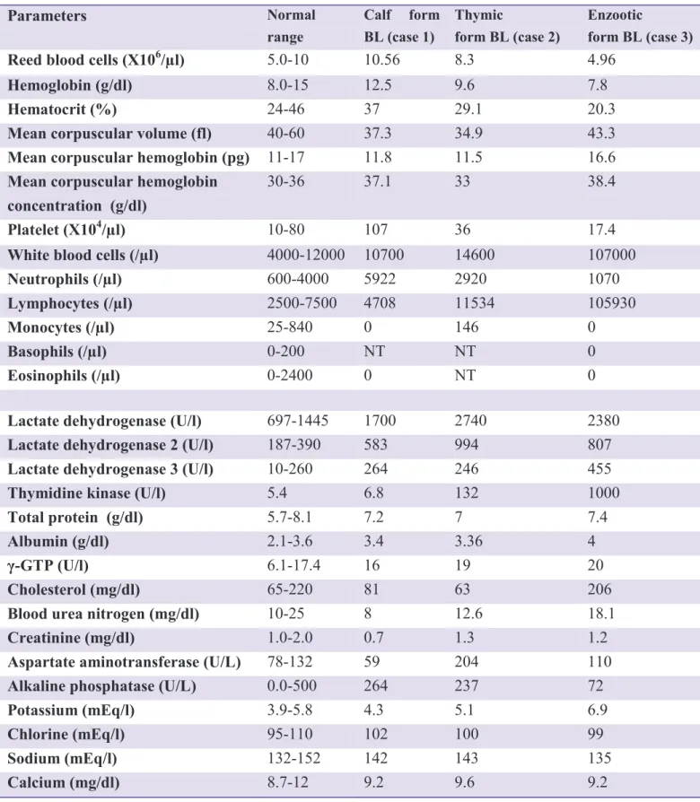
lameness, abdominal posture standing under, and swollen carpal and tarsal joints (Fig. 2-1). Rectal temperature was 39.9°C, heart rate was 120 bpm, and respiratory rate was 56 breaths/ minute. Swelling of peripheral lymph nodes, including superficial cervical, subiliac, parotid, mandibular, and mammary lymph nodes, was observed. Hematological examination did not reveal any abnormalities such as anemia or lymphocytosis (red blood cell count: 10.56 × 10⁶/μl; hemoglobin concentration: 12.5 g/dl; hematocrit: 37.0%; mean corpuscular volume 37.3fl; mean corpuscular hemoglobin concentration 31.7 g/dl; mean corpuscular hemoglobin 11.8 pg white blood cell count: 10,700/μl (neutrophils: 5,922/μl; lymphocytes: 4,708/μl)) (Table 2-1). Microscopic examination of peripheral lymphocytes was normal. Serum biochemical analysis showed low total cholesterol (81 mg/dl) and increased lactate dehydrogenase activity (LDH: 1,700 IU/l). LDH isozymes analysis showed slightly elevated activities for LDH-2 (583 IU/l) and LDH-3 (264 IU/l) compared to normal (Hoffman and Solter 2008). Serum creatinine 0.7 mg/dl; total protein7.2 g/dl; albumin 3.4 g/dl; sodium 142 mEq/l; chlorine 102 mEq/l; potassium 4.3 mEq/l and calcium 9.2 mg/dl were within normal range. Serum thymidine kinase (TK) activity was increased in the present case (6.8 IU/l) as compare to normal range (5.4 IU/l) (Table 2-1). Agar gel immunodiffusion assay for antibodies against bovine leukemia virus was negative. Cytology of fine needle aspirate from a superficial cervical lymph node revealed several large lymphoid cells with marked atypia and mitotic cells, suggesting the diagnosis of lymphosarcoma. Arthrocentesis of the swollen carpal and
28
stifle joints using a 23-gauge needle was also performed. Synovial fluid collected from each joint was yellow and cloudy, with 20 to 50 cells observed per high dry (X40) field in a stained smear with hemacolor® (Merck Chemicals, Darmstadt, Germany). Most cells were mononuclear, and mitotic cells were often observed.
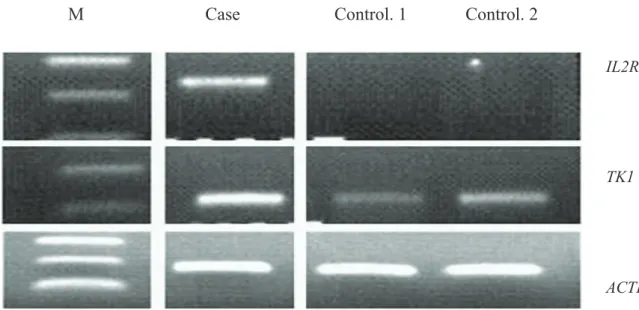
In the present case, the messenger RNA (mRNA) expression of IL2R and TK1 genes on fine needle aspirate sample taken from the swollen superficial cervical lymph nodes that contained tumor cells of the case animal have also been examined by RT-PCR. Both IL2R and TK1 genes mRNA were highly expressed in tumor tissue compared to that in control animals (Fig. 2-2).
The animal was euthanized and necropsied on day 10. Gross examination revealed marked swelling of systemic lymph nodes, including peripheral lymph nodes, and both abdominal and thoracic cavities. Discrete white masses of various sizes were also found in the kidneys, ribs, intracranial dura mater, compressed cerebrum, nasal septum, and frontal sinus. A yellowish brown gelatinous material that accompanied synovial villous hypertrophy periarticularly in both carpal and stifle joints, also observed (Fig.2-3).
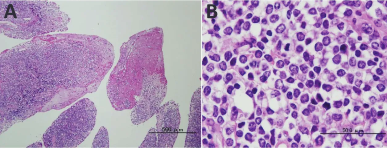
Histopathological examination revealed neoplastic lymphoid cells with large round nuclei and scant amounts of cytoplasm infiltration in the enlarged lymph nodes, liver, uterus, heart, and pulmonary pleura. The synovial membrane was infiltrated with numerous neoplastic lymphocytes and showed papillary projections consistent with macroscopic findings (Fig. 2-4). Neoplastic cells were also found in the leptomenig and perivascular spaces of the cerebrum. Immunohistochemical examination of the enlarged lymph nodes revealed that tumor cells stained positive for CD3 and negative for
BLA-29
36 antibodies, respectively. From these findings, the present case was classified as calf type B-cell bovine lymphosarcoma.
Case 2: Upon admission, rectal temperature was 39.0°C, heart rate 90 bpm, and
respiratory rate 26 breaths/min. Physical examination revealed a cervical mass measuring 30 × 20 × 20 cm (Fig. 2-6A), jugular vein distension, conjunctival hyperemia, and ruminal tympany. Tympany was caused by free gas, which was easily released using a nasal-gastric tube. Peripheral lymph node enlargement was not detected. The heifer also showed signs of depression and had a tendency to lie down on its side (Fig. 2-5B). Drooping of the upper eyelid, miosis and enophthalmos were observed on the right side of the face. Hematologic examination, showed microcytic and hypochromic red blood cells (RBC, 8.33 × 10⁶/μl; hemoglobin, 9.6 g/dl; hematocrit, 29.1%; mean corpuscular volume, 34.9 fl; and mean corpuscular hemoglobin, 11.5pg) and leukocytosis (WBC, 14,600/μl; neutrophils, 2,920/ μl; lymphocytes, 11,534/μl ; and monocytes: 146/μl) (Table 2-1). More than 80% of lymphocytes were morphologically atypical with wide cytoplasm, fine nuclear chromatin and nucleoli. The platelet counts (360,000/μl) were within the reference range. Serum creatinine, urea nitrogen, total protein, albumin, and serum calcium levels were normal (Table 2-1). Antibodies against BLV were not detected with agar-gel immunodiffusion test with commercial antigen (Kitasato Institute Research Center for Biologicals, Saitama, Japan). Serum biochemical analysis showed elevated aspartate aminotransferase (AST: 204 IU/l) and lactate dehydrogenase (LDH: 2,740 IU/l) activities. LDH isozyme analysis showed elevated activities for LDH-2 (994 IU/l) and LDH-3 (246 IU/l). Serum thymidine kinase (TK) activity was also increased (132.6IU/l) (Table 2-1). The findings of fine needle
30
aspiration cytology of the cervical mass revealed large lymphoblasts with mitoses present (Fig. 2-7A). These clinico-pathological findings strongly suggested that the present case was SBL, a thymic lymphosarcoma.
The mRNA expression of immunoglobulin associated alpha-1 (MB1) have also been examined as marker for the present case and the result demonstrates that, MB1 gene was highly expressed in the tumor tissue compared to those of control animals (Fig. 2-8).
The heifer was euthanized, and necropsy was performed on day 4 of illness. Gross examination revealed a solid mass (30 X 20 X 20 cm) in the cervical thymic region around the trachea, and it compressed the esophagus and trachea (Fig. 2-6A). Several small masses ranged from 1-8 cm in diameter were also found in the thoracic thymus, pleura, liver, uterus, ureters, and peri-renal tissue. Abdominal lymph nodes also showed slight enlargement. In addition, several small masses ranged from 0.5 to 1.5 cm in diameter were found in the mucosa of frontal sinus, and multiple extradural sites throughout the cranial vault, including cerebral dura mater, surrounding the pituitary gland tissue, and exterior dura mater of the medulla oblongata (Fig. 2-6B). The cerebral hemisphere showed extensive compression due to neoplastic masses (Fig. 2-6C). Histopathological examination revealed that the cervical mass was composed entirely of neoplastic lymphocytes with irregular nucleus and different in size. Similar neoplastic cells were also observed in small masses in other organs. Neoplastic cells were also in leptomeninges and perivascular space of the cerebrum (Fig. 2-7B). Immunohistochemical examination showed tumor cells positive for CD3 and negative for BLA-36 antibodies. Mild ischemic changes were observed in nerve cells of cerebral compression sites. The diagnosis of thymic lymphosarcoma was confirmed by these
31 pathological findings.
Case 3: The clinical signs of depression, emaciation, and tachypnea were observed in a
49 month-old Holstein cow (Fig. 2-9) on admission. Rectal temperature, heart rate, and respiratory rate were 40.5°C, 92 bpm, and 96 breaths/min, respectively. Swelling of multiple peripheral lymph nodes, including superficial cervical (R: 12 × 7 × 3cm, L: 15 × 9 × 3 cm), subiliac (R: 18 × 5 × 3 cm, L: 12 × 3 × 3 cm), mandibular (R and L: diameter 4 cm), and mammary lymph nodes (R and L: diameter 8 cm) was observed. Several masses were identified in the pelvic cavity by rectal palpation. Cytological finding of FNA of the right subiliac lymph node revealed the presence of large lymphoid cells with obvious cellular atypia and several mitotic cells, indicating lymphosarcoma (Fig. 2-10).
Hematologic examination showed microcytic and normochromic anemia and lymphocytosis [RBC: 4.96 × 10⁶/μl, hemoglobin: 7.8 g/dl, hematocrit; 20.3%, mean corpuscular volume: 43.3 fl, mean corpuscular hemoglobin concentration: 38.4 g/dl, WBC: 107,000/μl, neutrophils: 1,070/μl (1%), and lymphocytes: 105,930/μl (99%)] (Table 2-1). More than 90% of lymphocytes in peripheral blood were microscopically atypical with indented nuclei and finely stippled chromatin. Serum biochemical analysis showed increased LDH activity (2,380 IU/l). LDH isozymes analysis showed elevated activities for LDH-2 (807 IU/l) and LDH- 3 (455 IU/l). Extremely higher activities of serum TK (1,000 IU/l) were recorded compared with normal cattle (Table 2-1). Antibodies against BLV were detected by agar-gel immunodiffusion test (Kitasato Institute Research Center for Biologicals, Saitama, Japan).
Result of the RT-PCR that was performed on tumor tissues has demonstrated higher expression in the mRNA of MB1, IL2R and TK1 genes (Fig. 2-11).
32
The cow was euthanized and necropsy was performed on day 15. Gross examination revealed swelling of systemic lymph nodes, including superficial cervical, mandibular, mammary, medial, iliac, and renal lymph nodes. Furthermore, the spleen was enlarged and swollen (70 × 20 × 8 cm) (Fig. 2-12). Yellowish white tissue was found in the sternal bone marrow. Histopathological examination revealed neoplastic lymphoid cell infiltration in the enlarged lymph nodes, liver, spleen, bone marrow, uterus (especially in the endometrium), and lamina propria of the urinary bladder, abdomen and intestine. Immunohistochemical examination showed that tumor cells within enlarged lymph nodes were stained positive for BLA-36 and negative for CD3 antibodies. These findings suggested that the tumor cells were B-cell origin. Data of case 4 and 5, which used as control animal for RT-PCR not shown.
DISCUSSION
In this chapter, the mRNA expression of some genes such as IL2R, MB1 and TK1 have been measured using the RT-PCR assay in three different clinical cases of bovine leukosis. In case 1 (calf form BL), abnormalities such as multiple joint swelling and infiltration of many neoplastic cells in to the synovial membrane and in leptomenig and perivascular of the cerebrum was observed. Even though tumor cells often infiltrate many organs in calf for BL (Ohshima et al. 1980). Infiltration into joints and periarticular tissue is quite rare. Only one case of ataxia by tibiotrasal joint infiltration of tumor cells in calf form bovine leukosis has been reported (Oliver-Espinosa et al. 1994). This was a rare clinical case of calf form BL that accompanied with multiple joints swelling.
33
several masses were recorded in frontal sinus, cerebral dura mater, surrounding pituitary gland tissue and exterior dura mater of medulla oblongata. Involvement of the central nervous system (CNS) is a common clinical observation in EBL (Burton et al. 2010). However, the brain and spinal cord are usually not directly affected by the thymic form of lymphosarcoma (Alexander et al. 1996; Angel et al. 1991; Braun et al. 2007; Dungworth et al. 1964; Hatfield et al. 1986; Ohshima et al. 1980; Parodi et al. 1989). One exception was a clinical case of thymic lymphosarcoma with metastases causing spinal cord compression and pelvic limb paresis in a heifer (Holmes et al. 1990). To the best of my knowledge, macroscopic brain involvement has not been reported in thymic form BL. Horner’s syndrome was the other rare clinical sign that observed in this case. It results from interruption of ocular sympathetic pathways, from the midbrain close to pituitary gland through the spinal cord down to T1-T3 spinal segments, up the vagosympathetic trunk, and cranial cervical ganglion next to the tympanic bulla (Pace et al. 1997; Paquette 2010; Reede et al. 2008). Specific causes include traumatic lesions to the basisphenoid region, cervical trauma, abscesses, tumors, or space-occupying lesions in the anterior aspect of the thorax. In cattle, it has been associated with abscesses and cranial tumors, including adenocarcinoma and squamous cell carcinoma (Divers and Peek 2008; Guard et al. 1984; Pace et al. 1997). In the present case, ocular sympathic pathway close to pituitary gland might be affected by compression of extradural masses found in the cranial vault. Another possibility is unilateral damage of ocular sympathic pathway by cervical tumor caused unilateral Horner’s syndrome; however, the real cause was not clarified. And finally this case can be concluded as a rare clinical case of thymic lymphosarcoma that accompanied by brain involvement and Horner’s syndrome in a Holstein heifer.
34
In case 3 (Enzootic form BL), extremely higher activity of TK, which has recently been shown as BL marker, than normal cattle have been observed (Sakamoto, L et al. 2009). Also activity of LDH and LDH isozymes like LDH2, and 3 were higher in the present case than reference value (Kaneko et al. 1997). Higher activities of TK, LDH and LDH isozymes suggest aggressive proliferation of tumor cells in lymphoid organs and peripheral blood.
In this chapter, the mRNA expression of some genes known as biomarker for hematopoietic neoplasms in human medicine was also measured as a preliminary study for clinical diagnosis of certain types of BL. In case 1 and 3 (calf and enzootic from BL) over expression in the mRNA of IL2R and TK1 genes were observed. IL2R is a heterotrimeric protein expressed on the surface of immune cells, including lymphocytes and natural killer cells, and is the receptor for interleukin 2 (Nelson and Willerford 1980; Voss al. 1992). Close association of aberrant expression of IL2R with the infection of human T-cell leukemia virus (HTLV-1) was reported (Suzuki et al. 1987). IL2R is thought to be directly or indirectly activated by viral products, and the aberrant expression of gene might be involved in some stages of HTLV-1-infected lymphocytes (Maeda et al. 1985; Yodoi et al. 1983). TK is a cellular enzyme involved in a DNA synthesis salvage pathway, and its levels have been shown to correlate directly with the proliferative activity of tumor cells (Hallek et al. 1992; Kallender et al. 1987). Increased TK expression is often associated with increased expression of cell proliferation markers (Mao et al. 2002; Oudrad et al. 2002; Wu et al. 2000). Both IL2R and TK1 gene overexpression have been reported in several human leukemia cases (Erber and Mason 1988; Kristesen et al. 1994); however, there are no reports available for bovine leukosis. MB1 mRNA was also expressed in case 2 and 3 (thymic and enzootic form BL) as
35
compare to that in control animal. MB1 is a well-known B-cell specific gene, and it has been reported that it is a useful marker for B-cell neoplasms in human (Mason et al. 1995). This gene also plays a key role in B-cell development, stabilization, and function (Pike et al. 2004). MB1 in vitro over-expression in BLV-induced bovine B-cell lines has been also reported (Youn et al. 1994). Increased tumor cell proliferation in neoplastic lymph nodes may contribute to the overexpression of these genes.
In conclusion, results of the three clinical cases of BL demonstrate the usefulness of IL2R, TK1 and Mb1 genes as biomarker for clinical diagnosis of bovine leukosis. However, more BL cases should be examined to confirm the validity of using the expression of these genes as biomarkers of bovine leukosis. Detail of gene expression in different stages of the disease should be also clarified by using more reliable quantitative real-time PCR assay.
36
Table 2-1: Hematological and biochemical profiles of calf, thymic and enzootic form BL
Parameters Normal range Calf form BL (case 1) Thymic form BL (case 2) Enzootic form BL (case 3)
Reed blood cells (X106/μl) 5.0-10 10.56 8.3 4.96
Hemoglobin (g/dl) 8.0-15 12.5 9.6 7.8
Hematocrit (%) 24-46 37 29.1 20.3
Mean corpuscular volume (fl) 40-60 37.3 34.9 43.3
Mean corpuscular hemoglobin (pg) 11-17 11.8 11.5 16.6
Mean corpuscular hemoglobin concentration (g/dl)
30-36 37.1 33 38.4
Platelet (X104/μl) 10-80 107 36 17.4
White blood cells (/μl) 4000-12000 10700 14600 107000
Neutrophils (/μl) 600-4000 5922 2920 1070
Lymphocytes (/μl) 2500-7500 4708 11534 105930
Monocytes (/μl) 25-840 0 146 0
Basophils (/μl) 0-200 NT NT 0
Eosinophils (/μl) 0-2400 0 NT 0
Lactate dehydrogenase (U/l) 697-1445 1700 2740 2380
Lactate dehydrogenase 2 (U/l) 187-390 583 994 807
Lactate dehydrogenase 3 (U/l) 10-260 264 246 455
Thymidine kinase (U/l) 5.4 6.8 132 1000
Total protein (g/dl) 5.7-8.1 7.2 7 7.4
Albumin (g/dl) 2.1-3.6 3.4 3.36 4
γ-GTP (U/l) 6.1-17.4 16 19 20
Cholesterol (mg/dl) 65-220 81 63 206
Blood urea nitrogen (mg/dl) 10-25 8 12.6 18.1
Creatinine (mg/dl) 1.0-2.0 0.7 1.3 1.2
Aspartate aminotransferase (U/L) 78-132 59 204 110
Alkaline phosphatase (U/L) 0.0-500 264 237 72
Potassium (mEq/l) 3.9-5.8 4.3 5.1 6.9
Chlorine (mEq/l) 95-110 102 100 99
Sodium (mEq/l) 132-152 142 143 135
37
Fig. 2-1: Photograph of case 1 (calf form BL); emaciation, depression, lameness, standing under, swollen carpal and tarsal joints (arrows), and enlargement of peripheral lymph nodes (arrowheads) were recorded on day 9.
38
M Case Control. 1 Control. 2
Fig. 2-2: RT-PCR analysis of IL2R (230bp), TK1 (203bp) and ACTB (187bp) genes in lymph node tissue of case 1 (calf form BL) and two control animals. M: denotes DNA ladder.
IL2R
TK1
39
Fig. 2-3: Yellowish brown gelatinous material similar to synovial villous hypertrophy (arrows) observed periarticularly in the carpal joint of case 1 (calf form BL).
40
Fig. 2-4: Histopathological abnormality of the synovial membrane of the carpal joint of case 1 (calf form BL). (A) The synovial membrane was infiltrated with neoplastic cells and showed papillary projections (hematoxylin & eosin X100). (B) The neoplastic cells were large lymphoid cells with obvious atypia (hematoxylin & eosinX 400).
41
Fig. 2-5 A; Photograph of case 2 (thymic form BL) with cervical enlargement (arrow) and tympany. B; Heifer had tendency to lie down on its side.
42
Fig. 2-6: Post-mortem findings of the thymus, frontal sinus and cerebral hemisphere of case 2 (thymic form BL); A, a 30 x 20 x 20 cm mass, which compress esophagus and trachea, and caused tympany in this case. B, several masses (indicated by arrows) were observed in frontal sinus, and multiple extradural sites throughout the cranial vault. C, cerebral hemisphere shows compression by neoplastic masses (indicated by arrows).
A
B
C
Esophagus43
Fig. 2-7: Photomicrograph of cytological and histopathological findings of case 2 (thymic form BL). A, cytological findings of cervical mass, shows large lymphoblast with mitotic cells. B and C, histopathology of cerebrum, neoplastic lymphoid cells were in leptomeninges (B) and perivascular spaces (C). Bar=25μm
A
B
44
M Case C 1 C 2
MB1
ACTB
Fig. 2-8: RT-PCR analysis of the immunoglobulin associated alpha1 (MB1)(214bp), and actin beta (ACTB) (187bp) mRNA in the thymic masse of case 2 (thymic form BL) and lymph nodes of the two other control animals.
M: DNA ladder Case: thymic case C1: control 1 C2: control 2
45
Fig. 2-9: The photograph of case 3 (enzootic form BL), a 49 month-old Holstein cow with clinical signs of depression, emaciation and tachypnea.
46
Fig. 2-10: Cytological finding of fine needle aspiration of the lymph node of case 3 (enzootic form BL). The cell population is composed of middle to large sized lymphoid cells that have indented nuclei, finely stippled chromatin and scant amounts of cytoplasm (X400).
47
M Case Control.1 Control.2
Fig. 2-11: RT-PCR analysis of the IL2R (230bp), TK1 (203bp), MB1 (214bp), and ACTB (187bp) genes in lymph node tissues corresponding to case 3 (enzootic form BL) and two control animals. M denotes DNA ladder
IL2R
Mb1
Tk1
48
Fig. 2-12: Post-mortem finding of case 3 (enzootic form BL) revealed an enlarged and
49 Chapter 3
Evaluation of Gene Expression in Peripheral Blood Cells as a Potential Biomarker for Enzootic Bovine Leukosis
INTRODUCTION
Bovine leukosis/lymphosarcoma (BL) is one of the most common neoplastic diseases of cattle, and has been classified into two types according to pathological, epizootiological and clinic-pathological findings. These include sporadic bovine leukosis, which has unknown cause and enzootic bovine leukosis (EBL), which is associated with the bovine leukemia virus (BLV) (Bendixen 1965; Piper et al. 1975). The vast majority of animals with BLV remain persistently affected with no sign of infection and approximately 29% of cattle infected with BLV develop persistent lymphocytosis (PL), while fewer than 5% of animals affected by BLV develop lymphosarcoma (Ferrer 1979). Clinical signs of cattle affected by EBL are general malaise, decreased milk production, enlarged superficial lymph nodes, anorexia, abomasal ulceration, cardiac lesion and exophthalmos (Angelos and Thurmond 2008; Reed 1981). Findings that lead to suspicions of EBL include lymphocytosis, enlargement of peripheral lymph nodes and the presence of neoplastic lymphocytes in peripheral blood (Garry 2008). In general, fine needle aspiration (FNA) cytology of primary neoplasms or neoplastic lymph nodes can lead to a definitive diagnosis of EBL, but the sensitivity and specificity of FNA are not confidence (Washburn et al. 2007). Furthermore, diagnosis can be difficult for which lacks appearance of atypical lymphocytes and enlargement of lymph nodes (Garry 2008). Thus, more reliable biomarkers are recently required to diagnose EBL. With respect to bovine leukemia, higher activity of serum lactate dehydrogenase (LDH) and LDH isozymes have been
50
used as biomarkers to diagnose lymphosarcoma (Ishihara et al. 1980), even though LDH is not necessarily more specific for EBL, and is expressed in other diseases as well (Garry 2008). In addition, higher serum thymidine kinase activity has recently demonstrated potential as a biomarker for clinical diagnosis of EBL (Sakamoto, L et al. 2009), but this requires a radioimmunoassay test.
Genomic biomarkers are increasing in popularity for diagnosis of certain diseases within the field of human medicine. For example, the Wilms’ tumor 1 (WT1) gene is used as a biomarker due to its high expression levels in hematological malignancies and various cancers and low levels in normal tissues (Oji et al. 1999; Ueda et al. 2003). Additionally, high B-cell lymphoma/leukemia protein 2 (BCL2) activities have been found in mature peripheral B-cell neoplasms, such as those in B-cell chronic lymphocytic leukemia (Adachi et al. 1990; Schena et al. 1992). Although veterinary medicine would benefit greatly from similar methods and markers for clinical diagnosis of EBL, there has been little information available. In chapter 2, expression of some genes such as IL2R, TK1 and MB1 was examined as a preliminary study and the results demonstrated overexpression of the mentioned genes in single cases of BL. Thus, the present study evaluated mRNA expression levels of several target genes using quantitative reverse transcription polymerase chain reaction (qRT-PCR). Specifically, I analyzed interleukin 2 receptor (IL2R), WT1, thymidine kinase 1 (TK1), cytochrome P450 family 1-subfamily B- polypeptide 1 (CYP1B1), BCL2, phosphodiesterase isoform 7B (PDE7B), cyclin-dependent kinase inhibitor 2A (CDKN2A), tumor suppressor protein P53 (P53), E3 ubiquitine ligase 2 (MDM2), chitinase 3 like 1 (YKL-40) and immunoglobulin associated alpha-1(MB1), to diagnose clinical cases of EBL in cattle.