眼 窩 筋 炎 を 伴 っ た 肺 結 核 の 1 例
1中村 和芳
1堀尾 雄甲
2山中 徹
は じ め に 眼窩筋炎は 20 ∼ 30 歳代に好発する外眼筋に限局した 炎症性疾患である1)。原因不明の特発性と,結核,サル コイドーシス,甲状腺機能亢進症などに伴う続発性に分 けられる。特発性眼窩筋炎はステロイドによく反応する とされている2)。今回,ステロイド反応性が不良であり, 結核の治療により改善したことより肺結核に続発した眼 窩筋炎と考えられた症例を経験したので報告する。 症 例 症 例:34 歳,男性。 既往歴:特記すべきことなし。 喫煙歴:なし。アルコール:機会飲酒。 主 訴:全身 怠感,労作時息切れ(MRC grade 2)。 現病歴:2008 年 1 月頃より右眼の違和感を自覚して いた。同年 5 月に右眼球突出,右眼痛,上方視の複視を 生じ,頭部 MRI で右下直筋優位に外眼筋の腫大を認め た。甲状腺機能が正常であり,膠原病を否定され,サル コイドーシスに特徴的な両側肺門リンパ節腫脹や眼所見 がなく,ACE 正常からサルコイドーシスも否定的であっ たことより特発性眼窩筋炎と診断された(Fig. 1a, b)。 なお初診時,眼窩筋炎の原因として結核は鑑別に挙がっ ていなかった。コハク酸メチルプレドニゾロンナトリウ ム 1 日 1 g 点滴静注 3 日間によるステロイドパルス療法 を 7 日間隔で 2 回施行し,以後プレドニゾロン換算 30 mg から開始し,漸減した。ステロイド治療により眼痛 は軽快したが,複視,眼球突出は残存し,ステロイド反 応性が不良であった。ステロイドパルス療法後より軽度 の全身 怠感を自覚していたが,同年 11 月より増強し たため 12 月 3 日受診。画像所見より肺結核を疑われ 12 月 11 日入院となった。 初診時現症:身長 173.0 cm,体重 65 kg,体温 36.5℃, 血圧 130/79 mmHg,脈拍 72 ⁄分,SpO2 98%(room air),右眼球突出軽度,眼裂の左右差あり,その他胸部聴診所見 を含め異常なし。 初診時検査所見(Table):血算・生化学検査ともに異 常なし。QFT 陽性(ESAT-6:0.63,CFP-10:0.33),HIV 抗体陰性であった。なお喀痰は誘発したが採取されなか った。 1球磨郡公立多良木病院呼吸器科,2国立病院機構熊本南病院呼 吸器科 連絡先 : 中村和芳,球磨郡公立多良木病院呼吸器科,〒 868 _ 0598 熊本県球磨郡多良木町多良木 4210 (E-mail : kazu1124@pj9.so-net.ne.jp)
(Received 30 Nov. 2011/Accepted 26 Mar. 2012)
要旨:症例は 34 歳,男性。2008 年 1 月頃より右眼の違和感を自覚していた。2008 年 5 月に右眼球突出, 複視,右眼痛を生じ特発性眼窩筋炎と診断され,ステロイドパルス療法を 2 コース施行後,ステロイ ド減量中であった。治療により眼痛は軽快したが,複視,眼球突出は残存した。同年 11 月より全身 怠感が増強したため,12 月 3 日胸部エックス線・CT 施行。画像所見より右上葉の活動性肺結核を 疑われた。気管支鏡下擦過,洗浄液で抗酸菌塗抹陰性であったが,気管支鏡後の喀痰で結核菌核酸増 幅検査陽性で肺結核と診断。2HREZ/7HR にて肺の結核病巣も縮小し,眼球突出も軽快した。眼窩筋 炎は外眼筋に限局した炎症性疾患である。原因不明の特発性と,結核,サルコイドーシス,甲状腺機 能亢進症などに伴う続発性に分けられる。本症例は眼筋の穿刺による結核菌の分離や肉芽腫性病変の 証明による眼結核症の確定診断はなされていないが,結核に続発した可能性が示唆された。眼の異常 をきたした場合にも胸部の精査を行う必要があると考えられた。 キーワーズ:眼窩筋炎,眼球突出,複視,結核,ステロイド反応性
Table Laboratory fi ndings on admission Hemogram WBC RBC Hb Neu Lym Mono Eos Plt Biochemistry TP Alb BUN Crea Na K Cl T-Bil AST ALT 9000 /μl 548×104 /μl 16.3 g/dl 77.9 % 15.5 % 4.2 % 0.8 % 22.3×10⁴ /μl 7.0 g/dl 4.7 g/dl 14.5 mg/dl 0.89 mg/dl 142 mEq/l 4.6 mEq/l 97 mEq/l 0.71 mg/dl 18 IU/l 21 IU/l LDH 178 IU/l ALP 225 IU/l γγ-GTP 26 IU/l Na 140 mEq/l K 4.8 mEq/l Cl 97 mEq/l CRP 0.0 mg/dl Infection
QFT (ESAT-6 : 0.63 IU/mL, CFP-10 : 0.33 IU/mL) Aspergillus antigen (−) ββ-D glucan <5.0 pg/ml, endotoxin <0.8 pg/ml Thyroid function TSH 1.155 U/ml Free-T3 3.14 pg/ml Free-T4 1.10 pg/ml Collagen disease related test Anti-nuclear antigen : negative Various auto-antibodies : all negative
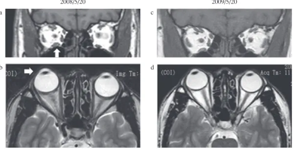
Fig. 1 Magnetic resonance image of orbital muscle
a, b: At presentation, right inferior rectal muscle was enlarged and infl amed.
c, d : After treatment with anti-tuberculosis therapy, obvious reduction in the muscle swelling and exophthalmos was reduced which was accompanied by improvement in the patient’s symptom. a b c d 2008/5/20 2009/5/20 初診時画像所見:胸部単純エックス線写真にて右上肺 野縦隔側に辺縁整な結節影を認めた。 4 月 3 日の検診時 も同病変を指摘できたが,増大傾向であった。胸部単純 CT では右 S1に径 25 mm 大の壁の厚い空洞病変,その周 囲に小葉中心性粒状影を認めた。縦隔リンパ節腫脹や胸 水貯留は認めなかった(Fig. 2a,b)。 臨床経過(Fig. 3):2008 年 12 月 12 日気管支鏡施行。 右上葉(B1)からの集痰,ブラッシング,気管支洗浄液 では抗酸菌塗抹陰性であった。気管支鏡後の喀痰で塗抹 陰性,結核菌培養陽性,結核菌 _ 核酸増幅検査陽性で肺 結核と診断した。12 月 15 日より抗結核薬 4 剤(イソニ アジド 300 mg,リファンピシン 450 mg,エタンブトール 750 mg,ピラジナミド 1200 mg)の投与を開始した。な お眼窩筋炎に対し,プレドニゾロン換算 5 mg 内服中で あり,リファンピシンによる血中濃度低下をきたすため プレドニゾロン 10 mg に増量した。画像上も結核病変は 縮小し,2009 年 1 月 16 日退院となった。抗結核薬を開 始後,右の複視は軽度残存するものの眼球突出は軽快し
Fig. 3 Clinical course
INH : isoniazid, RFP : rifampicin, EB : ethambutol, PZA : pyrazinamide mPSL : methyl prednisolone, PSL : prednisolone, BF : Broncho Fiberscopy
5/22 5/30 mPSL 1g 3TD PSL BF 30 20 17.5 15 10 5 10 5 INH+RFP+EB+PZA INH+RFP 2008 2009 4/3 5/20 12/3 2/20 6/19 9/18 onset of exophthalmos
and orbital pain QFT positive PSL off
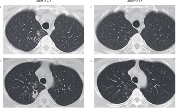
Fig. 2 Chest CT scan shows cavity and centrilobular shadows in apex of right lung at presentation (a, b).
9 months after anti-tuberculosis treatment these lesions almost disappeared and scarred (c, d). a b c d 2008/12/3 2009/9/18 た(Fig. 1c,d)。抗結核薬による副作用もなく,感受性 検査も薬剤耐性を認めずピラジナミド,エタンブトール は 2 カ月間で終了した。眼窩筋炎に対しステロイド内服 中であったためイソニアジド,リファンピシンは 7 カ月 間内服(計 9 カ月間)し, 9 月 18 日に治療を終了した。 治療終了時,結核病巣は著明に縮小した(Fig. 2c,d)。 なおステロイドは 6 月 19 日に中止した。 考 察 眼窩筋炎は外眼筋に限局した炎症性疾患であり,原因 不明の特発性と,結核,サルコイドーシス,甲状腺機能 亢進症などに伴う続発性に分けられる。
特発性眼窩筋炎は 1903 年 Gleason によって報告され た3)。その原因については諸説あるが,本疾患は除外診 断であり,眼窩筋炎が疑われる症例の評価においては, その原因として悪性腫瘍や感染症を見逃さないよう適切 なステップを踏む必要がある4)。特発性眼窩筋炎の臨床 症状は,急激な眼痛,眼球運動障害,結膜充血・浮腫, 眼球突出であり,画像(CT や MRI)にて外眼筋の肥厚が 認められる。一般に特発性眼窩筋炎のステロイド反応性 は良好とされるが,奏効率は 50% 程度にすぎないとの報 告もあり5),特発性眼窩筋炎と臨床症状が類似している
Mucosa-associate lymphoid tissue(MALT)タ イ プ 悪 性 リ ンパ腫はステロイド治療によく反応するとされている。 後に詳述するが,結核性眼窩筋炎の病初期にはステロイ ドに反応するとの報告6)もあり,ステロイドによる診断 的治療で特発性眼窩筋炎と診断するのは危険である7)。 続発性眼窩筋炎の原因として,甲状腺機能亢進症,肉芽 腫性疾患(結核8),梅毒,サルコイドーシス),膠原病お よびその類縁疾患(SLE,RA,ウェゲナー肉芽腫症,シ ェーグレン症候群)などが挙げられる。原因となる感染 症に関して,本邦ではインフルエンザB9),サイトメガロ ウイルス10)によると思われる報告があるが,われわれが 検索した限りでは,本邦での結核に続発した眼窩筋炎の 報告はなかった。本症例では,眼窩筋炎の原因として膠 原病,サルコイドーシス,甲状腺機能異常症は否定した ものの,結核を鑑別に挙げておらず,特発性眼窩筋炎と 診断された。ステロイドパルス療法後,経口ステロイド, ステロイド点眼により眼痛は軽快,眼球突出は軽度改善 したが,特発性眼窩筋炎にしてはステロイド反応性が不 良であった。 眼窩筋炎をはじめとした眼結核は,肺外結核の中でも 稀であり,眼結核は全身結核患者の 0.6%∼18% に存在す る11)。眼結核の発生機序として以下の 4 つの機序がある とされている11)。①最も多いパターンである血行性進展, ②眼瞼や結膜には,初感染原発巣として発症,③隣接臓 器からの直接浸潤や患者の喀痰による二次感染,④アレ ルギー反応と考えられる発症様式をとるフリクテン性角 結膜炎やイールズ病である。結核に続発した眼窩筋炎は, 隣接臓器からの直接浸潤や血行性進展により発症すると されるが,後者は稀とされる12)。本症例においては,頭 部造影 MRI では隣接臓器および脳結核は指摘できず,血 行性進展をきたしたと考えられた。 眼結核は,眼内結核と眼外結核に大別され,結核に続 発した眼窩筋炎は,眼外結核に分類される。眼内結核に おいて結核菌感染を証明することは,ほとんどの場合不 可能である。眼外結核については病理組織学的あるいは 微生物学的に結核の証明を試みるが,ルーチンとして施 行する検査としては侵襲が大きすぎるため眼外結核にお いても臨床的に診断を進めることが多い8)。 結核性眼窩筋炎の臨床症状で最もよくみられるものは 潜在性かつ進行性の片側性の眼球突出であり,熱感を伴 わない眼瞼の浮腫,結膜浮腫,結膜充血などを呈する8)。 その臨床経過は,急性発症する特発性眼窩筋炎とは対照 的に典型的には緩徐,慢性進行型であり,症状の持続期 間は 1 カ月から 12 年に及ぶとされる13)。本症例は今回, 急激な眼痛と眼球突出で発症し特発性眼窩筋炎と診断さ れた。しかし,既述したように症状が長期化しており, 慢性眼窩筋炎の急性増悪のような発症パターンをとった のではないかと推察する。 結核性眼窩筋炎をはじめとした眼結核の治療は,抗結 核薬の投与が基本であり,肺結核と同様に 6 ∼ 9 カ月の 抗結核薬投与が必要である14)。網膜静脈炎や結節性脈絡 網膜炎には有効なことが多い。 Oliveria らは当初,特発性眼窩筋炎と診断され,ステ ロイド投与中に結核性眼窩筋炎と診断された 1 例を報告 している6)。左外直筋の腫脹を伴った 17 歳男性は,特発 性眼窩筋炎と診断され,ステロイド(プレドニゾロン換 算 1 mg/kg)を投与された。初期のステロイドに対する 反応は良好であったが,ステロイド減量中に眼症状の再 燃を認め,最終的に結核性眼窩筋炎と診断された。Oli-veria らはステロイドが有効であったのは,ステロイドが 初期の炎症反応のみを抑制した可能性があると述べてい る。本症例でもステロイドの抗炎症作用により外眼筋の 炎症がある程度改善したと考えられるが,抗結核薬投与 がなされず眼球突出が残存したと推察する。抗結核薬投 与後は,複視,眼球突出は残存するものの明らかに改善 した(Fig. 1c, d)。この治療反応性は眼窩筋炎が結核に 続発した可能性を示唆する。今回,仮にステロイドパル ス療法前に胸部単純エックス線写真・CT を施行してい たとすれば結核の診断が早期につき,一元的に考えて結 核に続発した眼窩筋炎と診断されたであろうと考える。 全身ステロイド投与,特にステロイドパルス療法前は胸 部単純エックス線写真撮影による活動性肺結核の除外が 必要である。このことを怠ったことが,結核の診断およ び治療の遅れにつながった。 結 語 眼窩筋炎を伴った肺結核の 1 例を経験した。眼筋から 結核菌の分離,肉芽腫性病変の証明はなされていないが, 眼球突出,眼痛,複視などの眼症状出現前に肺病変が認 められており,一元的に考えると肺結核に続発した眼窩 筋炎である可能性がある。眼窩筋炎の原因として結核が あることを忘れずに治療前に胸部の精査が必要である。 今回,診断・治療において大変お世話になりました国
立病院機構熊本南病院呼吸器科 坂本理先生に深謝申し 上げます。 なお本演題は第 32 回日本呼吸器内視鏡学会学術集会 (2009 年 5 月 29 日,東京)で発表した。 文 献 1 ) 矢崎俊二:外眼筋炎.「神経症候群Ⅰ」, 日本臨牀社, 東 京, 1999, 721 _723.
2 ) Hsuan JD, Selva D, McNab AA, et al.: Idiopathic Sclerosing Orbital Infl ammation. Arch Ophthalmol. 2006 ; 124 : 1244_ 1250.
3 ) Gleason JE: Idiopathic myositis involving the intraocular muscles. Ophthalmol Rec. 1903 ; 12 : 471_478.
4 ) Costa RM, Dumitrascu OM, Gordon LK, et al.: Orbital myositis: diagnosis and management. Curr Allergy Asthma Rep. 2009 ; 9 : 316_323.
5 ) 恭子, 久保田敏信, 廣瀬浩士:眼瞼型特発性眼窩 炎症(眼窩偽腫瘍)の臨床的特徴と治療効果. 臨床眼 科. 2006 ; 60 : 1283_1287.
6 ) Oliveria BF, Takay FC, Shida TM, et al.: Orbital tuberculosis diagnosed by immunohistochemistry, case report. Rev Inst Med Trop Sao Paulo. 2004 ; 66 : 291_294.
7 ) 久保田敏信:眼窩腫瘍・炎症性疾患の診断と治療 最 近の話題, 特発性眼窩炎症と特発性外眼筋炎. 神経眼 科. 2008 ; 25 : 459_465.
8 ) Chakraborti C, Samanta SK, Biswas J, et al.: Tuberculous Myositis of the Orbit. Asian J Ophthalmol. 2008 ; 10 : 398_400. 9 ) 今泉利雄:インフルエンザ B ウイルスの感染が疑われ た眼窩筋炎の1症例. 神経眼科. 1992 ; 9 : 217_220. 10) 保科幸次, 久保田浩, 三村 治:ステロイド離脱が困 難 で あ っ た 両 側 性 眼 窩 筋 炎 の 一 例. 日 本 眼 科 紀 要. 1994 ; 45 : 572_574.
11) Schlossberg D: Tuberculosis and Nontuberculosis Myco-bacterial Infections. 5th ed., McGraw-Hill Professional. 2005, 202_214.
12) Pillai S, Malone TJ, Abad JC, et al.: Orbital tuberculosis. Ophthalmol Ptast Reconstr Surg. 1995 ; 11 : 27_31. 13) Helm CJ, Holland GN: Ocular tuberculosis. Surv
Ophthal-mol. 1993 ; 38 : 229_256.
14) American Thoracic Society/Centers for Disease Control and Prevention/Infectious Diseases Society of America: Treatment of Tuberculosis. Am J Respir Crit Care Med. 2003 ; 167 : 603_662.
Abstract Ocular tuberculosis is rare. We report a case of
orbital myositis suspected to be infected with tuberculosis. In January 2008, a 34-year-old man experienced discomfort in the right eye. In May 2008, this patient developed right exophthalmos, diplopia, and pain in the right eye, and he was diagnosed with idiopathic orbital myositis. The patient under-went 2 courses of steroid pulse therapy; after which, the dos-age of steroids was reduced. The steroid treatment reduced the eye pain, but his diplopia and exophthalmos persisted. By November of the same year, his general malaise had increased, and chest X-ray radiography and computed tomography were performed on 3rd December. On the basis of the imaging results, we suspected active pulmonary tuberculosis of the right upper lobe. The smear made by using the sample obtained after bronchial brushing was negative for acid-fast bacilli, but a Mycobacterium tuberculosis nucleic acid amplifi cation test of the post-bronchoscopic sputum yielded positive results. Therefore, the patient was diagnosed with pulmonary tuber-culosis. After the 2HREZ/7HR regimen of treatment, the extent of the tuberculosis lesions of the lung was reduced and the exophthalmos and eye pain were alleviated. Orbital myo-sitis is infl ammation of the extraocular muscles and can be
either idiopathic, without a known etiology, or secondary to conditions such as tuberculosis, sarcoidosis, or hyperthyroid-ism. Our patient was not defi nitively diagnosed with tuber-culosis of the eye. A defi nitive diagnosis of tubertuber-culosis of the eye would require detection of granulomatous lesions in the eye or isolation of Mycobacterium tuberculosis by punc-turing the eye muscles; however, our fi ndings suggested the possibility that it was secondary to tuberculosis. We think that a careful examination of the chest should be performed for patients with ocular abnormalities.
Key words: Orbital myositis, Exophthalmos, Diplopia,
Tu-berculosis, Response to steroid therapy
1Respiratory Department, Taragi Municipal Hospital, 2
Respira-tory Department, National Hospital Organization Kumamoto-minami National Hospital
Correspondence to: Kazuyoshi Nakamura, Respiratory De-partment, Taragi Municipal Hospital, 4210 Taragi, Taragi-machi, Kuma-gun, Kumamoto 868_0598 Japan.
(E-mail: kazu1124@pj9.so-net.ne.jp) −−−−−−−−Case Report−−−−−−−−