Other uses, including reproduction and distribution, or selling or licensing copies, or posting to personal, institutional or third party
websites are prohibited.
In most cases authors are permitted to post their version of the article (e.g. in Word or Tex form) to their personal website or institutional repository. Authors requiring further information
regarding Elsevier’s archiving and manuscript policies are encouraged to visit:
http://www.elsevier.com/authorsrights
Original article
Relationship between coronary artery stenosis and cardio-ankle vascular index (CAVI) in patients undergoing cardiovascular surgery
Masafumi Kanamoto
a,*, Naoki Matsumoto
b, Tatsuya Shiga
c, Fumio Kunimoto
a, Shigeru Saito
daGunma University Hospital Intensive Care Unit, Japan
bDepartment of Anesthesiology, Gunma Prefectural Children’s Medical Center, Japan
cDepartment of Anesthesiology, Gunma Prefectural Cardiovascular Center, Japan
dDepartment of Anesthesiology, Gunma University Hospital, Japan
a r t i c l e i n f o
Article history:
Received 2 August 2012 Accepted 7 November 2012 Available online 27 February 2013
Keywords:
Anesthesia Atherosclerosis
Cardio-ankle vascular index Cardiovascular surgery
a b s t r a c t
Background: The cardio-ankle vascular index (CAVI) was developed as an indicator of arterial wall stiffness, and it is less influenced by blood pressure (BP). We investigated the relationship between the CAVI and coronary artery disease (CAD), and evaluated the effects of rapid changes in BP induced by anesthetics on CAVI.
Materials and methods:We measured the CAVI in 76 patients before and after the administration of anesthetics for elective cardiovascular surgery. The patients were assigned to groups with or without CAD (0VD). The CAD group was then divided into 3 subgroups based on the number of stenotic vessels (1VD, 2VD, and 3VD). We compared the CAVI between CAD and 0VD, and changes in BP during the induction of anesthesia. All data were analyzed using Stat View 5.0 software.
Results:Systolic BP significantly decreased from 14521 to 10720 mmHg, whereas CAVI was not altered after the administration of intravenous anesthetics. Changes in BP and in pre-anesthetic CAVI (pre-CAVI) did not correlate. The pre- and post-anesthetic values for the CAVI (post-CAVI) in the 0VD and CAD groups were 8.341.01 and 8.441.39, and 9.951.22 and 10.121.56, respectively. Both values were higher in the CAD, than in the 0VD group (P<0.05).
Conclusion:The CAVI is independent of BP and reproducible regardless of the induction of anesthesia and is significantly higher in patients with CAD. The CAVI might be able to predict atherosclerosis and cor- onary artery stenosis in patients undergoing cardiovascular surgery.
CopyrightÓ2013, SciBioIMed.Org, Published by Reed Elsevier India Pvt. Ltd. All rights reserved.
1. Introduction
Atherosclerosis is a major contributor to cardiovascular diseases that accounts for a considerable portion of mortality and morbidity rates.1,2The degree of atherosclerosis in a coronary artery is usually evaluated by invasive coronary angiography (CAG). Coronary CT imaging is non-invasive, but it requires transferring patients to a specific location, and this modality is not routinely available at all institutions.
Brachial-ankle pulse wave velocity (baPWV) is a simple method of assessing early arteriosclerosis. However, blood pressure (BP) influences measured baPWV values, and complicates the inter- pretation of measured values among patients with unstable blood
pressure.3,4The novel cardio-ankle vascular index (CAVI) is inde- pendent of BP because BP is adjusted based on the stiffness parameter b, which can be rapidly measured without using specialized techniques.5e9 Reports indicate that CAVI correlates with coronary atherosclerosis,10e12atherosclerotic complications in patients with diabetes,13,14intimal thickness of the carotid artery,15 left ventricular diastolic function,16,17 and the degree of athero- sclerosis in patients on hemodialysis.18However, a direct correla- tion between the CAVI and coronary artery stenosis in patients undergoing cardiovascular surgery has not been described, and the effects of rapid BP changes on the CAVI has not been evaluated.
Blood pressure often rapidly changes in patients who undergo cardiovascular surgery, especially during peri-operative periods such as anesthesia induction.
Here, we measured the CAVI in patients undergoing elective cardiovascular surgery to determine its relationship to pre- operative coronary angiography (CAG) findings. We also deter- mined the effects of BP changes induced by anesthetics in the
*Corresponding author. Gunma University Hospital Intensive Care Unit, Department of Anesthesiology, 3-39-15 Showamachi, Maebashishi, Gunma 371- 8511, Japan.
E-mail address:[email protected](M. Kanamoto).
Contents lists available atSciVerse ScienceDirect
Journal of Cardiovascular Disease Research
j o u r n a l h o m e p a g e : w w w . e l s e v i e r . c o m / l o c a t e / j c d r
0975-3583/$esee front matter CopyrightÓ2013, SciBioIMed.Org, Published by Reed Elsevier India Pvt. Ltd. All rights reserved.
http://dx.doi.org/10.1016/j.jcdr.2013.02.001
operating room by measuring CAVI immediately before and 3 min after the induction of anesthesia (before tracheal intubation). Cor- onary artery stenosis was preoperatively evaluated in all patients.
2. Materials and methods 2.1. Subjects
All patients provided written informed consent to participate in this study, which was approved by the Ethics Committee of the Gunma Prefectural Cardiovascular Center.
Between September 2010 and March 2011, 98 patients under- went CAG before elective cardiovascular surgery at Gunma Pre- fectural Cardiovascular Center. We excluded 22 of them because of a low ankle-brachial index (ABI<0.9) and chronic atrialfibrillation that could affect the CAVI.5,10,15The remaining 55 male and 21 fe- male patients were included in this study.
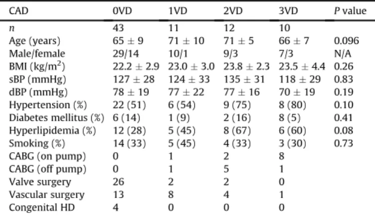
The patients were assigned to groups according to the number of stenotic vessels identified by CAG. The ratio (%) of luminal nar- rowing was determined according to the American Heart Associa- tion criteria.19 Significant stenosis was defined as narrowing of 75% of the vessel diameter (CAD group). The 0VD group comprised 43 patients without significant stenosis of the coronary arteries, and the 1VD, 2VD, and 3VD groups comprised 11, 12, and 10 patients with significant 1-, 2-, and 3-vessel stenosis, respec- tively. The elective cardiovascular procedures included coronary artery bypass grafting (on- or off-pump), valve replacement, vascular replacement (of the aortic arch or abdominal aorta), and radical surgery for congenital heart diseases (Table 1).
2.2. CAVI measurements
We measured the CAVI using a Vasera VS-1500 (Fukuda Denshi Co. Ltd., Tokyo, Japan). In brief, cuffs were applied to the bilateral upper arms and ankles, with the patient lying supine and the head placed in the midline position. To detect brachial and ankle pulse waves using cuffs, a low cuff pressure of 50 mmHg was applied to minimize the effect of cuff pressure on hemodynamics, and BP was measured thereafter. We applied a scale conversion for convenient comparison with PWV. We calculated CAVI as follows:
an
ð2
r
=D
PÞ lnðPs=PdÞPWV2o þb;WherePs is systolic BP,Pd is diastolic BP, PWV is pulse wave ve- locity,DPisPsePd,ris blood density, and a and b are constants.
Blood pressure was obtained using an upper arm cuff.
Pulse wave velocity was obtained by dividing the estimated vascular length by the time required for the pulse wave to propa- gate from the aortic valve to the ankle and measured using upper arm and ankle cuffs. We determined scale conversion constants (a, b) to match the CAVI with the aortic PWV method established by Hasegawa and coworkers.20 The scale conversion constants allowed the conversion of massive amounts of extant PWV data into CAVI. All these measurements and calculation systems were assembled in the hardware, and values were automatically calcu- lated by the Vasera.
2.3. Anesthetic management
After measuring thefirst CAVI value, anesthesia was induced with propofol (1e2 mg/kg) or midazolam (0.1e0.15 mg/kg) and fentanyl (4e6mg/kg) or remifentanil (0.5mg/kg/min). Rocuronium (0.8 mg/kg) was then administered, and the patients were mask- ventilated with 100% oxygen. A second CAVI value was measured 3 min later and immediately before endotracheal intubation.
2.4. Statistical analysis
All data are expressed as meanstandard deviation (SD). The sample size was calculated before starting the study based on the notion that an increase of 1 in the CAVI is important. The power of the sample size to detect a 20% difference between 0VD and CAD with a 5% probability of a type II error was 80%. Systolic BP and the CAVI were compared before and after the induction of anesthesia using a paired t-test. Patients’ characteristics and pre/post- anesthesia CAVI values in the 0VD and CAD group were compared using an unpairedt-test. A one-way ANOVA and Fisher’s test were used for multiple comparisons in 0VD, 1VD, 2VD, and 3VD CAVI values. Values ofP<0.05 were considered to indicate sta- tistical significance in all comparisons. All data were analyzed using Stat View 5.0 software (Abacus Concepts Inc., Berkeley, CA, USA).
3. Results
Table 1 shows the clinical characteristics of the 76 patients.
Systolic and diastolic BP before the induction of anesthesia, body mass index, and ratios of complications (hypertension, diabetes mellitus, and hyperlipidemia) did not significantly differ between the 0VD and CAD groups.
Systolic BP significantly decreased from 145 21 to 10720 mmHg with a rate of change of 260.1% (Fig. 1a), whereas the CAVI did not change after the induction of anesthesia (Fig. 1b).
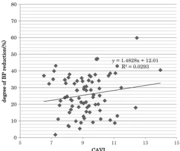
The change in BP did not correlate with pre-CAVI values (Fig. 2).
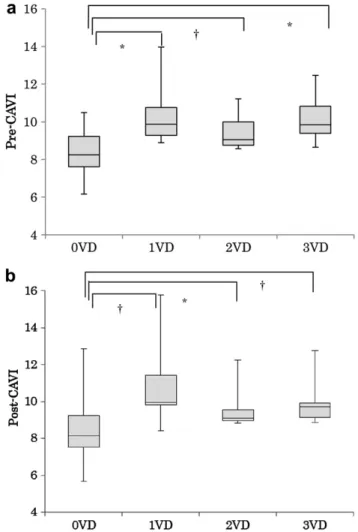
The pre- (pre-CAVI) and post- (post-CAVI) anesthesia values for CAVI were 8.341.01 and 8.441.39 in the 0VD group, 9.951.22 and 10.121.56 in the CAD group (Fig. 3a and b), and 10.271.46 and 10.752.01, 9.560.98 and 9.560.98, and 10.071.19 and 10.11.44 in the 1-, 2- and 3VD groups, respectively (Fig. 4a and b).
These values were significantly higher in all three groups with CAD than with 0VD (P<0.05).
4. Discussion
The CAVI has recently served as a parameter of atherosclerosis in various diseases since not only can it be measured rapidly and non- invasively, but it is also independent of BP and quite reproducible.5e
9Some studies have indicated that CAVI could be a useful predictor of coronary atherosclerosis in individuals with risk factors for Table 1
Patients’characteristics and types of elective surgery according to coronary artery disease.
CAD 0VD 1VD 2VD 3VD Pvalue
n 43 11 12 10
Age (years) 659 7110 715 667 0.096
Male/female 29/14 10/1 9/3 7/3 N/A
BMI (kg/m2) 22.22.9 23.03.0 23.82.3 23.54.4 0.26
sBP (mmHg) 12728 12433 13531 11829 0.83
dBP (mmHg) 7819 7722 7716 7019 0.19
Hypertension (%) 22 (51) 6 (54) 9 (75) 8 (80) 0.10 Diabetes mellitus (%) 6 (14) 1 (9) 2 (16) 8 (5) 0.41 Hyperlipidemia (%) 12 (28) 5 (45) 8 (67) 6 (60) 0.08
Smoking (%) 14 (33) 5 (45) 4 (33) 3 (30) 0.73
CABG (on pump) 0 1 2 8
CABG (off pump) 0 1 5 1
Valve surgery 26 2 2 0
Vascular surgery 13 8 4 1
Congenital HD 4 0 0 0
Values are shown as meansSD or number of patients.Pvalue is 0VD vs. CAD. 0VD, 1VD, 2VD, and 3VD, no, 1-, 2- and 3-vessel disease, respectively. BMI, body mass index; s/dBP, systolic/diastolic blood pressure; CABG, coronary artery bypass graft surgery; Congenital HD, congenital heart disease.
M. Kanamoto et al. / Journal of Cardiovascular Disease Research 4 (2013) 15e19 16
cardiovascular disease, and indicate the severity of coronary atherosclerosis. Nakamura et al reported that the CAVI increases with an increasing number of stenosed vessels and established a CAVI cutoff of 8.91 for the presence of coronary stenosis.10Izuhara et al similarly found that CAVI is independently associated with the severity of coronary atherosclerosis.15Horinaka et al and Miyoshi et al found other evidence that the CAVI is a good predictor of coronary artery disease.11,12Sairaku et al reported that the CAVI is higher in patients with acute coronary disease than in those with stable angina pectoris.21
The presence and extent of coronary artery disease is usually determined by coronary angiography, although it is invasive and associated with an increased risk of complication. Coronary artery computed tomography (coronary CT) is non-invasive, and it can detect coronary atherosclerosis with high sensitivity. However, these methods require specialized devices and operators, as well as a large space. Consequently, these modalities are not routinely available at all institutions. The extent of atherosclerosis has been estimated using PWV, but this can be influenced by blood pressure,
and it is not very reproducible.3,4Because it is independent of BP, highly reproducible, easy to apply, and does not require special techniques, the potential of CAVI as a novel parameter of athero- sclerosis has recently become recognized.
We measured CAVI in patients before and after the induction of anesthesia for elective cardiovascular surgery to determine whether CAVI is independent of changes in BP induced by anes- thetics, and to define the relationship between the CAVI and cor- onary artery stenosis. Changes in BP induced by anesthetics did not influence CAVI in the present study, and the CAVI was significantly higher in patients with coronary artery disease. To our knowledge, this is thefirst study to measure CAVI before and after the induction of anesthesia in the operating theater and to determine the effects of acute changes of BP on CAVI.
We found that CAVI was consistently independent of BP in all patients examined. The decrease in blood pressure after the administration of anesthetics is thought to be considerable in pa- tients with a high CAVI (that is, with advanced atherosclerosis.) However, the present study found no statistically significant cor- relation. Anesthesia induction provoked similar BP changes in pa- tients with and without CAD. Patients with CAD are generally thought to have more atherosclerosis and hypertension than healthy individuals. However, our patients were scheduled for elective surgery, and their pre-operative BP was carefully managed.
This pre-operative background might explain the minor difference in BP values.
Although the CAVI significantly increased in patients with cor- onary artery disease, it was not related to the severity of CAD as reported.10,15This might be because of the limited number of pa- tients with multiple coronary stenosis who elected to undergo surgery in the present study. A larger-scale study of patients with more severe disease might be able to detect a correlation between CAVI and the number of diseased coronary arteries. The present results suggest that coronary stenosis can exist at least in one branch when patients have a high CAVI.
In addition, this study has several limitations. Firstly, we excluded patients with ABI<0.9 because their CAVI was considered inaccurate. However, CAD is closely related to peripheral arterial disease, and a low ABI is an independent marker of an additive risk for CAD.22Therefore, ABI might predict coronary atherosclerosis, Fig. 2.Relationship between systolic BP reduction and CAVI. Degree of reduction in systolic BP and CAVI do not significantly correlate (R¼0.17,P¼0.13). The degree of BP reduction (rate of change (%)) is calculated; (1epost-anesthesia BP/pre-anesthesia BP)100.
Fig. 1.(a) Comparison of systolic blood pressure (sBP) between pre- and post- anesthesia. Systolic BP significantly decreased after administering intravenous anes- thetics. Systolic BP: pre vs. post-anesthesia: 14521 vs. 10720 mmHg. *P<0.01.
Rate of change (%) was calculated as: (1 e post-anesthesia BP/pre-anesthesia BP)100. (b) Comparison of CAVI between pre- and post-anesthesia. CAVI did not significantly change after administering intravenous anesthetics. CAVI pre vs. post- anesthesia; 9.041.36 vs. 9.161.68. *P¼0.61. Rate of change (%) was calculated as: (1epost-anesthesia CAVI/pre-anesthesia CAVI)100.
especially when the value is low. Measuring both the CAVI and the ABI in patients with CAD might be valuable where possible, since measurements of these two parameters are reliable, and they seem to be mutually compensational; that is, earlier atherosclerosis is detected by CAVI, whereas ABI detects established and advanced atherosclerosis. Secondly, we also excluded patients with atrial fibrillation. However, patients with CAD might have atrialfibrilla- tion. Another study of such patients might provide more extensive information about the association between atherosclerosis and CAD.
The present study demonstrated that CAVI could indicate cor- onary artery stenosis in patients with heart disease. Applying this finding to patients without heart disease might be presumptuous.
However, if coronary artery stenosis can be indicated by the CAVI in patients with coronary risk factors such as hypertension, diabetes, hyperlipidemia, and a smoking habit, physicians could prepare more effective intra- and post-operative management strategies to prevent the onset of serious cardiovascular events.
We concluded that the CAVI could predict atherosclerosis and coronary artery stenosis in patients undergoing cardiovascular
surgery, and that it was reproducibly independent of BP regardless of the effects of anesthetics.
Conflicts of interest
All authors have none to declare.
Acknowledgment
The authors wish to thank Forte (Tokyo, Japan) for assistance with manuscript preparation in English. This study was supported in part by grants to Dr. Kunimoto and Dr. Saito from the Japanese Ministry of Education, Culture, Sports, Science and Technology.
References
1. World Health Organization. The World Health Report 2001 Annex Table 2.
Deaths by causes, sex and mortality stratum in WHO regions, estimates for 2000. Available from:http://www.who.int/whr/2001/main/en/annex/annex2.
htm. Last Accessed 08.05.02.
2. Murray CJ, Lopez AD. Alternative projections of mortality and disability by cause 1990e2020: Global Burden of Disease Study.Lancet. 1997;349:1498e1504.
3. Yamashina A, Tomiyama H, Takeda K, et al. Validity, reproducibility, and clin- ical significance of noninvasive brachial-ankle pulse wave velocity measure- ment.Hypertens Res. 2002;25:359e364.
Fig. 4.(a) CAVI before induction of anesthesia (pre-CAVI) and number of CAD (*P<0.01,yP<0.05). Pre-CAVI and number of CAD are not significantly related (1- vs.
2- and 3VD,P¼0.17 andP¼0.72, respectively; 2- vs. 3VD,P¼0.28). (b) CAVI after induction of anesthesia (post-CAVI) and number of CAD (*P<0.01,yP<0.05). Post- CAVI and number of CAD are not significantly related (1- vs. 2- and 3VDP¼0.08 andP¼0.41, respectively; 2- vs. 3VD,P¼0.30).
Fig. 3.(a) CAVI before induction of anesthesia (pre-CAVI) in 0VD vs. CAD. Pre-CAVI is significantly higher in CAD than in 0VD (*P<0.01). (b) CAVI after induction of anes- thesia (post-CAVI) in 0VD vs. CAD. Post-CAVI is significantly higher in CAD than in 0VD (*P<0.01).
M. Kanamoto et al. / Journal of Cardiovascular Disease Research 4 (2013) 15e19 18
4. Imanishi R, Seto S, Toda G, et al. High brachial-ankle pulse wave velocity is an independent predictor of the presence of coronary artery disease in men.
Hypertens Res. 2004;27:71e78.
5. Shirai K, Utino J, Otsuka K, Takata M. A novel blood pressure-independent arterial wall stiffness parameter; cardio-ankle vascular index (CAVI).
J Atheroscler Thromb. 2006;13:101e107.
6. Kubozono T, Miyata M, Ueyama K, et al. Clinical significance and reproducibility of new arterial distensibility index.Circ J. 2007;71:89e94.
7. Ibata J, Sasaki H, Kakimoto T, et al. Cardio-ankle vascular index measures arterial wall stiffness independent of blood pressure.Diabetes Res Clin Pract.
2008;80:265e270.
8. Konishi R, Kimura S, Kawagoe I, Kanai M, Mitsuhata H. Assessment of the quality of the newly developed rapid oscillometric blood pressure measure- ment.Masui. 2010;59:1058e1062.
9. Kim B, Takada K, Oka S, Misaki T. Influence of blood pressure on cardio-ankle vascular index (CAVI) examined based on percentage change during general anesthesia.Hypertens Res. 2011;34:779e783.
10. Nakamura K, Tomaru T, Yamamura S, Miyashita Y, Shirai K, Noike H. Cardio- ankle vascular index is a candidate predictor of coronary atherosclerosis.Circ J.
2008;72:598e604.
11. Horinaka S, Yabe A, Yagi H, et al. Comparison of atherosclerotic indicators between cardio ankle vascular index and brachial ankle pulse wave velocity.
Angiology. 2009;60:468e476.
12. Miyoshi T, Doi M, Hirohata S, et al. Cardio-ankle vascular index is indepen- dently associated with the severity of coronary atherosclerosis and left ven- tricular function in patients with ischemic heart disease.J Atheroscler Thromb.
2010;17:249e258.
13. Wakabayashi I, Masuda H. Association of acute-phase with arterial stiffness in patients with type 2 diabetes mellitus.Clin Chim Acta. 2006;365:230e235.
14. Wakabayashi I, Masuda H. Relationships between vascular indexes and atherosclerotic risk factors in patients with type 2 diabetes mellitus.Angiology.
2008;59:567e573.
15. Izuhara M, Shioji K, Kadota S, et al. Relationship of cardio-ankle vascular index (CAVI) to carotid and coronary arteriosclerosis. Circ J. 2008;72:
1762e1767.
16. Sakane K, Miyoshi T, Doi M, et al. Association of new arterial stiffness parameter, the cardio-ankle vascular index, with left ventricular diastolic function.J Atheroscler Thromb. 2008;15:261e268.
17. Mizuguchi Y, Oishi Y, Tanaka H, et al. Arterial stiffness is associated with left ventricular diastolic function in patients with cardiovascular risk factors: early detection with the use of cardio-ankle vascular index and ultrasonic strain imaging.J Card Fail. 2007;13:744e751.
18. Ichihara A, Yamashita N, Takemitsu T, et al. Cardio-ankle vascular index and ankle pulse wave velocity as a marker of arterialfibrosis in kidney failure treated by hemodialysis.Am J Kidney Dis. 2008;52:947e955.
19. Austen WG, Edwards JE, Frye RL, et al. A reporting system on patients evalu- ated for coronary artery disease. Report of the Ad Hoc Committee for Grading of Coronary Artery Disease, Council on Cardiovascular Surgery, American Heart Association.Circulation. 1975;51:5e40.
20. Hasegawa M. A fundamental study on human aortic pulse wave velocity.Tokyo JikeikaiIka Daigaku Zasshi. 1970;85:742e760.
21. Sairaku A, Eno S, Hondo T, et al. Head-to-head comparison of the cardio-ankle vascular index between patients with acute coronary syndrome and stable angina pectoris.Hypertens Res. 2010;33:1162e1166.
22. Koji Y, Tomiyama H, Ichihashi H, et al. Comparison of ankle-brachial pressure index and pulse wave velocity as markers of the presence of coronary artery disease in subjects with a high risk of atherosclerotic cardiovascular disease.
Am J Cardiol. 2004;9:868e872.