T
he gastrointestinal (GI) tract is the most com- monly involved site of extranodal non-Hodgkin’s lymphomas. The predominant subtypes of lymphomas are different according to the site of the GI tract: mu- cosa-associated lymphoid tissue (MALT) lymphoma for the stomach (46.2%) [1], follicular lymphoma for the duodenum (38%) [2], and diffuse large B-cell lym- phoma (DLBCL) for the small and large intestines (41%) [3]. DLBCL of the small intestine is a rare dis- ease but sometimes causes abdominal pain, intestinalobstruction or perforation. Our studies focusing on clinicopathological features of primary intestinal DLBCL revealed that perforation was one of the inde- pendent poor prognostic factors in addition to the pre- dominance of activated B-cell (ABC) phenotype [4,5].
A20, also known as tumor necrosis factor alpha-in- duced protein 3 (TNFAIP3), is located on chromosome band 6q23 and negatively regulates the NF-κB pathway, which is dysregulated by several types of genetic alter- ations including oncogenic mutations of MALT1 and CARD11 [6]. Deletion of A20 (TNFAIP3) was reported
CopyrightⒸ 2018 by Okayama University Medical School.
http ://escholarship.lib.okayama-u.ac.jp/amo/
Original Article
A20 (TNFAIP3) Alterations in Primary Intestinal Diffuse Large B-cell Lymphoma
Masayoshi Fujiia,b, Katsuyoshi Takataa*, Shih-Sung Chuangc*, Tomoko Miyata-Takataa, Midori Andod, Yasuharu Satoa, and Tadashi Yoshinoa
aDepartment of Pathology, Okayama University Graduate School of Medicine, Dentistry and Pharmaceutical Sciences, Okayama 700-8558, Japan, bDepartment of Pathology, St. Mary’s Hospital, Himeji, Hyogo 670-0801, Japan,
cDepartment of Pathology, Chi-Mei Foundation Hospital, Tainan 710, Taiwan,
dDepartment of Pathology, Kagawa Prefectural Central Hospital, Takamatsu 760-8557, Japan
The gastrointestinal (GI) tract is the most frequently involved site of extranodal non-Hodgkin lymphomas, and diffuse large B-cell lymphoma (DLBCL) is the most common subtype occurring in the GI tract. TNFAIP3 (A20) genetic alterations were reported to be involved in DLBCL’s pathogenesis and a portion of GI-DLBCL cases har- bor this alteration. However, the frequency and clinicopathological relations focusing on small and large intes- tinal DLBCL are unclear. Here, we examined A20 deletion and protein expression and analyzed the clinicopath- ological features of 52 cases of primary intestinal DLBCL. The most frequently involved site was the ileocecal region (75%), followed by small bowel (13.5%) and large intestine. Immunohistochemically, the ileocecal cases expressed BCL6 (p=0.027) and MUM1 (p=0.0001) significantly more frequently than the small intestinal cases.
Six of 47 cases (13%) had A20 heterozygous deletion, whereas all 6 heterozygously deleted cases had detectable A20 protein expression. In summary, A20 abnormality was less prevalent among intestinal DLBCLs with some discordancy between gene deletion and protein expression. Although the A20 alteration status did not affect any clinicopathological characteristics in this series, further studies exploring alterations of A20 and other NF-κB components in primary intestinal DLBCL are needed.
Key words: primary intestinal diffuse large B-cell lymphoma, cell of origin, A20, TNFAIP3, heterozygous deletion
Received March 2, 2017 ; accepted August 10, 2017.
*Corresponding author. Phone : +81-86-235-7150; Fax : +81-86-235-7156 E-mail : [email protected] (K. Takata) or [email protected].
org.tw (Chuang SS) Conflict of Interest Disclosures: No potential conflict of interest relevant
to this article was reported.
in several types of lymphoma such as DLBCL of the ABC phenotype, MALT lymphoma, Hodgkin lym- phoma, and Epstein-Barr virus (EBV)-associated lym- phoma. A20 is a dual-function enzyme that adds and subtracts ubiquitin moieties to deactivate and degrade receptor-interacting protein (RIP), an essential media- tor of the proximal TNFR1 signaling complex [7]. A20 restricts toll-like receptor-induced NF-κB signals by deubiquitylation and also regulates MAP kinase signal- ing cascades. Further, A20 restrains inflammatory responses and acts as an anti-apoptotic factor, regulat- ing influences on immune homeostasis [8].
Although approx. 20% of DLBCL cases were reported to carry abnormalities in the A20 gene [9], the frequency of A20 alterations and its clinicopathological significance in intestinal DLBCL are not characterized.
In the present study, we investigated A20 deletion and protein expression and analyzed their significance in 56 cases of primary intestinal DLBCL.
Materials and Methods
Patient selection. Fifty-eight surgically resected intestinal DLBCL samples (obtained from 1990 to 2012, Chi-Mei Foundation Hospital, Taiwan and affiliated hospitals) were used in this study, and they are the same cases series used in the report by Lu et al. [5]. We applied strict inclusion criteria for primary intestinal lymphoma according to the American Joint Committee on Cancer Staging Manual, which is a modification of the Ann Arbor system. We excluded cases of secondary involvement from other organs in this study. The intes- tinal site was classified according to the criteria of Koch et al.: duodenum, small intestine, ileocecum, colon, and rectum [10]. The histologic diagnosis of each case was made according to the criteria by the current World Health Organization classification [11].
A total of 56 formalin-fixed paraffin embedded tis- sue (FFPET) samples were used for tissue microarray, and two whole resected FFPETs were used for immuno- histochemical (IHC) and genotypic studies. Our study was approved by the Ethics Committee and Institu- tional Review Board of Okayama University Hospital (ID no. 493) and Chi-Mei Medical Centre (ID no.
10102-016).
FISH analysis for A20. A20 deletion was investi- gated by dual-color fluorescence in situ hybridization (FISH) on FFPETs using s spectrum orange-labeled A20
probe (BAC clone RP11-783B20) and spectrum green- labeled centromeric probe for chromosome 6 (CEP6) (Vysis/Abbott Molecular Laboratories, Des Plaines, IL, USA) according to the manufacturers’ instructions [12].
The cells were scored only when 2 internal positive con- trol signals (CEP6) were present, and the signal ratio of A20 and CEP6 was calculated to evaluate the A20 status.
In DLBCL, the threshold for determining A20 homozy- gous deletions was the fraction of signals ranging from 20% to 60%, and that for heterozygous deletions was from 60% to 80% as described [13].
IHC analysis. IHC staining was performed using an automated Bond Max autostainer (Leica Biosystems, Melbourne, Australia) and a Ventana XT autostainer (Ventana Medical Systems, Tucson, AZ, USA). The clones and dilutions of the primary antibodies used for this study were as follows: A20 (EPR2663, [1 : 100], Epitomics, Burlingame, CA, USA), CD20 (L26, [1 : 200], Novocastra Laboratories, Newcastle Upon Tyne, UK), CD10 (56C6, [1 : 50], Novocastra), BCL6 (D8, [1 : 250], Santa Cruz, CA, USA), MUM1 (MUM1p, [1 : 50], Dako, Glostrup, Denmark), BCL2 (Bcl-2, [1 : 40], Dako), c-MYC (9E10, [1 : 50], Santa Cruz).
In accord with previous reports, tumors that were comprised of at least 20% of A20-positive cells were scored as positive [14]. When the internal positive con- trol cells were not clearly positive for A20, the sample was classified as “indeterminate” or “equivocal.” In the
“undetermined” groups, the tumor cells were negative, and in the “equivocal” groups, the tumor cells were weakly positive.
Statistical analysis. Overall survival (OS) was measured from the date of diagnosis to death from any cause. Event-free survival (EFS) was not available because of paucity of data of most cases. Survival curves were generated by the Kaplan-Meier method, and the value was compared by log-rank test. Fisher’s exact test or chi-squared test was used. We considered p-values <0.05 significant.
Results
Patient characteristics. Clinical data was avail- able for 52 of the 58 patients, including 29 men (56%) and 23 women (44%). The median age was 64 years (range, 23-87 years). The tumor sizes ranged from 2.4 to 16 cm with a mean size of 8.4 cm. The most fre-
quently involved site was the ileocecal region (39 of 52 patients, 75%), followed by the small bowel (jejunum and ileum, 7 of 52 patients, 13.5%) (Table 1). Two patients had multiple sites of tumors: one involving the small intestine and colon, and the other involving the duodenum, small intestine and ileocecal region. The median follow-up time was 21 months (range, 0.2-210 months).
Pathological features. According to the DLBCL tumor morphology, IHC findings were evaluable for 52 samples, and the cell-of-origin was determined using Hans’ algorithm. Sixteen cases (31%) were GCB-type, and 36 (69%) were non-GCB-type (Figs.1 and 2).
Ileocecal samples were significantly more frequently positive for BCL6 (p=0.027) and MUM1 (p=0.0001)
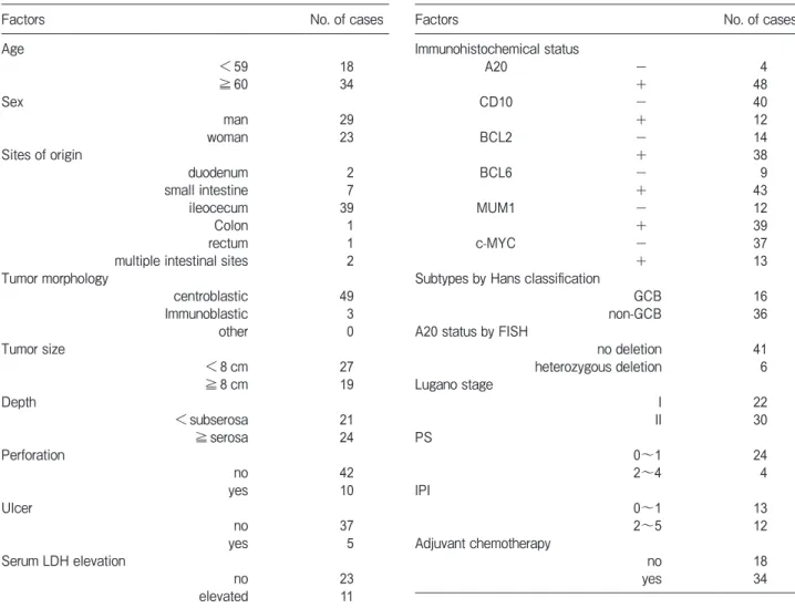
Table 1 Clinicopathological features of 52 Primary Intestinal DLBCL cases
LDH, lactate dehydrogenase; PS, performance status; IPI, International Prognostic Index.
Factors No. of cases
Age <59 18
≧60 34
Sex man 29
woman 23
Sites of origin
duodenum 2
small intestine 7
ileocecum 39
Colon 1
rectum 1
multiple intestinal sites 2 Tumor morphology
centroblastic 49
Immunoblastic 3
other 0
Tumor size
<8cm 27
≧8cm 19 Depth
<subserosa 21
≧serosa 24 Perforation
no 42
yes 10
Ulcer
no 37
yes 5
Serum LDH elevation
no 23
elevated 11
Factors No. of cases
Immunohistochemical status
A20 - 4
+ 48
CD10 - 40
+ 12
BCL2 - 14
+ 38
BCL6 - 9
+ 43
MUM1 - 12
+ 39
c-MYC - 37
+ 13
Subtypes by Hans classification
GCB 16
non-GCB 36
A20 status by FISH
no deletion 41
heterozygous deletion 6
Lugano stage
I 22
II 30
PS 0~1 24
2~4 4
IPI 0~1 13
2~5 12
Adjuvant chemotherapy
no 18
yes 34
+
+ +
-
-
-
GCB type non-GCB type
CD10 MUM1
GCB type BCL6
MUM1 +/‒ non-GCB type
12 cases 27 cases
4 cases 40 cases
9 cases 31 cases
52 cases
Fig. 1 Immunohistochemical subtypes according to Hans crite- ria.
compared to the small intestinal cases. Thirty-eight of the 52 (73%) samples were positive for BCL2, and 13 of 15 (26%) were positive for MYC. Six (12%) cases were BCL2 and MYC double-expressing DLBCL cases.
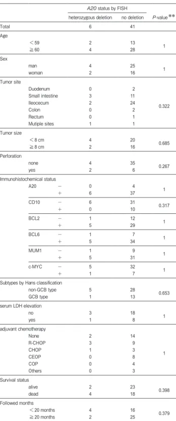
A20 deletion. Forty-seven of the 52 samples showed evaluable FISH signals. Six of 47 cases (13%) had A20 heterozygous deletion (Fig.3), whereas there
was no case with homozygous deletion. The clinico- pathological features of these six A20-deleted cases are summarized in Table 2. The median age of the 6 patients (4 men, 2 women) was 63 (range 51-81 years).
The most frequently involved site was the ileocecal region (four cases, 67%) with a mean tumor size of 8.2 cm. Perforation was detected in 2 cases (33%), which led to death in both cases within 7 months. In a median follow-up time of 9 months, 4 patients (67%) were dead. In a univariate analysis, there were no sig- nificant differences between A20-heterozygously deleted and non-deleted cases including OS (p=0.63). The clinicopathological features of these 6 heterozygous A20 deletion cases and the 41 deletion-negative cases are summarized in Table 3.
IHC findings and their correlation with clinico- pathological characters and prognosis. Forty-six of 52 samples were evaluable for the A20 IHC analysis.
Forty-two samples (91%) were positive, and the remain- ing 4 samples (9%) were negative. All 6 cases with a heterozygous A20 deletion were positive for A20 IHC.
Of the six A20-heterozygously-deletion cases, 5 were non-GCB-type and one was GCB-type. The median age of the 4 patients (2 men, 2 women) with negative A20 IHC was 63 (range 61-83 years). The ileocecal region (3 cases, 75%) was the most involved site, with a mean tumor size of 7.0 cm. Of these 4 patients, there was no
20µm
A B C D E
Case no.1
Case no.53
Fig. 2 Tissue array samples (2 cases) of hematoxylin-eosin stain (A) and immunostains (B, A20; C, CD10; D, BCL6; E, MUM1), (Olympus BX51).
5µm
Fig. 3 FISH analysis of DLBCL case performed with a combina- tion of A20 probe (orange) and chromosome 6 centromeric probe (green). Two green signals (yellow arrowheads) and one red signal (white arrows) are seen in one cell, indicating heterozygous dele- tion of the A20 gene (Olympus IX71).
intestinal perforation or mortality in a median fol- low-up time of 57 months. Of these A20 IHC-negative patients, 2 were non-GCB-type and 2 were GCB-type.
The adjuvant chemotherapies were performed with the regimens of R-CHOP (rituximab, cyclophospha- mide, doxorubicin, vincristine and prednisone) (12 cases, 39%), CHOP (4 cases, 13%), CEOP (etoposide substituted for doxorubicin) (8 cases, 26%), and COP (4 cases, 13%). Other regimens included R-hyper CVAD (rituximab with hyperfractionated cyclophos- phamide, vincristine, doxorubicin and dexametha- sone, 1 case, 3%), rituximab, etoposide and cyclophos- phamide (1 case, 3%), rituximab and cyclophosphamide (1 case, 3%). The clinicopathological factors were not significantly different among the six A20-deleted, four A20 IHC-negative, and the remaining 41 patients, probably because the sample sizes of the former 2 groups were too small.
Discussion
We examined A20 deletion and protein expression using primary intestinal DLBCL cases. CARD11 and MYD88 mutations are involved in the NF-κB pathway alterations including A20, and these are frequent events in non-GCB type DLBCL. We found that 6 of 52 cases (13%) showed A20 heterozygous deletion, but all these cases were positive for A20 protein expression. On the other hand, 4 of the 52 cases were negative for A20 pro- tein expression, whereas no A20 deletion was observed.
We demonstrated that A20 deletion did not necessarily cause A20 protein down-expression [13]; some cases with heterozygous deletion showed A20 IHC positivity although cases with homozygous deletion showed A20 IHC negativity. It has also been reported that not only A20 deletion but also A20 promoter hypermethylation could contribute to A20 down-expression [15]. Therefore, in the 4 present A20 non-deleted and IHC-negative cases, the IHC negativity might have been caused by promoter hypermethylation.
We hypothesized that only A20 haplo-insufficiency from heterozygous deletion might cause lymphoma- genesis in B-cell lymphoma. Honma et al. reported that monoallelic A20 inactivation as well as biallelic A20 inactivation was frequently found in ABC-phenotype DLBCLs and mantle cell lymphomas, and they demon- strated that A20 induced resistance to apoptosis and increased colony formation ability in human EB-LCL
Table 2 Clinicopathological features of A20 heterozygous deleted cases
Case no.
AgeSexTumor site
Tumor size (cm)
PerforationImmunohistochemical status
Serum LDH elevation
Adjuvant chemotherapy
Survival status Followed month
A20CD10BCL2BCL6MUM1c-MYCHans classification 151woman
Small bowel (jejunum)
6.0none+---+-non-GCBnoR-CHOPalive11 2065womanIleocecal region5.0none+-+++-non-GCBnoCHOPalive135 2881manIleocecal region5.5none+-+++-non-GCBnoR-CHOPdead23 3260manMultiple (jejunum and colon)6.0yes+-++--GCBNDR-CHO dead7 3759manIleocecal region10.5none+-+++-non-GCBelevatednonedead6 3978manIleocecal region16.0yes+-++++non-GCBNDnonedead7 LDH, lactate dehydrogenase;ND, Not Done;CHOP, cyclophosphamide, doxorubicin, vincristine and predonisone;R-CHOP, Rituximab and CHOP.
(Epstein Barr virus lymphoblastoid cell line) even with a partial knockdown [15]. These results might support the role of A20 gene in our cohort.
We also speculated that not only A20 heterozygous deletion but also other gain-of-function mutations such as CARD11 and MYD88 which are frequently found in ABC-type DLBCLs could affect the lymphomagenesis.
Although the cell types were different, Wolfrum et al.
reported that pro-atherosclerotic NF-κB target gene was elevated and atherosclerosis was increased in A20 hap- lo-insufficient mice, so that A20 resulted in NF-κB deregulation [16]. This result might support the rela- tionship between A20 and NF-κB.
Because we had only paraffin-embedded samples for tissue microarray, our study was strictly limited to immunohistochemical and FISH analyses. We could not obtain further data including those from a real-time reverse transcription-polymerase chain reaction (RT-PCR), mutational analysis, or methylation-spe- cific PCR analysis. In order to clarify the specific mech- anisms in the NF-κB pathway, some DLBCL-derived cell lines (probably established from pleura or lymph node) must be used. The gastrointestinal tract is one of the specific extranodal sites with special immune sys- tems, and it is strongly affected by antigen stimulation and other cytokines compared to other organs. When the above-mentioned types of cell lines are used, con- clusions that differ from those observed with intestinal DLBCL (as in the present study) might be obtained. To the best of our knowledge, there are no DLBCL cell lines derived from intestine, and we suspect that it might be difficult to demonstrate the functional status of A20 in primary intestinal DLBCLs.
There were some discrepancies between our present FISH and IHC results, and we speculate that a FISH analysis could be a better way for determining the A20 genetic status compared to immunohistochemical anal- yses. Giulino et al. reported the first immunohisto- chemical findings for A20 [14]. They observed that an A20-biallelic mutation case (1 case) was negative for A20 IHC, whereas cases with A20 mutation and/or monoallelic deletion frequently retained reactivity toward A20 IHC.
Based on comprehensive gene expression analyses, DLBCLs are subclassified into GCB, non-GCB, and unclassified types, and there are significant prognostic differences between the GCB type and the non-GCB type [17]. It is thus important to classify the cells-of-or-
Table 3 Clinicopathological features of A20 heterozygous deleted cases vs. deletion negative cases*
A20 status by FISH
heterozygous deletion no deletion P-value**
Total 6 41
Age
<59 2 13 1
≧60 4 28
Sex
man 4 25 1
woman 2 16
Tumor site
Duodenum 0 2
0.322
Small intestine 3 11
Ileocecum 2 24
Colon 0 2
Rectum 0 1
Mutiple sites 1 1
Tumor size
<8cm 4 20 0.685
≧8cm 2 16
Perforation
none 4 35 0.267
yes 2 6
Immunohistochemical status
A20 - 0 4 1
+ 6 37
CD10 - 6 31 0.317
+ 0 10
BCL2 - 1 12 1
+ 5 29
BCL6 - 1 7 1
+ 5 34
MUM1 - 1 9 1
+ 5 31
c-MYC - 5 32 1
+ 1 7
Subtypes by Hans classification
non-GCB type 5 28 0.653
GCB type 1 13
serum LDH elevation
no 3 18 1
yes 1 8
adjuvant chemotherapy
None 2 14
1
R-CHOP 3 9
CHOP 1 3
CEOP 0 8
COP 0 4
Others 0 3
Survival status
alive 2 23 0.398
dead 4 18
Followed months
<20months 4 16 0.379
≧20months 2 25
LDH, lactate dehydrogenase; CHOP, cyclophosphamide, doxorubicin, vincristine and predonisone; R-CHOP, Rituximab and CHOP; CEOP, cyclophosphamide, etoposide, vincristine and predonisone; COP, cyclophosphamide, vincristine and predonisone.
*Insufficient 5 cases by FISH analysis were excluded.
**Fisherʼs Exact test.
igin by immunohistochemistry in routine practice. In primary gastrointestinal DLBCLs, the frequency of the non-GCB type was reported to be 6-14% in the small intestine, and 57% in the colon [18,19]. In the present series, approx. 70% of the cases were non-GCB type.
This relatively higher prevalence of the non-GCB type might be due to the higher proportion of ileocecal sam- ples in this study and/or to genetic factors, as DLBCL cases in Taiwan, regardless of tumor sites, have shown a relative high frequency (72.5%) of the non-GCB phe- notype [20].
We have reported that perforation, high Performance Status (>2), and no adjuvant chemother- apy were independent poor-prognosis factors for pri- mary intestinal DLBCL [4,5]. In addition, tumor size
>8 cm was a new independent poor-prognosis factor (p=0.03, 95% CI: 1.11-8.34) (Table 1). To the best of our knowledge, no study has demonstrated that the tumor size of intestinal DLBCL could be an indepen- dent predictor of prognosis. The Lugano classification does not refer to the tumor size in intestinal lympho- mas [21]. Large-sized tumors might carry a higher risk of perforation, and thus large tumor size could be a poor-prognosis factor.
In conclusion, we observed that A20 abnormality was less prevalent among intestinal DLBCLs than DLBCLs at other anatomic sites. We also observed some discordancy between gene deletion and protein expression. Although the A20 alteration status did not affect any clinicopathological characters in this series, further studies exploring alterations of A20 and other NF-κB components in primary intestinal DLBCL are needed.
References
1. Castro FA, Jansen L, Krilaviciute A, Katalinic A, Pulte D, Sirri E, Ressing M, Holleczek B, Luttmann S and Brenner H; GEKID Cancer Survival Working Group: Survival of patients with gastric lymphoma in Germany and in the United States. J Gastroenterol Hepatol (2015) 30:1485-1491.
2. Yoshino T, Miyake K, Ichimura K, Mannami T, Ohara N, Hamazaki S and Akagi T: Increased incidence of follicular lym- phoma in the duodenum. Am J Surg Pathol (2000) 24: 688-693.
3. Matysiak-Budnik T, Jamet P, Fabiani B, Nion-Larmurier I, Marjanovic Z and Ruskoné-Fourmestraux A; Groupe Dʼétude des Lymphomes Digestifs (GELD); Federation Francophone de Cancerologie Digestive (FFCD), Dijon, France: Primary intestinal B-cell lymphoma: a prospective multicentre clinical study of 91 cases. Dig Liver Dis (2013) 45:947-952.
4. Chuang SS, Ye H, Yang SF, Huang WT, Chen HK, Hsieh PP,
Hwang WS, Chang KY, Lu CL and Du MQ: Perforation predicts poor prognosis in patients with primary intestinal diffuse large B-cell lymphoma. Histopathology (2008) 53: 432-440.
5. Lu YH, Chang ST, Yang SF, Weng SF, Huang WT, Hsieh PP, Hsu JD, Tsou MH and Chuang SS: Primary Intestinal Diffuse Large B-cell Lymphoma in Taiwan Showed a Relative Higher Rate of Perforation and EBV Association. Appl Immunohistochem Mol Morphol (2016) 24: 541-549.
6. Compagno M, Lim WK, Grunn A, Nandula SV, Brahmachary M, Shen Q, Bertoni F, Ponzoni M, Scandurra M, Califano A, Bhagat G, Chadburn A, Dalla-Favera R and Pasqualucci L: Mutations of multiple genes cause deregulation of NF-kappaB in diffuse large B-cell lymphoma. Nature (2009) 459:717-721.
7. Wertz IE, OʼRourke KM, Zhou H, Eby M, Aravind L, Seshaqiri S, Wu P, Wiesmann C, Baker R, Boone DL, Ma A, Koonin EV and Dixit VM: De-ubiquitination and ubiquitin ligase domains of A20 downregulate NF-kappaB signaling. Nature (2004) 430:694-699.
8. Lee EG, Boone DL, Chai S, Libby SL, Chien M, Lodolce JP and Ma A: Failure to Regulate TNF-Induced NF-κB and Cell Death Responses in A20-Deficient Mice. Science (2000) 289: 2350- 2354.
9. Paik JH, Go H, Nam SJ, Kim TM, Heo DS, Kim CW and Jeon YK: Clinicopathologic implication of A20/TNFAIP3 deletion in diffuse large B-cell lymphoma: an analysis according to immuno- histochemical subgroups and rituximab treatment. Leuk Lymphoma (2013) 54: 1934-1941.
10. Koch P, del Valle F, Berdel WE, Willich NA, Reers B, Hiddemann W, Grothaus-Pinke B, Reinartz G, Brockmann J, Temmesfeld A, Schmitz R, Rübe C, Probst A, Jaenke G, Bodenstein H, Junker A, Pott C, Schultze J, Heinecke A, Parwaresch R and Tiemann M; German Multicenter Study Group:
Primary gastrointestinal non-Hodgkinʼs lymphoma: I. anatomic and histologic distribution, clinical features, and survival data of 371 patients registered in the German Multicenter Study GIT NHL 01/92. J Clin Oncol (2001) 19: 3861-3873.
11. Stein H, Warnke RA, Chan WC, Jaffe ES, Chan JKC, Gatter KC and Campo E: Diffuse large B-cell lymphoma, not otherwise spec- ified; in WHO Classification of Tumours of Haematopoietic and Lymphoid Tissues, 4th Ed. IARC Press, Lyon (2008) pp233-237.
12. Iqbal J, Sanger WG, Horsman DE, Rosenwald A, Pickering DL, Dave B, Dave S, Xiao L, Cao K, Zhu Q, Sherman S, Hans CP, Weisenburger DD, Greiner TC, Gascoyne RD, Ott G, Müller- Hermelink HK, Delabie J, Braziel RM, Jaffe ES, Campo E, Lynch JC, Connors JM, Vose JM, Armitage JO, Grogan TM, Staudt LM and Chan WC: BCL2 Translocation Defines a Unique Tumor Subset within the Germinal Center B-Cell-Like Diffuse Large B-Cell Lymphoma. Am J Pathol (2004) 165:159-166.
13. Ando M, Sato Y, Takata K, Nomoto J, Nakamura S, Ohshima K, Takeuchi T, Orita Y, Kobayashi Y and Yoshino T: A20 (TNFAIP3) deletion in Epstein-Barr virus-associated lymphoproliferative disor- ders/lymphomas. PLOS ONE (2013) 8: e56741.
14. Giulino L, Mathew S, Ballon G, Chadburn A, Barouk S, Antonicelli G, Leoncini L and Liu YF: A20 (TNFAIP3) genetic alterations in EBV-associated AIDS-related lymphoma. Blood (2011) 117: 4852- 4854.
15. Honma K, Tsuzuki S, Nakagawa M, Tagawa H, Nakamura S, Morishima Y and Seto M: TNFAIP3/A20 functions as a novel tumor suppressor gene in several subtypes of non-Hodgkin lympho- mas. Blood (2009) 114:2467-2475.
16. Wolfrum S, Teupser D, Tan M, Chen KY and Breslow JL: The protective effect of A20 on an atherosclerosis in apolipoprotein
E-deficient mice is associated with reduced expression of NK-kappaB target genes. Proc Natl Acad Sci USA (2007) 104:
311-317.
17. Hans CP, Weisenburger DD, Greiner TC, Gascoyne RD, Delabie J, Ott G, Müller-Hermelink HK, Campo E, Braziel RM, Jaffe ES, Pan Z, Farinha P, Smith LM, Falini B, Banham AH, Rosenwald A, Staudt LM, Connors JM, Armitage JO and Chan WC: Confirmation of the molecular classification of diffuse large B-cell lymphoma by immunohistochemistry using a tissue microarray. Blood (2004) 103: 275-282.
18. Mitchell KA, Finn WG and Owens SR: Differences in germinal centre and non-germinal center phenotype in gastric and intestinal diffuse large B-cell lymphomas. Leuk Lymphoma (2008) 49: 1717- 1723.
19. Connor J and Ashton-Key M: Gastric and intestinal diffuse large B-cell lymphomas are clinically and immunophenotypically differ- ent. An immunohistochemical and clinical study. Histopathology (2007) 51: 697-703.
20. Chang ST, Chen SW, Ho CH, Kuo CC, Sakata S, Takeuchi K and Chuang SS: Immunophenotypic and genetic characteristics of diffuse large B-cell lymphoma in Taiwan. J Formos Med Assoc (2016) 115: 961-967.
21. Rohatiner A, dʼAmore F, Coiffier B, Crowther D, Gospodarowicz M, Isaacson P, Lister TA, Norton A, Salem P, Shipp M and Somers R: Report on a workshop convened to discuss the pathological and staging classifications of gastrointestinal tract lymphoma. Ann Oncol (1994) 5: 397-400.