Colorectal cancer creening at multidetectorrow computed tomography: detection of flatand polypoid lesions with a dedeicad workstation
坂本 崇
1
Contents
Abstract ... 3
Publication list ... 5
Acknowledgements ... 6
Abbreviations ... 7
- Chapter I - ... 8
Clinical Role of CT Colonography 1. Background 2. Clinical Application 3. Techniques and protocols 4. Summary - Chapter II - ... 16
Improved image quality at screening CT colonography - Optimization of the preprocessing for CT colonography 1. Introduction 2. Materials and Methods 3. Results 4. Discussion 5. Conclusion - Chapter III - ... 34 Evaluation of colonic dilatation by CT colonography: the influence of
antispasmodics and the patient’s body size
1. Introduction
2
2. Materials and Methods 3. Results
4. Discussion 5. Conclusion
- Chapter IV - ... 43 Detection of Flat Colorectal Polyps at Screening CT Colonography in Comparison with Conventional Polypoid Lesions
1. Introduction
2. Materials and Methods 3. Results
4. Discussion 5. Conclusion
General conclusions ... 62
References ... 63
3
Abstract
Background and Purpose: In recent years, computed tomography colonography (CTC) has been clinically applied to screen for colorectal cancers, and perceived as highly diagnostic, because of the widespread use of multidetector-row CT and increased accuracy of analysis software. In addition, CTC, which yields objective and
reproducible results, is a high-potential screening method, in that it is a low-invasive test associated with mild distress, and can be easily normalized. Thus, the purposes of this study are: 1) to compare the detectability of colorectal lesions among 3 different colon cleansing techniques; 2) to evaluate the effect of the use of antispasmodics on colonic dilatation; and 3) to evaluate the detection capability and usefulness of CTC in the screening of flat- and polypoid lesions by comparing CTC- and optic colonoscopy findings.
Materials and Methods: In the first basic study, three preprocessing methods were
compared: polyethylene glycol on the previous day, polyethylene glycol on the same
day, and a bowel-cleansing tablet on the previous day. In the second basic study, the
subjects were 83 patients who underwent CTC screening (40 without antispasmodics,
43 with antispasmodics). Volume rendering images obtained in the supine and prone
positions were used for evaluation. In the third clinical study, we evaluated the CTC
detection capability for flat colorectal polyps with a flat surface and a height not
exceeding 3mm by comparing to conventional polypoid lesions according to the polyp
diameter. Four types of reconstruction images including multiplanar reconstruction,
volume rendering, virtual gross pathology, and virtual endoscopic images were used for
visual analysis. We compared the abilities of the 4 reconstructions for polyp
4
visualization.
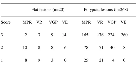
Results: Visual assessment scores were also significantly different between the three methods (p<0.05). The lumen was visualized more clearly when the volume of residual colonic contents was less. Performing polyethylene glycol on the previous day allows blind areas to be reduced with high precision. In the second basic study, Colonic dilatation was significantly greater with antispasmodics than without (p<0.05). In the third clinical study, Detection sensitivity for flat polyps was 31.3%, 44.4%, 87.5% for lesions measuring 2-3 mm, 4-5 mm, and 6 mm, respectively; the corresponding sensitivity for polypoid lesions was 47.6%, 79.0%, 91.7%. Virtual endoscopic imaging showed best visualization among the 4 reconstructions.
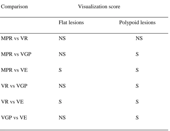
Conclusion: The virtual endoscopic imaging showed highest visualization score for detection of both flat- and polypoid colorectal polyps. CTC using 64-row
multidetector CT is useful for colon cancer screening to detect the lesions measuring 6
mm or more, although the detection of flat lesions is still challenging.
5
Publication list
1. Takashi Sakamoto, Katsuhiko Mitsuzaki, Daisuke Utsunomiya, Katsuhiko Matsuda, Sadahiro Yamamura, Joji Urata, Megumi Kawakami and Yasuyuki Yamashita
Detection of flat colorectal polyps at screening CT colonography in comparison with conventional polypoid lesions
Acta Radiol. 53(7): 714-9 2012.
2. Tam ura, Y., Utsunomi ya, D., Sakamoto, T., Hi rai, T., Ni shiharu, T., Urat a, J ., Yam ashita, Y.,
Reduction of contrast material volume in 3D angiography of the brain using MDCT.
AJR Am J Roentgenol. 195(2): 455-8 2010
3. Mitsuz aki , K ., S akamoto, T ., Matsuda , K., Fukunaga, K., Suga, M., Yoshida, K., Kudo , K., Fujim oto, T., Tada, S., and J oji , Urat a ,.
Initial experiences in CT colonography (CTC) for colon cancer screening.
Journal of Gastroenterological Cancer Screening. 49(1): 42-54 2011
4. Mitsuz aki, K., Tada, S., Matsuda, K., Fuku naga, K., Wat anabe, Y., Suga, M ., S akamoto, T ., Joji, Urat a., Kudo, K., Yoshida, K., Uehara, M., Eguchi, H., Kamio, T., Oda, Y.,
The Current Status and Issues of the Screening for Colorectal Cancer with CT Colonography.
Stomach and Intestine. 47(1): 55-65 2012
6
Acknowledgements
These academic investigations were done during from 2008 to 2012 at department of Diagnostic Radiology, Graduate School of Medical Sciences, Kumamoto University.
I wish to extend my sincere thanks to Dr. Daisuke Utsunomiya, Department of Diagnostic Radiology, Graduate School of Medical Sciences, Kumamoto University for general guidance and constructive instructions.
I am deeply grateful to Professor Yasuyuki Yamashita, chairman of the Department of Diagnostic Radiology, Graduate School of Medical Sciences, Kumamoto University.
Of course, I owe a great deal to the colleagues of Radiology, Dr. Urata, RT.
Kawakami, RT. Matsuda, for their cooperation and helps in my works.
Finally, the greatest gratitude is offered to Dr. Mitsuzaki who motivated me to be
engaged in scientific study.
7
Abbreviations
2D: two-dimensional 3D: three-dimensional
ACR: American College of Radiology CT: computed tomography
CTC: computed tomography colonography KV: kilovolt
OC: optic colonoscopy
PPV: positive predictive value mAs: milliamperesecond
MPR: multiplanar reconstruction
MDCT: multidetector-row CT
NPV: negative predictive value
PEG: polyethylene glycol
VC: virtual colonoscopy
VE: virtual endoscopy
VGP: virtual gross pathology
VR: volume rendering
8
- Chapter I -
Clinical Role of CT Colonography
1. Background
2. Clinical Application 3. Techniques and protocols
4. Summary
1. BACKGROUND
Colorectal cancer is common malignancy all over the world, and is
preventable if detected early at screening examinations. Optic colonoscopy (OC) is a conventional method for screening of colorectal lesions; however, it is relatively invasive and operator-dependent. Computed tomography colonography (CTC) has been introduced as an alternative technique. It is also known as virtual colonoscopy (VC), is a useful technique for the evaluation of the entire colon. It has potential advantages over conventional colonoscopy because of its minimally invasive nature and no need for sedation and recovery time. The examination is based on a helical, thin-section CT of the cleansed and distended colon. Data evaluation is performed with commercially available CTC post-processing software with simultaneously available multiplanar two-dimensional (2D) and virtual endoscopy (VE)
three-dimensional (3D) image displays. Also, CTC can be a valuable tool for the
identification of extracolonic findings [1, 2]. Yee and colleagues [3] evaluated
extracolonic findings in 500 subjects who underwent CTC, and they reported that
clinically important extracolonic findings were identified in 9% of patients (45/500).
9
CTC was first popularized by Vining and colleagues [4] at the Bowman Gray School of Medicine in 1994, where they used helical CT data to provide 3D images, simulating the endoluminal view of traditional OC. They called the technique CTC [5].
The term of “CTC” was later adopted by the American College of Radiology (ACR) because of its more accurate description of the test [6]. Early studies of CTC were performed in the supine position, using prior-generation single- or dual-row CT scanners and 2D imaging. Incorporation of supine and prone positions,
multidetector-row CT (MDCT) scanners, and both 2D and 3D imaging has allowed for steady improvement in resolution and detection [6, 7]. The use of the supine- and prone positions in a single CTC test allows for better displacement of fluid and stool to reduce areas of the colon that may be obscured by retained fluid or poorly distended.
The clinical use of CTC has been the detection of colon cancer and the surveillance of
colorectal polyps, although CTC had not been widely used on prior-generation MDCT
with 16- or less detectors. After the introduction of newer-generation MDCT with 64-
or more detectors, its use has been growing in popularity among physicians and patients
for cancer screening. As opposed to OC, CTC does not require conscious sedation or
endoscopy, making it less invasive and less time-consuming. Currently, there are
several clinical indications for CTC. They include evaluation of the colon after an
incomplete or unsuccessful conventional colonoscopic examination and evaluation of
the colon proximal to an obstructing neoplasm [8-10]. Another potential indication for
CTC is in the evaluation of frail and elderly patients or patients who would have an
increased risk with conventional colonoscopy. The use of CTC to monitor patients
after surgery for colorectal cancer is currently under investigation [11, 12]. In addition,
CTC may contribute to colorectal screening by providing a safe, effective, and rapid
examination that can be used to evaluate the entire colon for clinically significant
lesions.
10
2. CLINICAL APPLICATION
CTC has developed an important role for the evaluation of the colorectal lesions. In some situations it may be a safer method to visualize the colon effectively, or it may be the only available option when other techniques have failed. CTC
requires the volumetric acquisition of data using helical CT and is now achieved with MDCT with 64 detectors or more. Post-processing of CT data sets is performed on a computer workstation with specific software to generate axial images, multiplanar reconstruction (MPR) views, and 3D images of the colon. (Fig. 1-4)
Before the patient is scanned, however, several preparatory steps are required to produce an optimally diagnostic study. A well-cleansed colon with good distention is essential to achieve a high-quality study for polyp and cancer detection. A poorly prepared colon may be the cause of both false-negative and false-positive findings [13, 14]. Standard CTC protocol requires scanning the patient in supine- and prone
positions, which allows segments of colon with poor cleansing or suboptimal distention in one position to be reevaluated in the opposing position with potentially improved distention and cleansing. The rationale for using various colonic cleansing regimens for CTC is discussed. Positive labeling of residual material and electronic subtraction of tagged material are potential strategies to reduce and possibly eliminate the need for purgatives that would further increase patient acceptance of CTC compared with other techniques. The two distending agents for CTC (room air and carbon dioxide) are discussed, along with practical tips for administration and the role of antispasmodic drugs.
The ability of CTC to detect colorectal polyps has been tested in many previous
studies[13, 15, 16]. CTC appeared to be promising in high-risk populations; with a
reported sensitivity greater than 90% for polyps ≥10 mm. Recent results in the low
11
prevalence population were more heterogeneous and less impressive (34–93.8%) [15, 17-19]. This wide range of results may be likely caused by differences in patient selection, examination and evaluation techniques, and reader experience [20, 21].
Also, the detection of flat colorectal polyps is still challenging.
Thus, we consider that the appropriate preparation and the validation of the clinical role of CTC in the large number of patients are critically important.
3. TECHNIQUES AND PROTOCOLS
State-of-the-art CTC requires MDCT and a high-end computer workstation with advanced graphic software that displays 2D and 3D views of the colon. Patients typically are scanned in a craniocaudal direction in both supine and prone positions.
Scanning in the supine and left lateral decubitus positions has been proposed as an alternative to supine and prone scanning whenever patients cannot lie prone.
Gryspeerdt and colleagues [22] found improvements in colonic distention using either supine/prone or supine/left lateral decubitus. Fewer breathing artifacts were noted with left lateral decubitus imaging in elderly patients.
Intravenous contrast material is not administered routinely for screening CTC.
Disadvantages of the use of intravenous contrast include increased invasiveness, the possibility of contrast reactions, higher radiation dose, increased interpretation times, and higher cost. Intravenous contrast should not be administered if patients undergo oral stool and fluid tagging because of the potential difficulty in differentiating an enhancing lesion from tagged material. Morrin and colleagues [23] found that administration of intravenous contrast significantly improved reader confidence for assessment of bowel wall conspicuity and for the detection of medium-sized polyps (6–
9 mm) in suboptimally cleansed colonic segments. When the diagnosis of colon
12
cancer is already established or is suspected based on initial imaging, intravenous contrast should be administered for CTC for staging purposes.
A typical CTC protocol using MDCT consists of a collimation of 0.625 to 2.5 mm with a gantry rotation time of 0.5 seconds resulting in a scan time of less than 10 seconds. MDCT scanners have enabled subcentimeter collimation without
compromising z-axis coverage. The volumetric data set is used for traditional 2D axial images and MPR as well as to produce 3D endoluminal views. Motion artifact from peristalsis and respiration is decreased or eliminated with MDCT because scan times are significantly shortened. Several studies have demonstrated that thinner reconstructions allow increased sensitivity for small polyps (< 6mm) and improve specificity in both phantom and human datasets [24-27]. Lui and colleagues [24] performed a study in 25 patients and found increased specificity for polyps 5 mm or larger using a slice
thickness of 1.25 mm x 1 mm when compared with thicker slices (5 mm x 2 mm) using a 4-row MDCT.
The ACR practice guidelines for the performance of CTC recommend use of MDCT with a slice collimation of 3 mm or less and a reconstruction interval of 1.5 mm or less [28]. In a recent consensus study a maximum acceptable slice thickness of 3 mm or less was recommended by 88% (22/25) of selected CTC experts [29].
According to the ACR guidelines, we suggest that 64-row MDCT should be appropriate for CTC.
CTC has several limitations such as ionizing radiation and preparation.
Because of the intrinsic high contrast between the intraluminal gas and the soft tissue of
the colonic wall, dropping the milliamperesecond (mAs) should be performed. Macari
and colleagues [30] performed CTC in 105 subjects using 50 mAs, 120 kilovolt (kV),
and a 1.25-mm slice thickness with a 1-mm reconstruction interval. Sensitivity of
CTC for the detection of polyps 6 to 9 mm and 10 mm or larger were 70% and 93%,
13
respectively. To obtain diagnostic image quality, the preparation such as colon cleansing and colonic dilatation is especially important. However, the appropriate preparation techniques have been undefined.
4. SUMMARY
For the appropriate performance of CTC in clinical practice, the optimal colonic preparation i.e. bowel cleansing and sufficient colonic distention should be critical. The first purpose of this thesis was to optimize the technique in patient preparation (Chapter II and III). The second purpose was to evaluate the detection capability and usefulness of screening CTC with 4-different reconstruction methods; i.e.
MPR, volume rendering (VR), VE, and virtual gross pathology (VGP) images (Chapter IV).
14
Fig.1 Volume Rendering Image Fig.2 Virtual Endoscopy
Fig.3 MultiPlanar Reconstruction Image
15
Fig.4 Virtual Gross Pathology
Virtual endoscopy image (Fig.2) shows polypoid lesion (arrow). MultiPlanar
Reconstruction image (Fig.3) shows polypoid lesion of uniform soft tissue density
(arrow).
16
- Chapter II –
Improved image quality at screening CT colonography – Optimization of the preprocessing for CT colonography
1. Introduction
2. Materials and Methods 3. Results
4. Discussion 5. Conclusion
1. INTRODUCTION
The introduction of CTC using MDCT has made it possible to acquire
high-resolution images over a wide range in a shorter time. Due to the improvements of image processing techniques, the application of CTC as a screening examination has been extensively investigated, mainly in the United States and Europe. [15, 31]
In Japan, on the other hand, the combined use of fecal occult blood testing and total colonoscopy is recommended for screening, and as a result, few studies have been conducted to evaluate the usefulness of CTC as a screening examination. In order for CTC to be employed for screening, bowel preparation protocols that are easy to perform and also ensure accurate examination results must be established. Obtaining images with a consistent level of image quality under conditions in which there is a minimal volume of residual liquid or solid matter in the colon is the most important factor in improving visualization of the intestinal lumen and thus ensuring accurate diagnosis in clinical practice (Fig. 1a. 1b).
In the present chapter, we evaluated the diagnostic accuracy and clinical
usefulness of CTC by comparing different bowel-preparation protocols.
17
2. MATERIALS AND METHODS Study group
Of 450 patients who underwent total colonoscopy between February 2009 and September 2010, 151 patients who agreed to undergo CTC examination before
colonoscopy were enrolled in this study. Three different bowel preparation protocols were developed and randomly employed for CTC examination. In the group A, a bowel cleansing agent (polyethylene glycol [PEG]) was administered on the day before the examination, and CTC was performed on the following day. In the group B, the bowel-cleansing agent was administered in the morning of the day of examination, and CTC was performed in the afternoon. In the group C, a bowel-cleansing agent
(Monobasic sodium phosphate monohydrate : Visiclear® Combination Tablets, Zeria Pharmaceutical, Japan [Bowel cleansing tablets]) was administered on the day before the examination, and CTC was performed on the following day (Fig. 2).
Group A and B received 2 liters of the bowel cleansing agent, group C received bowel cleansing tablets 50tablets and 2 liters of the water, which is required for
colonoscopy. In addition, water-soluble iodinated contrast medium (sodium
amidotrizoate and meglumine amidotrizoate, Gastrografin) was used for fecal tagging.
When the remaining amount of bowel cleansing agent had fallen to 380 mL, 20 mL of Gastrografin was added, and the total volume of 400 mL of this mixture was then administered.
The study group included 74 men and 77 women with an average age of 49.1±
9.8 years (age range: 25 to 79 years). The group A included 45 patients and the group
B included 60 patients and the group C included 46 patients. The age of the patients
was 50.3±12.5 years in the group A, 51.0±8.3 years in the group B and 45.3±7.7 years
in the group C. The body weight of the patients was 60.3±12.4 kg in the group A and
63.7±11.2 kg in the group B and 61.7±10.2 kg in the group C. The target lesions were
polyps measuring 2 mm or more in diameter detected by colonoscopy. All patients
18
gave informed consent to undergo CTC examination, which involves X-ray exposure, requires the injection of an antispasmodic agent immediately before colonoscopy as well as the administration of iodinated contrast medium, and may cause a bloated sensation or abdominal pain.
CTC
After bowel preparation was completed, an antispasmodic agent (Sesden, 10 mg) was injected intramuscularly 10 minutes before CTC. CTC was then performed using a 64-row MDCT system (Aquilion 64, Toshiba, Japan). Total colonoscopy was performed after CTC. Image analysis was performed at an image workstation
(Ziostation System N610, Version 1.21b, Amin, Japan).
Data Acquisition
A tube voltage of 120 kV, a tube current of 100 mA, and a gantry rotation speed of 0.5 s/rot were employed for scanning. The slice collimation was 0.5 mm 64 slices, the helical pitch was 0.83, and the couch-top movement speed was 27 mm/rot in all patients.
The patient was placed on the couch top in the left lateral decubitus position
and received the antispasmodic agent (Sesden, 10 mg) by intramuscular injection in
order to suppress intestinal peristalsis before CTC was started. Rectal examination
was performed to check for the presence of lesions in the anus, and a 12-EG Nelaton
catheter for insufflation was then introduced via the anus. Air or carbon dioxide was
administered with the patient in the left lateral decubitus or prone position using an
automatic insufflator with a pressure measurement function. After a scout view was
obtained to confirm sufficient dilatation of the colon, image acquisition was performed
during breath-holding (at end expiration) in the prone and supine positions. The scan
19
range extended from the bottom of the diaphragm to the inferior edge of the pubis in both positions, with scanning performed in the foot-to-head direction during
breath-holding for 7 s to 10 s. Image reconstruction was performed with a slice thickness of 0.5 mm for the acquired helical data. Images reconstructed at 0.5-mm intervals (900 to 1000 slices) were then transferred to the image workstation.
Total Optic Colonoscopy
After CTC examination was completed, the antispasmodic agent (Sesden, 10 mg) was again injected intramuscularly 10 minutes before total colonoscopy and a sedative (Horizon, 10 mg) was injected intravenously immediately before total
colonoscopy. The endoscopist (who was qualified as a supervising physician and had performed more than 5000 colonoscopic examinations) performed colonoscopy (CF endoscope, Fujinon Toshiba, Japan). The colonoscope was first advanced into the cecum and then withdrawn toward the anus while checking for the presence of polyps in each region. The findings such as the region, size, and macroscopic morphological characteristics of the polyps were then recorded on the designated form. The size of the polyps was measured using biopsy forceps inserted via the forceps channel, with the forceps pressed against the polyps closed (2 mm in diameter) and open (5 mm in
diameter). Biopsy specimens were generally obtained for polyps measuring 6 mm or more in diameter and polyps characterized as depressed plaques measuring 5 mm or less in diameter.
Image Interpretation
Two physicians (the endoscopist who was qualified as a supervising physician
and a board-certified radiologist) interpreted the colonoscopic images independently in
a blinded manner. In the event of disagreement regarding the diagnostic findings, the
20
final diagnosis was reached by discussion and consensus. The patient data was selected at the workstation, and images comparable to those obtained by barium enema were generated using the VR method to check for insufficient dilatation, deformation of the walls, and mucosal irregularities. Dissected colon images (VGP images) were then used to identify the regions containing suspected lesions. In the dissected colon
images, the presence of elevated lesions and morphological abnormalities were checked by observing the image of each semilunar fold. All of the regions containing
suspected lesions were checked and then evaluated in diagnosis confirmation mode.
In diagnosis confirmation mode, the regions containing suspected lesions were observed using VE and MPR images in order to determine whether or not lesions were present.
When a lesion was found, its region and size were evaluated. With regard to size, the maximum diameter of the lesion was measured in two-dimensional MPR images. In addition, the VE images were observed from two directions (from the oral end and from the anal end) to check for the presence of lesions. When a suspected lesion was found, the region including the suspected lesion was checked in the VGP image again. The final diagnosis was then established based on the findings identified in the VE, VGP, and MPR images. The images obtained with the patient in the prone and supine positions were interpreted in the manner described above to check for lesions.
Study Items
The results obtained by total colonoscopy were used as the gold standard, and
the diagnostic results obtained using the CTC images were compared between the three
groups for lesions measuring 2 mm or more in diameter. The colon was divided into
six regions: the cecum, the ascending colon, the transverse colon, the descending colon,
the sigmoid colon, and the rectum. The presence and the location of colonic lesions
were visually assessed on the CTC images by the 2 observers. The sensitivity,
21
specificity, and diagnostic accuracy were then calculated in each group.
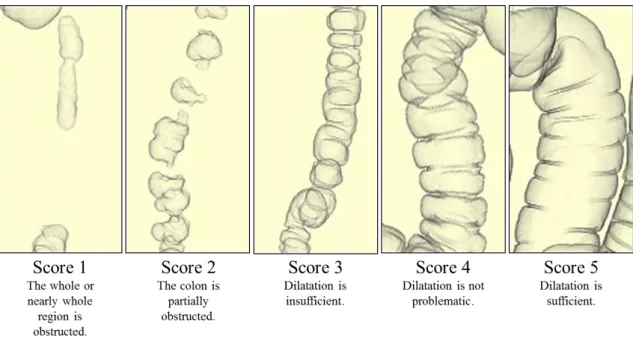
Next, the volume of residual liquid in the colon following each of the bowel preparation protocols was evaluated. The volume of residual liquid in each region was evaluated by visual assessment of the VE and MPR images and then rated using a 5-grade scale: 0%, 25%, 50%, 75%, and 100% (Fig. 3). For visual assessment,
statistical analysis was performed using the Kruskal-Wallis test. A p value of less than 0.05 was considered to be statistically significant.
3. RESULTS
Of the 151 patients who underwent total colonoscopy and CTC, fecal tagging could not be performed due to iodine hypersensitivity in 15 patients.
Polyps measuring 2 mm or more in diameter were found in 61 (40.4%) of the 151 patients. The total number of polyps that were detected by colonoscopy was 123 lesions: 91 lesions measuring 2-4 mm and 32 lesions measuring 5 mm or more. Of these polyps, the final diagnosis was established by histopathological examination of biopsy or surgical specimens in 20 patients (25 lesions). The numbers of patients and types of polyps were as follows: 4 patients with a hyperplastic polyp (6 lesions) and 16 patients with tubular adenoma (19 lesions).
Comparison of Accuracy Rates among the 3 Groups
The accuracy rates in each group are shown in Table 1. In the group A, the sensitivity, specificity, positive predictive value (PPV), negative predictive value (NPV) and diagnostic accuracy were 80.0%, 83.1%, 42.1%, 96.4%, and 82.6%, respectively.
In the group B, the sensitivity, specificity, PPV, NPV and diagnostic accuracy were
50.0%, 87.8%, 47.3%, 88.9%, and 81.0%, respectively. In the group C, the sensitivity,
specificity, PPV, NPV and diagnostic accuracy were 58.3%, 76.7%, 22.5%, 94.1% and
22
74.8%, respectively. A significant difference was observed in sensitivity between the group A and the group B, group C (p<0.05). A significant difference in PPV was observed between the group A, group B and the group C (p<0.05).
Comparison of Detection Rates According to the Size of the Lesion among the 3 Groups
The detection rates according to the size of the lesion in each group are shown in Table 2. In the group A, the sensitivity and PPV for all lesions were 75.8% and 37.9%, respectively. The sensitivity was 68.4% (13/19) for lesions measuring 2-4 mm and 85.7% (12/14) for lesions measuring 5 mm or more. The sensitivity for lesions measuring 5 mm or more was 85.7%, showing good results. The PPV was 28.8% for lesions measuring 2-4 mm and 57.1% for lesions measuring 5 mm or more. The larger the lesion, the higher the PPV. In the protocol B group, the sensitivity and PPV for all lesions were 49.2% and 47.8%, respectively. The sensitivity was 44.4% (24/54) for lesions measuring 2-4 mm and 72.7% (8/11) for lesions measuring 5 mm or more, showing good results. The PPV was 44.4% for lesions measuring 2-4 mm and 61.5%
for lesions measuring 5 mm or more. In the protocol C group, the sensitivity and PPV for all lesions were 53.8% and 16.3%, respectively. The sensitivity was 50.0% (10/20) for lesions measuring 2-4 mm and 66.6% (4/6) for lesions measuring 5 mm or more, showing good results. The PPV was 12.8% for lesions measuring 2-4 mm and 50.0%
for lesions measuring 5 mm or more. In all protocols, the tendency was observed that the larger lesions had the higher PPV.
Evaluation of the Volume of Residual Liquid in the Colon in Each Group
The visual evaluation results for the volume of residual liquid in each region of
the colon are shown in Fig 4. For the images obtained with the patient in the prone
23
position, the ratings in the protocol A were 4.48 in the cecum, 4.68 in the ascending colon, 4.31 in the transverse colon, 4.44 in the descending colon, 4.28 in the sigmoid colon, and 4.86 in the rectum, with an overall rating of 4.51, while the ratings in the protocol B were 3.63 in the cecum, 3.91 in the ascending colon, 3.03 in the transverse colon, 3.50 in the descending colon, 2.65 in the sigmoid colon, and 4.21 in the rectum, with an overall rating of 3.49. The ratings in the protocol C were 4.00 in the cecum, 4.34 in the ascending colon, 3.07 in the transverse colon, 4.13 in the descending colon, 3.65 in the sigmoid colon, and 4.52 in the rectum, with an overall rating of 4.06.
Significant differences were observed between the three groups in all regions (p<0.05).
For the images obtained with the patient in the supine position, the ratings in the protocol A were 4.48 in the cecum, 4.66 in the ascending colon, 4.62 in the transverse colon, 4.00 in the descending colon, 4.53 in the sigmoid colon, and 4.75 in the rectum, with an overall rating of 4.51, while the ratings in the protocol B were 4.13 in the cecum, 3.91 in the ascending colon, 3.88 in the transverse colon, 2.26 in the descending colon, 3.20 in the sigmoid colon, and 3.80 in the rectum, with an overall rating of 3.53.
The ratings in the protocol C were 4.13 in the cecum, 3.82 in the ascending colon, 3.88 in the transverse colon, 3.30 in the descending colon, 3.91 in the sigmoid colon, and 4.26 in the rectum, with an overall rating of 3.96. Significant differences were observed between the 3 groups in all regions (p<0.05).
Bowel preparation protocol A resulted in a smaller volume of residual liquid as
compared with protocol B and protocol C.
24
4. DISCUSSION
The key element of a high quality CTC examination is a well-prepared clean, and well-distended colon. Residual stool and fluid may lead to a false-negative or false-positive diagnosis. Therefore, CTC, at present, requires full bowel preparation, just like colonoscopy and double contrast barium enema examination [32].
The mechanism of action and delivery of the three most commonly used cathartic agents for bowel cleansing differ, explaining their relative advantages and disadvantages in various patient populations. PEG is an osmotically balanced electrolyte lavage solution, which results in minimal water and electrolyte absorption and secretion [33]. Because PEG does not result in internal fluid shifts, it has the advantage of being safe in the vast majority of patients and provides a gentle colonic lavage [34].
The goal of colon preparation is to cleanse the colon and to provide contrast for fecal and fluid tagging. This preparation increases the specificity of CTC by tagging residual or adherent fecal material.
In the assessment of residual liquid with the bowel preparation protocol
employing PEG on the previous day (group A), the average scores were 4.51 for the
images obtained with the patient in the prone position and 4.51 for the images obtained
with the patient in the supine position. In the assessment of residual liquid with the
protocol employing PEG on the same day (group B), the average scores were 3.49 for
the images obtained with the patient in the prone position and 3.53 for the images
obtained with the patient in the supine position. In the assessment of residual liquid
with the protocol employing bowel cleansing tablets on the previous day (group C), the
average scores were 4.06 for the images obtained with the patient in the prone position
and 3.96 for the images obtained with the patient in the supine position. Visualization
of the lumen was improved as the volume of residual liquid decreased, with significant
25
differences observed between groups A and C and group B (p < 0.05).
For subjects who received PEG on the previous day (group A), the sensitivity, specificity, PPV, and NPV were 80.0%, 83.1%, 42.1%, and 96.4%, respectively. For subjects who received PEG on the same day (group B), the sensitivity, specificity, PPV, and NPV were 50.0%, 87.8%, 47.3%, and 88.9%, respectively. For subjects who received bowel cleansing tablets on the previous day (group C), the sensitivity, specificity, PPV, and NPV were 58.3%, 76.7%, 22.5%, and 94.1%, respectively. A significant difference was observed in sensitivity between group A and groups B and C (p < 0.05).
In the assessment according to the size of the lesion, the protocol employing PEG on the previous day showed excellent results for lesions measuring 4 mm or less in diameter, and a significant difference was observed in sensitivity as compared with the other protocols.
For lesions measuring 5 mm or more in diameter, which are considered the targets for detailed examination, no significant differences were observed between the protocols employing PEG on the previous day and PEG on the same day. Since the protocol employing bowel-cleansing tablets resulted in large amounts of cellulose residue, false positives were increased. As a result, the time required for image interpretation was increased and the examination accuracy (PPV) was reduced.
Patients are required to ingest 2L of PEG the before the study. Although PEG is an effective agent for cleansing the bowel, it is not ideal for CTC because it often results in excessive retained fluid in the colon and is considered a „„wet prep.‟‟ Excess fluid in the colon limits the diagnostic ability of CTC but is not a limitation during OC, because fluid can be removed at the time of the procedure.
PEG on the previous day was found to minimize blind areas due to the presence of
residual liquid in the colon.
26
5. CONCLUSION
The bowel preparation protocol in which the patient received PEG on the
previous day of the CTC examination was found to minimize blind areas due to the
presence of residual liquid in the colon. It is therefore considered that this is the most
effective bowel preparation protocol for CTC examinations with highest detection
capability. With regard to the detectability of lesions measuring 5 mm or more in
diameter (which are the targets for detailed examination), PEG on the same day also
showed good sensitivity and can be considered as an optional bowel preparation
technique.
27
Table.1 Comparison of Accuracy Rates of CTC in the three Groups Diagnostic accuracy of CTC in the three groups
Sensitivity Specificity PPV NPV Accuracy (%) (%) (%) (%) (%)
Group A 80.0* 83.1 42.1 96.4 82.6 (PEG on the previous day)
Group B 50.0 87.8 47.3 88.9 81.0 (PEG on the same day)
Group C 58.3 76.7 22.5** 94.1 74.8 (Bowel cleansing tablets on the previous day)
PPV: Positive predictive value, NPV: Negative predictive value
*A significant difference in sensitivity was observed between group A and groups B and C.
**A significant difference in PPV was observed between groups A and B and group C.
28
Table.2 Comparison of Detection Rates According to the Size of the Lesion between the Three Groups
Detection rates according to the size of the lesion Size of lesion GroupA GroupB GroupC
5 mm Number of lesions 12/14 8/11 4/6 Sensitivity (%) 85.7 72.7 66.6 PPV (%) 57.1 61.5 50.0
2 to 4 mm Number of lesions 13/19 24/54 10/20 Sensitivity (%) 68.4 44.4 50.0 PPV (%) 28.8 44.4 12.8
Overall Number of lesions 25/33 32/65 14/26 Sensitivity (%) 75.8 49.2 53.8 PPV (%) 37.9 47.8 16.3
PPV: Positive predictive value
PEG on the previous day resulted in the highest detectability of small lesions measuring 4 mm or less in diameter.
However, no significant differences were seen in the detectability of lesions measuring
5 mm or more in diameter (which are the targets for detailed examination) between PEG
on the previous day and PEG on the same day.
29
Fig.1a Optimal conditions in which there is almost no residual liquid in the colon
Fig.1b Less favorable conditions in which there is a larger volume of
residual liquid in the colon (yellow arrows)
30
Bowel cleansing agent (2000 mL) +
Water-soluble iodinated contrast medium (20 mL)
Bowel cleansing agent (2000 mL) +
Water-soluble iodinated contrast medium (20 mL)
Bowel cleansing tablets (50 tablets) + water (2000 mL) +
Water-soluble iodinated contrast medium
Day before the examination Day of the examination 19:00 0:00 9:00 12:00
Group A
Group B
Group C
Group A : bowel cleansing agent was administered around 19:00 after dinner on the night before the examination.
CTC was performed in the morning (10:30 to 12:00) of the following day.
Group B: bowel cleansing agent was administered in the morning (9:00 to 10:00) on the day of the examination.
CTC was performed in the afternoon (14:00 to 15:00) on the same day.
Group C : The bowel cleansing tablets were administered around the same time as for Group A
Tagging method: Bowel cleansing agent (1620mL) +
bowel cleansing agent(380mL) + Water-soluble iodinated contrast medium (20 mL)
Fig.2 CTC bowel preparation protocols
(PEG on the previous day)
(PEG on the same day)
CTC
CTC
(bowel cleansing tablets on the previous day )