• 検索結果がありません。

Experimental study on active oxygen species to warm ischemic lung. - Availability of GSH, SOD, and alloprinol -

N/A
N/A
Protected

Academic year: 2021

シェア "Experimental study on active oxygen species to warm ischemic lung. - Availability of GSH, SOD, and alloprinol -"

Copied!
11
0
0

読み込み中.... (全文を見る)

全文

(1)

Acta Med. Nagasaki 34: 177-187

Experimental study on active oxygen species to warm ischemic lung. - Availability of GSH, SOD, and alloprinol -

Hideki TANIGUCHI

The Fist Department of Surgery Nagasaki University School of Medicine Received for publication, December 26, 1987

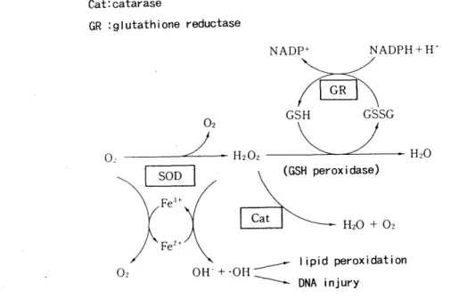
SUMMARY : Ischemic-reperfusion injury caused by oxygen-drived free radicals is one of the great problems to be solved for successful lung transplantation and preservation. This study was performed to clarify the effect of the free radical scavenger, reduced glutathione (GSH), superoxide dismutase (SOD), and alloprinol for warm ischemic lung, and to elucidate changes of active oxygen species production in neutrophils.

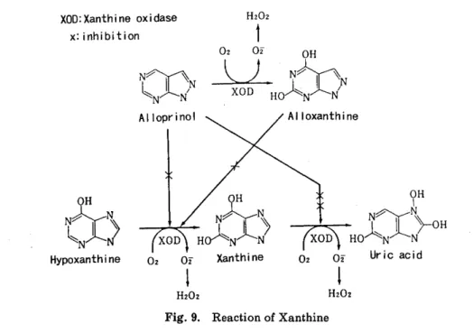
<Method > Sixty-three mongrel dogs underwent hilar stripping following left thoracotomy. The left pulmonary artery, pulmonary vein, and left main bronchus were clamped for 2-3 hours. To reduce the free-oxygen radicals, alloprinol (30mg/kg /day, for 3 days peros), GSH (50mg/kg, I. V.) administration and perfusion through

the  pulmonary.  artery  with  4℃  Euro‑Collins'solution  including  GSH(1mg/l),  SOD

(15mg/ml), and alloprinol (20mg/l) were performed. At the time of right pulmonary artery occlusion test, the blood gas analysis of arterial blood and measurement of left pulmonary arterial pressure were done on the pre-ischemic period, immediately after and one hour after declamping.

At the same time, active oxygen species production in neutrophils was evaluated using flow cytometric procedure.

<Results> 1) The good pulmonary gas-changing function remained after ischemic period by administration and perfusion with free radical scavenger GSH, SOD, and alloprinol.

2) Changes of pulmonary arterial pressure at right pulmonary artery occulision test was not statistically significant.

3) Production of active oxynene species in neutrophils was increased after ischemic period.

In conclusion, all of GSH, SOD, and alloprinol are effective in elminating ische- mia-reperfusion injury to warm ischemic lung.

INTRODUCTION

Since 1965, clinical application of lung trans- plantation has been made in 3 patients in Japan.1) 2) However, its outcome failed to obtain long survival due to immunologic rejec- tion. On the contrary, in Europe and the united states of America there are quite a few pro-

longed survivors following transplantation including renal transplantation with an did of development of cyclosporine A.3)

Therefore, reperfusion injury is a great

concern to success of clinical lung transplanta-

tion except for immunologic response. There

are many reports in the pathogenesis of reper-

f usion syndrome. Great concern is focused on

an activation of active oxygen species 4) 5) 6) to

(2)

prevent damage to warm ischemic donor lung.

The purpose of this study is to clarify the role of free radical scavenger such as GSH, SOD and alloprinol to elimenate reperfusion injury at transplantation by means of an evaluation of inhibited generation of free radical in peripheral neutrophils.

MATERIAL AND METHOD

1) Animal

Sixty-three Mongrel dogs weighing from 8 to 14 kg in body weight were used. All dogs were supplied from Laboratory Animal Center for Biomedical Reserch, Nagasaki University School of Medicine.

2) Experimental Method

Dogs were anesthetized with 25mg/kg of pentobarbital sodium, intubated intratracheally and ventilated with Harvard Ventilator, 250- 350m1 in tidal volume, 12-14/min in respira-

tory rate. Cannulation into the femoral artery was performed to measure the systemic blood pressure continuously. Left thoracotomy was made at the 5th ICS. The left main pulmonary artery and left main bronchus were isolated from the surrounding tissue. A procedure of hilar stripping , which is composed of dividing branches of the vagus, bronchial artery and lymphatic channels.

After heparization of 100mg/kg, the left main pulmonary artery, vein and left main bronchus were separately clamped to make a model of warm ischemic lung for 2- 3 hours.

After ralease of clamps with elapsed time of 2- 3 hours blood flow was recirculated. In in- fusion groups prior to restart of blood flow, a small catheter (1.9 mm I.D.) was introduced into the pulmonary artery to monitor the infusion pressure of 40cmH2O and also a small hole was made in the left atrial wall so as to allow perfusate to flow out.

At the time of completion of perfusion prior to restoration of blood circulation, the monitoring catheter was withdrawn and the left atrial wall was repaired.

At the first hour following blood recircula- tion, PA pressure was measured and blood samples from PA, PV and femoral A were pre-

pared for blood gas analysis and assessment of free radical in neutrophils.

3) Measurement of generation of free radical in neutrophils.

The measurement was in accordance with modified Bass' method 7 ) (Fig. 1) by using Spectrum III (ORTHO Co. ). As a stimulator to neutrophils, phorbol myristate acetate (PMA) was used.

Fig. 1. Principle of evaluation of active oxygen species production

Measurement steps>

1) 0.1ml Blood samples were taken, 1.9m1 of 0.5M 2-7-dichlorofluorescin diacetate (DCFH-

DA) was added, incubated at 37°C for 15 min.

2) 0.5ml of EDTA adjusted to 24mM with PBS were added to prevent aggregation of neutrophils.

3) and incubated at 37°C for 25 min in addi- tion to 10u1 of PMA (15,ug/ml)

4) washed with PBS and centrifused at 1300 rpm for 10 min.

5) hemolysed with lysing solution of 0.87%

NH4 Cl

6) washed with PBS and centrifused at 1300 rpm for 10 min

7) the pellet is suspended with 10% gel-Hanks'

(3)

solution

8) analysed with the use of spectrum III. As a negative control, the peripheral blood from a dog was used and the positive zone (13%) was adjusted to be 2-5% of B-start at the resting phase and generation of active oxygen species was calculated as being 13% after stimulation with PMA.

Experimental groups

The groups in the experiment were divided into 7.

group I : simple warm ischemia (n= 13) group II : intravenously given GSH (50mg/kg) at induction of anesthesia and prior to recircu- lation (n=8)

group III : orally given alloprinol (30mg/kg) 3 days before the experiment (n=10)

group IV : perfusion group with Euro-Collins' solution (E-C solution) (n=8)

group V : perfusion group with E-C solution +GSH (1mg/ml) (n=8)

group VI : perfusion group perfused with E-C solution+SOD (15mg/l) (n=8)

group VII : group perf used with E-C solution + Alloprinol (20mg/l) (n=8). (Alloprinol suppli- ed from Jap Welcome Co.)

Statistical analysis

The values were expressed as the means ± standard deviation (SD). The difference among means was determined to be significant with Wilcoxon-t test.

RESULTS 1) PA pressure (Table 1, Fig. 2)

The PA pressures in all groups were elevated after temporary contralateral PA blockage.

There was no statistically significant diffe-

Fig. 2. Pulmonary artlrial pressure after rt PA occulusion test

rence among the groups.

2) arterial Pa02 (Table 2, Fig. 3)

The levels of pa02 in group II, III were supe- rior to that in group I and also those in group V, VI, VII were better than in group IV imme- diately and one hour after restoration of blood circulation. Statistically significant differences were calculated in group II immediately after and one hour after and in group III immediately after recirculation (P<0.05) and in group III one hour after recirculation (p<0.01) and as compared with the control in group V, VII one hour after (p<0.05) respectively.

3) arterial PaCO2 (Table 3, Fig. 4)

The levels of PaCO2 were increased by tempo- rary contralateral PA block test. Its grade of group II III and Group V VI VII was depressed as compared with that of group I and IV, in particular there is significant difference bet- ween group II , V and the control groups.

Table 1. Pulmonary arterial pressure (mmHg)

group I group II group III group IV group V group VI group VII pre-ischemia 19.7± 9.0 21.7± 4.1 18.4± 4.8 19.5± 2.8 19.0± 4.0 20.8± 5.7 21.6± 4.7

r. PA occ. pre. 30.1± 6.5 30.4± 3.8 29.6± 6.2 27.9± 3.3 27.9± 4.1 29.9± 7.3 31.3± 5.2

immediately after 20.6± 7.8 18.9± 4.2 19.8± 5.6 19.8± 2.9 18.9± 3.8 22.1± 7.9 21.1± 3.9

r. PA occ. im. after 31.1±11.7 27.4± 5.6 28.3± 4.7 33.1± 4.2 29.3± 4.6 31.4± 7.3 30.6± 6.1

1 Hr after 22.6± 8.0 20.6± 4.6 20.8± 5.6 20.0± 3.8 19.0± 3.1 23.9± 5.4 20.6± 3.0

r. PA occ. -1Hr after 31.4±12.2 27.0± 5.1 28.9± 5.5 30.4± 5.6 26.3± 6.0 34.0± 6.8 29.5± 6.7

(4)

Table 2. Blood gas alalysis-Pa02- (mmHg)

group I group II group III group IV group V group VI group VII pre-ischemia 424.9± 75.5 455.8± 48.4 473.5± 58.3 469.5± 46.3 441.0±113.2 462.8± 32.1 459.7± 67.2

r. PA occ. pre. 223.4±109.3 230.4±106.0 312.6±164.0 275.3±103.5 249.0± 90.6 222.8±126.3 177.3±112.3 immediately after 357.7± 97.4 495.5±102.8 438.6±136.6 452.7±136.3 440.8±132.6 399.7±100.7 459.9±141.9 r. PA occ. im. after 86.4± 44.2 225.2±118.3 236.4±166.0 193.5±113.9 252.3±138.9 230.4±109.7 294.7±151.9

1 Hr after 155.3± 88.8 356.2±173.2 390.0±119.1 340.3± 85.3 373.0±104.2 354.4±116.6 367.5±184.7 r. PA occ. -1Hr after 92.0± 30.8 182.6± 97.6 215.0±100.0 113.6± 47.3 237.6±155.2 198.9±148.0 232.5±184.5

Fig. 3. Pa02 after rt PA occulusion test

Fig. 4. PaC02 after rt PA occultsion test

Table 3. Blood gas analysis-pressure (mmHg)

group I group II group III group N group V . group VI group VII pre-ischemia 26.9± 9.3 17.9± 3.3 18.9± 4.3 19.2± 2.1 18.4± 5.0 18.2± 5.5 18.2± 3.1

r. PA occ. pre. 25.7± 6.9 26.1± 6.7 22.2± 6.5 25.4± 4.3 25.5± 8.4 24.7± 6.4 24.8± 6.8 immediately after 23.8± 6.4 19.9± 4.7 19.5± 5.0 23.3± 7.6 19.4± 4.4 20.3± 5.1 19.2± 5.1 r. PA occ. im. after 33.2± 7.3 24.5± 6.1 23.9± 8.4 28.4± 6.5 26.2± 7.1 25.3± 5.9 26.1± 8.5 1 Hr after 29.2± 8.7 22.1± 5.7 22.4± 6.3 27.3± 6.4 22.2± 4.0 23.2± 4.4 23.9± 7.5 r. PA occ. -1Hr after 36.0± 5.1 25.8± 5.6 27.0± 8.8 31.5± 4.9 25.1± 6.0 26.8± 7.3 25.5± 8.5

4) Intrapulmonary shunt (Table 4, Fig 5) The intrapulmonary shunting rate was calculated as the following equation8), Qs/Qt

=0 .03 (PAO2 -PaO2) /4.5+0.003 (PAO2 - Pa02 ), wherein Fi02 = 1, Sa02 = 100%, Ca02 - Cv02 =4.5 vol%.

The intrapulmonary shunting rates were figured out during performing temporary contralateral PA block test Intrapulmonary

shunt lessened in groups in which protective drugs for storaged lungs were used, in parti- cular, were of great benefit in group II imme- diately and one hour after recirculation (P 0.01).

5) Generation of free radical in neutrophils (Table 5, Fig. 6)

Fig. 7 revealed a cytogram of Spectrum III , in

(5)

Table 4. Blood gas analysis-shut ratio- (%)

group I group II group III group IV group V group VI group VII pre-ischemia 23.32±2.97 23.04±4.10 22.53±5.22 20.32±3.67 22.27±3.73 23.20±5.33 25.03±4.17 immediately after 28.10±1.36 23.17±4.63 25.23±4.75 24.71±4.63 21.92±5.64 22.96±4.34 20.12±6.20

1 Hr after 27,80±0.99 24.95±3.67 24.94±1.46 27.47±1.52 22.46±6.55 23.96±5.80 22.65±8.15

Fia. 5. Intrapulmonary shunt Fig. 6. Generation of free radical in neutrophils Table 5. Production of active oxygene species by PMN (%)

group I group II group III group IV group V group VI group VII pre-ischemia (PA) 72.7±12.0 73.1±'16.8 67.3±15.7 71.0±12.9 77.8±12.4 79.2±10.7 72.0±13.3 pre-ischemia (PV) 72.9±12.1 73.1±14.8 64.7±12.3 69.8±14.1 80.9± 5.5 79.9±11.1 70.7±10.7 immed. after (PA) 83.0± 9.3 86.9± 6.6 73.6± 9.8 82.0±14.8 81.3±10.3 89.3± 4.8 80.9±11.9 immed. after (PV) 81.9± 9.6 86.4± 4.3 76.0±12.4 80.7±14.1 81.7± 9.8 87.5± 6.4 83.7±11.8 1 Hr after (PA) 81.5±10.7 90.3± 3.1 78.1± 7.2 83.1±12.4 80.9±13.9 85.6± 6.9 82.8± 8.9 1 Hr. after (PV) 81.1±10.2 92.9± 1.8 80.1± 5.9 77.5±16.6 86.7±11.5 87.0± 6.7 79.9±13.5

which the ordinate represented the. size of cells obtained by the foward light scatter method, on the other hand, the abscissa present- ed inner structure of cells obtained by the 90 degree light scatter method.

Fig. 8-A demonstrated a histogram at the resting states and Fig. 8-B showed a histogram after stimulation by PMA. It showed that neutophils distributed regularily. In every group, generation of free radical was accerelat- ed after ischemia. However, there was no significant difference in generation of free radical. between the blood sumpling from

pulmonary arteries and veins.

As compared with the control, generation of free radical one hour after recirculation was pronounced in group II , although there was no definite difference in the other groups.

DISCUSSION

Since lung transplantation has first been

made in clinical application by HAY9), scores

of patients were reported. However, cyclo-

sporin offered insight into the significant

effect on suppression of immune response. In

(6)

based on increased permeability of the vessel wall and subsequent edema. Furthermore cytotoxic ferment breaks away from the inside

of cells to the blood.

On the other hand, it is accepted that reper- fusion injury is mainly caused by free radicals.

4)-6) 10) 11)

WEISS 10) suggested that main damage to cellular structure and function is associated with action of free radicals. HAGLUND et al 11) suggested that organ damage of storage lungs is due to not only ischemia but reoxygenation.

JACKSON et a112) reported that free radicals allow destruction of protein, membrane and nucleic acid and causes stagnation, incomplete reexpansiori of donor lung, thickness of alveo- lar septum and fibrosis in the pulmonary parenchym.

A damage to a donor lung caused by free redicals offers a great problem on ischemia

Fig., 7. Cotogram of spectum I

Fig. 8-A Histogram of spectum III (Resting)

consequence Copper reported prolonged survi- vors with lung allograft in clinical use.

To expect an adequate function of lung allografts immediately after lung transplanta- tion, reperfusion injury should be minimized during storage of a donor lung. MCCORD4) ex- plains that ischemic damage to a donor -lung is

Fig. 8-B. Histogram of spectrum III (PMA stimulation)

during storage of a donor lung and restoration of blood circulation after completion of transplantation procedure.

Lung edema which takes place in early stage

after lung transplantation is referred as

reimplantation response 13) 14) and main cause

is ischemia and denervation. However, it still

(7)

remains unsettled.

<Alloprinol>

In 1968 Mcconn 15) pointed out that most important source of free radicals is xanthine oxidase system as shown in Fig. 916), when the reactions of ATP, ADP, AMP to hypoxanthine progress, 02 supply by blood recirculation makes 02 - by a reaction of hypoxanthine to Xanthine. It needs for Xanthine oxidase (XOD) which contains abundantly in organs.

Alloprirol is an inhibitor of XOD, which is used for gout.

There are some reports on the effects of alloprinol with respect to the heart4) 17) 18) small interstine4) 19) and liver4 ), although it is scanty concerning the lung.

HASImvIOTO 20) reported that XOD activities in various organs are as follows, 19.5munit/g in the brain, 240.0 'in the liver, 185.1 in the lung, 26.3 in the heart, 225.4 in the spleen, 122.3 in the kidney 630.4 in the pancreas 708.0 in the small intestine 95.2 in the colon 147.2 in the adrenal grand respectively. KINASEWITZ 21) also cited that alloprinol effectively inhibited increased permeability of vessel walls in the pulmonary capillary. From the facts, alloprinol is one of the effective inhibitors against ischemia and reperfusion injury.

It is well known that the protective role of

alloprinol in ischemia and reperfusion injury is required for alteration to oxyprinol in vivo and it consumes some time interval. Therefore, it is recommended that alloprinol should be administered in advance.

In this study, it is substantiated that the use of alloprionl prior to 3 days is of great benefit to eliminate damage of ischemia and reperfusion injury to a donor lung as compared with the control and a result of group Vm which was intraoperatively perf used with protective drugs.

<GSH>

GSH acts as one of radical scavengers and cooperates with glutathione perioxidase (GSH- Px) to eliminate an action of active oxygen species22) 26 ), which deals with H202 in the cells and/or mitochondria 23) and plays a key role in acting as ground substance24) with an aid of NADPH and NADH actions (Fig. 10).

In the hypoxic circumstances, a shortage of ATP supply results in a decrease in GSH synthesis and a delay in dealing with active oxygen species. 25) It is a confirmed fact that hypoxia in the brain reduces the amount of GSH 27), and it is suggested that GSH plays an important role in removing active oxygen species.

On the other hand, it is well known that a

Fig. 9. Reaction of Xanthine

(8)

Fig. 10. Reaction of glutathione

shortage of GSH in the lung facilitates oxygen intoxication. 28)

DAwsoN29) explained a merit of the use of exogenous GSH in the experiments of isolated lung and/or isolated lung cells in the hypoxic-c ircumstances to prevent is chemic damage to pulmonary parenchym. It is also clarified that damage to the epithelial cells of the gut is protected by exgenous GSH, and administration of GSH contributes to elimination of active oxygen species. 30)

It is dubious as to whether intravenous administration of GSH is absorbed in the reticuloendothelial system or not. It is accepted that the half life of GSH is just 3 min and its action reduces to 90% at 10 min and during that time the content of GSH in the liver and kidney does not vary in rats. In human being the half life is 2 min when given intravenously and it is quickly metabolized.

When GSH is dissolved, it converts into three amino-acids of cystine, glycine and glutamic acid and is resynthesized to GSH in the liver and carried via blood circulation.

In this study, it is ascertained that GSH is of great help to reduce ill effects of ischemic damage and reperfusion injury on a storage lung. The reasons are due to direct protective

action of GSH and/or resynthesized GSH in the liver from amino-acids of dissolved GSH.

It is said that antioxidation by the help of GSH is established with GSH, intracellularily synthesized, which plays a key role in contri- buting to dealing with glutathione peroxidase (GSH-Px) in the plasma.') 32)

<SOD>

Since MCCORD et a133) discovered SOD in 1969, advances in basic study on SOD have been established. SOD includes the types of Mn, Fe and Cu, Zn. In mammalias, SOD exists in the Cu, Zn type in the cells and the Mn type in the mitochondria.

In the human body, SOD is distributed in varying patterns 34) (Table. 6). The main action is promotion of a reaction from 02 to H202.

Therefore, final product of 02 should be re- moved in humans.

SOD increases with advancing age. However, this tendency toward an increase in SOD is much more potent in neonates than in older patients35 ). In the experiment with the rat lung which has high resistance to oxygen activity, the following fact is clarified that type II alveolar cells increase in number and SOD is activated at the same time3s)

However, it is doubtful if intravenous

(9)

Table. 6. Cu, Zu-SOD and Mn-SOD in human organs (mg/kg)

Cu, Zn-SOD Mn-SOD

liver 490 390

lung 254 25

kidney (cortex) 164 142

RBC 157 0

uterus 148 26

brain 136 55

heart 119 97

muscle 107 52

thyroid 103 65

testis 76 22

spleen 71 22

lymph node 58 71

fatty tissue 8 3

administration of SOD is helpful to reduce 02 intoxication because of a short half time. It is reported that 55 % of SOD is excreted in the urine 2 hours after I. V. administration and 29

% accumulates in the renal cortex37 ). In this study, direct administration by perfusion was performed to maintain its action as long as possible in order to expect a role of SOD.

Needless to say, there are many devices to keep long-standing action of SOD. For example, it is a trial product of included SOD to lipo- some which shows a high affinity to the cell membrane containing a rich lipoprotein so that it is easy for SOD to be taken into the cells, and attention is paid to prolong half life time. FREEMAN reported that by using vascu- lar endothelium which is highly sensitive to oxygen intoxication, SOD included in liposome provided a 95% resistance to oxygen, and it is confirmed that in in-vivo experiment it made a half time of circulating SOD prolonged and achieved inhibition of a 100% 02 intoxication with an aid of high activity of SOD.

<Neutrophils>

The main source of active oxygen species is neutrophils. Ischemia causes active arachidon acid reaction and consequently free radicals are generated. In addition destructive product of fibrin, production of leukotrien B4, platelet activating factor (PAF) and so on activate neutrophile in the reaction circle of generation of free redicals.39)

In this study, the levels of free radicals were

kept high on account of stimulation of ische- mia at the time of start of recirculation and by activated neutrophils at one hour after recirculation. The method of measuring free redicals according to Bass7) by using flow cytometry is of great benefit in terms of simplicity and accuracy.

In the experiment regarding ischemic hearts 40) it is reported that, when recirculation was performed with f ree-neutrophil blood, the ischemic area in the heart was reduced and minimized and lessened the degree of infiltra- tion of neutrophils around the tissues by add- ing SOD, suggesting some parts of SOD to neutrophils.

In this study, the author emphasized that intravenous administration of GSH offered insight into the effect that given GSH accele- rates GSSG reaction circle by facilitating NADPH reaction, and consequently leads to an increase in 02 generation.

Needless to say, 02- generated in the body is eliminated by the help of GSH action, there- fore damage of O2 is minimized. On the other hand, in the perfusion groups, activation of 02 was lower than those in the other groups.

It is more likely for GSH to be not in contact with the membranes of neutrophils in the perfusion groups. It is different from in I.

V given groups.

As for the pressure of the pulmonary artery, it was a tendency to be lower in the groups in which protective drugs were used. However, it was of no statistical significance. It seems to be associated with the slight degree of reversi- ble alteration of perivascular edema and thrombosis, reflecting no ominous effects on pulmonary vascular resistance.

SUMMARY

The study aimed at an inhibition of free radicals to prevent warm ischemic damage to a donor lung.

1) SOD, GSH and alloprinol play a protective role in preventing warm ischemic damage and in eliminating alveolar-capillary block

2) These drugs were significantly of no use to

reduce the pressure of the pulmonary artery

following lung transplantation

(10)

3 ) Generation of active oxygen species in neutrophils increased by an addition to ischemia and start of recirculation, in particu- lar, that in the GSH-given group was much more prominent than those in the other groups.

ACKNOWLEDGEMENT

The author would like to thank Prof Dr Masao ToMYFA, First Department of Surgery, Nagasaki University School of Medicine for his advice and revision and thank Dr Katsunobu KAWAHARA and the doctors for the willing co-operation and technical assistance in this study.

I also appreciate for kindness of animal supply from the Labolatory Animal Center for Biomedical Reserch of Nagasaki University School of Medicine and thank to the members of Blood Transfusion Service of Nagasaki University Hospital. This work was partially supported by a Grant-in-Aid for general scien- tific reserch from the Ministry of Education of Japan.

REFFERENCE

1) Y.TsuJr : Experimental study on lung trans- plantation : Jap. J. transplant. : 1 : 236-240,

1966.

2) Y.HAYATA et al : Clinical manufestation of lung transplantation . Jap. J. Chest Disease : 27 : 99-102, 1968.

3) R.Y. CALNE, D.J.G. WHITE, S.THIRU, et al Cyclosporine A in patients receiving renal

allografts from cadaver doners : Lancet : 2

1323-1327, 1987.

4) J.M. MCCORD : Oxygen-derived free radicals in post-ischemic tissue injury : The new

Engl. J. of Med.: 312, 159-163, 1985.

5) S.L.MARKLAND : Oxygen toxicity and protec- tive systems : Clin. Toxicology : 23, 4-6 : 289-

298, 1985.

6) I. A. CLARK : Tissue damage caused by free o- xygen radicals : Pathology : 18 : 181-186, 1986.

7) D.A.BAss, J.W.PARCE, L.R.DECHATELET, et al : Flow cytometric studies of oxdative product

formation by neutrophils -A graded response

of membrane stimulation : The J. of Immuno-

logy : 130, 4 : 1910-1917, 1983.

8) Y.OxsAKr, T. IRIS : Shunt : Training of exami- nation for respiratory function : 86-91, 1983,

Chuugai shuppan Co.LTD,Tokyo.

9) J.D.HARDY et al : Lung homotransplantation

in man-Report of the initial case : JAMA 186 : 1065-1074, 1963.

10) A.J.WEIss : Oxygen, ischmia and inflamma- tion : Acta Phisio, Scand. : 548: 9-37, 1986.

11) U.HAGLUND, O.LuNDGREN : Intestinal ischemic and shock factors : Fed. Proc.:37: 2729-2733,

1978.

12) R.M.JACKSON : pulmonary oxygen toxicity : Chest : 88,6 : 900-905, 1988.

13) S.S.SIEGELMAN, S.B.P.SINHA, F.J.VErrH : Pul- monary reimplantation response : Ann.

Surg. : 177, 1 : 30-36, 1973.

14) M.G.EHRiE, J.D.CRAPO, N.C. DURHAM, et al : Reimplantation response in _ isografted rat

lungs. -Analysis of causal factors- : J.

Thorac. Cardiovasc. Surg.: 87: 702-711, 1984.

15) J.M. MCCORD, LFRIDovICH : The reduction of cytochrome C by milk Xanthine oxidanse : J. Biol. Chem. : 243 : 5753-5760, 1968.

16) Y. OOYANAGI : Introduction : Superoxide and Medicine : 1-22, 1981, Kyouritsu Shuppan

Co.LTD, Tokyo.

17) D.J.CHAMBERS, M.V.BRAIMBR.IDGE, D.J. HEARSE : Free radicals and Cardioplegia alloprinol

and oxypurinol reduce myocardial injury

following ischemic arrest : Ann. Thorac.

Surg.: 44 :291-297, 1987.

18) K. BANDO, M.TAGO, S. TERAMOTO : Prevention

of free radical induced myocardial injury by alloprinol -Experimental study in cardiac

preservation and transplantation : J. Thorac.

Cardiovasc. Surg.: 95 :465-476, 1988.

19) D.A.PARKS, G.B.BULKLEY, D.N.GRANGER et al :Ischemic injury in the cat intestine

Gastroenterelogy : 82 : 9-15, 1982.

20) S. HASIaMOTO : A new spectrophotametric as- say method of Xanthine oxidase in crude

tissue homogenate : Anal. Biochem. : 62 :

426-435, 1974.

21) G.T.KINASEwrrz, L.J.GROOME, R.P. MARSHALL

et al : Effect of hypoxia on permeability of pulmonary endotherium of canine visceral

pleura : Appl. Physiol. : 61 : 554-560, 1986.

22) K.NISHIKIORu, H.MAENO : Glutathione for medi- cal treatment : Protein, Nucleic Acid, Enzyme

: 33, 9 : 1625-1631, 1988.

23) K.TAKAHASHI : Seren and glutathione peroxi- dase : Protein, Nucleic Acid, Enzyme : 33, 9 :

1945-1504, 1988.

24) N.S. KOSOwER, E.M. KosowER : Free radicals in Biology : (ed W.A.Pryor) : 12 : 58-84, 1976,

Achademic Press.

(11)

25) N.TATEISHI, T. HIGASHI, N. TANIGUCFII : Metabol- ic-map of glutathione : Protein, Nucleic Acid,

Enzyme : 33, 9 : 1358-1364, 1988.

26) T,YUSA : Free radical scavenger and pulma- nary disease : Annual Review of Resp. : 79-

87, 1988.

27) A.J.L.COOPER, W.A.PUISINsLLI, T.E.DJFFY ; Glutathione and ascorbate during ischemia

and postischemic reperfusion in rat brain

J. of Neurochemistry : 35, 5 : 1242-1245, 1980.

28) S.M.DNEKE, B.A.LYNCH, B.L.FANBuRG : Tran- sient depletion of lung glutathione by diethyl-

maleate enhances oxygen toxicity : J. Appl.

Physio l.: 58 : 571-574, 1985.

29) J.R. DAWSON, K. VAHAKANGAS, B. JERNSTRON et al : Glutathione conjugation by isolated lung

cell and isolated, perfused lung-Effect of

extracellular glutathione : Eur.J.Biochem.

138 : 439-443, 1984.

30) L.H.LASH, T.M.HAGEN, D.P.JONES : Exogenous glutathione products intestinal epitherial

cells from oxidative injury : Proc. Natl.

Acad. S ci. USA : 83 : 4641-4645, 1986.

31) M. INOUE : Membranous transportation of glutathione and its metabolism : Glutathione

: 43-66, 1985: Koudansha Co. LTD, Tokyo.

32) K.R.MADDIPATI, L.J.MARNETT : Characteriza-

tion of the major hydroperoxide reducing activity of human plasma. Purification and

properties of a selenium dependent gluta-

thione peroxide : J. Biol. Chem. : 262 : 17398-

17403, 1987.

33) J.M. MccouD, I. FRIDOVICH : SOD an enzimic function for erythrocuprein : J. Biol. Chem.

25 : 6049-6055, 1969.

34) S. MARKLUND : Distribution of Cu, Zn-SOD and Mn-SOD in human tissue and extracellu-

lar fluid : Acta Phisiol. Scand. :492 : 19-23, 1980.

35) Y. KOBAYASHI, T. Usul : Clinical meaning of superoxide dismutase : Jap. J. Metabolism : 15 : 23-32, 1978.

36) B.A.FREEMAN, B.J.MASON, M.C.WILLIAMS et al : Antioxididant enzyme activity in alveolar

type II cells after exposure of rats to hypero-

xia : Exp. Lung Res. : 10 : 203-222, 1986.

37) Y.SHIOKAWA : Free radical and anti-inflamma-

tory drugs. : Active Oxygen Species : 318-333, 1987: Ishiyaku shuppan Co.LTD, Tokyo.

38) B.A.FREEMAN, J.F.TuRRENs, Z.MIRZA, et al Modulation of oxidant lung injury by using

liposome intrapped superoxide dismutase

and catalase : Fed. Proc. : 44 : 2591-2595,1985.

39) E. OKABE, H. ITOH, Cardiac ischemia -Free radical as mediator of ischemia-reperfusion

injury.- : Jap. J. Clin. Med. : 46, 10 : 2190- 2195, 1988.

40) M.L.HESS, et al : Identification of Hydrogen peroxide and hydroxyl radicals as media-

tors of leukocyte-induced myocardial dys-

function : Adv. Myocardiol : 5 :159-175, 1985.

Fig.  1.  Principle  of  evaluation  of  active  oxygen         species production
Fig.  2.  Pulmonary  artlrial  pressure  after  rt  PA         occulusion test
Fig.  3.  Pa02  after  rt  PA  occulusion  test
Fig.  8-A  demonstrated  a  histogram  at  the  resting  states  and  Fig.  8-B  showed  a  histogram  after  stimulation  by  PMA
+4

参照

関連したドキュメント

Consistent with the results of echocardiographic and histo- logical measurement, the mRNA expression levels of these cardiac remodeling markers were significantly decreased

Histologic appearance varies markedly from area to area in the same case, varying from vascular granulation tissue heavily in filtrated with both plasma cells and lymphocytes to

This study was therefore designed to examine the induc- tion of CYP2E1 in genetically obese Zucker rats fed a normal diet (OB) and its effect on the disposition kinetics of CZX and

We describe a little the blow–ups of the phase portrait of the intricate point p given in Figure 5. Its first blow–up is given in Figure 6A. In it we see from the upper part of

In this, the first ever in-depth study of the econometric practice of nonaca- demic economists, I analyse the way economists in business and government currently approach

In this case, the extension from a local solution u to a solution in an arbitrary interval [0, T ] is carried out by keeping control of the norm ku(T )k sN with the use of

In the second computation, we use a fine equidistant grid within the isotropic borehole region and an optimal grid coarsening in the x direction in the outer, anisotropic,

Physiologic evaluation of the patient with lung cancer being considered for resectional surgery: Diagnosis and management of lung cancer, 3rd ed: American College of Chest