Original Article for the Journal of Heart Valve Disease
Novel method of assessing ascending aorta with a stenotic bicuspid aortic valve
Department of Cardiovascular Surgery
Hirosaki University, Graduate School of Medicine
Kaoru Hattori, Ikuo Fukuda, Kazuyuki Daitoku, Masahito Minakawa, Wakako Fukuda,
Yasuyuki Suzuki
Correspondence: Ikuo Fukuda M.D,Ph.D.
5 Zaifu-cho, Hirosaki, Aomori, 036-8562 Japan
Tel: +81 172 39 5073
Fax: +81 172 37 8340
E-mail: ikuofuku@hirosaki-u.ac.jp
Abstract
Background and aim of the study: Patients with bicuspid aortic valve (BAV) have an increased
risk of serious aortic complications such as aortic dissection, rupture and dilatation of the
ascending aorta. Previous findings have suggested that ascending aortic dilatation with a BAV
has a typical asymmetric configuration at the right-anterior aspect of the aorta. The study aim
was to quantify asymmetric configurations of the aorta using a three-dimensional (3D)
reconstruction tool.
Methods: A retrospective review was conducted of 52 patients (27 males, 25 females; mean age
69 ± 9 years) with aortic stenosis who presented with ascending aortic dilatation defined as an
aortic diameter >35 mm. Of these patients, 24 (46%) had a BAV and 28 (54%) had a tricuspid
aortic valve (TAV). A patient-specific 3D thoracic aortic model was reconstructed from
computed tomography (CT) data. Three-dimensional centerlines were automatically calculated.
The size of the ascending aorta was determined by calculating the cross-sectional area (in mm2)
of the vertical section against the centerline. The symmetry of the dilated aorta was evaluated as
the ellipticity of the maximum vertical section of the ascending aorta. The size and symmetry of
the ascending aorta, and background factors including pressure gradient, aortic valve area,
degree of regurgitation, ejection fraction and cardiovascular risk factors, were compared
between the BAV and TAV groups.
Results: Only age differed significantly between the groups (p = 0.003). The size and ellipticity
of the ascending aorta and the maximum cross-sectional area of the aortic arch were
significantly greater in the BAV group (p = 0.001 and p = 0.004, respectively).
Conclusion: The ascending aorta assessed using Mimics 3D reconstruction software was
frequently asymmetrically dilated in stenotic BAV, and the expansion progressed to the aortic
arch. It is believed that calculating the ellipticity of the vertical section against the centerline
offers an innovative means of quantifying aortic symmetry in three dimensions.
Bicuspid aortic valve (BAV) is a common congenital heart malformation that occurs in 0.5% to
2% of the population (1,2). Patients with a BAV are at increased risk of developing serious
aortic complications, including aortic dilatation, dissection and rupture after reaching adulthood
(2-4). Aortic complications (aortopathy) are associated in 10% - 35% of cases, and aortic
dissection occurs in about 4% of patients with BAV (3). Patients with BAV and a dilated aorta
have a nine-fold increased risk of aortic dissection (5). Histological examinations of aortas with
a BAV frequently demonstrate cystic medial degeneration, a characteristic histopathological
defect of the aortic media (3,5).
Ascending aortic dilatation associated with BAV sometimes progresses even after aortic valve
replacement (AVR). Yasuda et al. (6) reported that the ascending aortic diameter increased at an
annual rate of 0.18 ± 0.08 mm/m2 after AVR. Aortic dilatation has a typical asymmetric
configuration at the right-anterior aspect of the aorta (7). Valve-related hemodynamic
abnormalities might lead to the asymmetric distribution of wall shear stress, resulting in
asymmetric aneurysmal formation (8,9).
Although patients with a BAV require regular monitoring to prevent fatal cardiovascular events,
three-dimensional (3D) deformation of the aorta is difficult to quantify in routine computed
tomography (CT) images. Hence, it was postulated that asymmetry of the ascending aorta and
dilatation of the transverse aortic arch would be more significant in patients with BAV than with
TAV. This hypothesis was tested by comparing the 3D parameters of aortic dilatation in patients
with stenotic BAV and TAV.
Clinical material and methods
Patient selection
A retrospective review was conducted of data derived from 52 patients (27 male, 25 female;
mean age 69 ± 9 years; range: 44 to 81 years) with aortic stenosis (AS) who presented with
ascending aortic dilatation defined as an aortic diameter > 35 mm. All patients underwent AVR
at the authors’ department between January 2002 and December 2012. Twenty-five (48%) of the
patients were treated only by AVR, 27 (52%) were treated by concomitant surgeries that
included graft replacement of the ascending aorta (n = 16), coronary artery bypass grafting (n =
5), mitral valve surgery (n = 3), reduction aortoplasty (n = 3), annuloplasty of the tricuspid valve
(n = 2), and the maze procedure (n = 2) (single concomitant, n = 23; double concomitant, n = 4).
Patients with infective endocarditis and suboptimal CT images for the reconstruction of 3D
models using Mimics software were excluded from the study. Data were compared from 24
patients with BAV and 28 with TAV. The morphological type of BAV was defined according to
the Sievers’ classification. The cusp configuration was type 1 (R/L) in eight patients (33%), type
1 (R/N) in four patients (17%), type 1 (L/N) in three (13%), type 0 (ap) in eight (33%), and type
0 (lat) cusp fusion in one patient (4%) (Fig. 1).
The following parameters were compared to evaluate morphological changes of the aorta: size
of the thoracic aorta; symmetry of the ascending aorta; transvalvular pressure gradient (mmHg)
of the aortic valve; area of the aortic valve (cm2); degree of aortic regurgitation; left ventricular
ejection fraction (%); body surface area (m2); and cardiovascular risk factors including age,
gender, smoking history, hypertension, diabetes and dyslipidemia (defined as serum total
cholesterol > 219 mg/dl).
Imaging protocols and 3D reconstruction
The thoracic section of the aorta was reconstructed from axial CT data converted into DICOM
(Digital Imaging and Communications in Medicine) files. All 52 patients scheduled for
cardiovascular surgery were preoperatively assessed by CT using a whole body spiral CT
scanner (model TSX-310 B; Toshiba Medical Systems, Tochigi, Japan) according to standard
procedures in the authors’ department or affiliated hospitals. All CT slices measured 512 × 512
pixels, and the mean pixel size was 0.63 ± 0.083 mm. The patients were injected with 60-100 ml
of Iopamiron contrast material (Bayer Healthcare, Leverkusen, Germany) at a rate of 2.0-3.5
ml/s. Images were acquired during a single sustained breath-hold by the patient to reduce
respiratory-induced motion and associated artifacts. The slice thickness was between 1 and 5
mm. Images > 1 mm thick were further sliced to 1 mm using the image processing tools
provided with Mimics software.
The DICOM data of CT scans were imported into the software Mimics v16 (Materialise,
Belgium) to reconstruct patient-specific 3D models of the thoracic aorta using the algorithm
introduced by Doyle et al. (10). The region of interest on CT images was determined and a
thresholding technique was applied to form rudimentary segmentation. The new 3D masks were
manually edited using the tools provided with Mimics to smooth the surface and remove any
non-physiological bulges (Fig. 2).
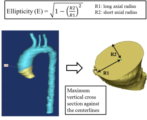
Measurement techniques
The following parameters were measured on the 3D reconstructed models in Mimics: 3D
centerlines, and the size, symmetry and ellipticity of the aorta. Three-dimensional centerlines,
equivalent to the central axis of the aorta, were automatically calculated. Aortic size was
determined by calculating the cross-sectional area (in mm2) of the maximum vertical
cross-section against the centerline (Fig.3). The symmetry of the dilated aorta was evaluated as
the ellipticity of the maximum vertical section of the ascending aorta calculated using the
formula:
Ellipticity (E) =
2
1 1 2
− R
R
Where R1 is the long axial radius and R2 is the short axial radius.
It was expected that, in symmetrical dilated aorta, R1 and R2 would be close to equal and the
ellipticity would be zero. On the other hand, in asymmetrical dilated aorta, the ratio of R1 and
R2 was expected to be larger because the vertical cross-section of asymmetric aorta was more
distorted (Fig. 4).
Statistical Analysis
All data were statistically analyzed using SPSS software (version 20; SPSS, Inc., Chicago, IL,
USA). Results were expressed as absolute numbers and ratios (%) or as means ± SD.
Categorical data between the two groups were compared using Person’s chi-squared test.
Continuous numeric variables including age, pressure gradient, aortic valve area, ejection
fraction, aortic cross-sectional area and aortic ellipticity were compared using the
Mann-Whitney U-test. A p-value < 0.05 was considered to be statistically significant.
Results
Patient characteristics
The characteristics of all patients are listed in Table I. Age differed significantly between the
BAV and TAV groups (64 ± 11 versus 73 ± 5 years; p = 0.003), whereas no inter-group
differences were noted in pressure gradient, aortic valve area, concomitant AR, ejection fraction,
body surface area, and other cardiovascular risk factors.
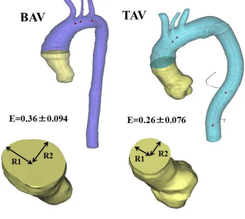
Aortic cross-sectional areas and ellipticity of the ascending aorta
The aortic cross-sectional areas and ellipticity of the maximum vertical section of the
ascending aorta are listed in TableⅡ. In the BAV and TAV groups, the ascending aorta area was
significantly larger (1540.9 ± 375.2 and 1226.8 ± 309.8 mm2, respectively; p = 0.001) and
significantly more elliptical (0.36 ± 0.094 and 0.26 ± 0.076, respectively; p = 0.001) in BAV
patients (Fig.5). The maximum cross-sectional area of the proximal aortic transverse arch was
also significantly larger in the BAV group (1150.5 ± 333.2 versus 940.0 ± 207.8 mm2; p =
0.004). These findings showed that patients with BAV had a more asymmetrically dilated
ascending aorta, and that such expansion frequently progressed to the proximal aortic arch.
Discussion
Quantitative assessment of 3D aortic configuration
The 3D configuration of the aorta is difficult to quantify conventionally, mostly because of the
3D complexity of the aortic configuration. As the aortic cross-sections visualized in
conventional axial CT images are not vertical to the central axis of the aorta, the measured
values included in the axial CT data do not always accurately represent the aortic status. In
addition, results include errors due to analog data manipulations. Some reports have described
how to evaluate aortic symmetry. For example, Lu et al. (7) quantified ascending aortic
symmetry by calculating the ratio of the greater to the lesser ascending aortic curvature on the
candy-cane sagittal view in 3D CT images. Doyle et al. (11) evaluated the symmetry of
abdominal aortic aneurysm (AAA) using a 3D reconstruction tool, which allows the automated
calculation of the 3D centerline of the aorta. These authors quantified abdominal aortic
symmetry by measuring the perpendicular distance from the proximal and distal points of the
centerline to a defined point on the centerline (11).
In the present study the aortic configuration was also evaluated using Mimics 3D
reconstruction software. The aortic size was determined by calculating the cross-sectional area
of the vertical section against the centerline, and symmetry was evaluated by calculating the
ellipticity of the maximum vertical section, which changes depending on the ratio of the long
and the short axial radii. It is believed that this technique allows the 3D quantitation of aortic
configurations. The present findings showed that ascending aortic dilatation was more
asymmetric and more frequently progressed to the proximal transverse arch when associated
with stenotic BAV than with TAV.
Asymmetric aortopathy
Dilatation of the ascending aorta associated with BAV has a typical asymmetrical
configuration at the right-anterior aspect of the aorta. In the present patients, the dilated
ascending aorta was found to be significantly more asymmetrical with stenotic BAV than that
with stenotic TAV. Finol et al. (12) showed that maximum wall shear stress (WSS) increases
concomitantly as aneurysms become more asymmetrical. These authors showed that, by using
computational AAA models, an asymmetric configuration remarkably increases the maximum
WSS at peak flow and induces secondary flow during late systole (12). When Doyle et al. (11)
studied the relationship between WSS and AAA asymmetry using finite element analysis (FEA)
software, they showed that AAA asymmetry, in addition to the maximum aneurysm diameter,
may be a good clinical predictor of AAA rupture (11,13). It has been reported that the posterior
wall tends to be the more highly stressed region and also the rupture site, even though the bulge
is predominantly in the anterior region of the AAA (13,14). As the anterior region of the
aneurysm bulges outwards, the posterior region is frequently constrained from radial expansion
by the spinal column, and this results in increased posterior WSS (11). It has been postulated
that the symmetry of the ascending aorta also is a risk factor for ascending aortic complications
associated with BAV. The novel method of quantifying aortic symmetry described in the present
study might reveal associations between aortic dilatation rates and the degree of asymmetry.
Previous studies have reported that asymmetric aortopathy associated with BAV mainly
originates in valve-related blood flow abnormalities (8,9). It was suggested that the most
prevalent flow abnormality associated with BAV is a clockwise-nested helical flow
accompanied by a peripheral skewing of the jet towards the right-anterolateral aspect of the
aorta (8). The right-anterior eccentric jet-stream caused asymmetrical WSS on the convexity of
the aorta (9). These flow abnormalities are most remarkable in the ascending aorta, with a large
proportion normalizing in the proximal descending aorta (15).
Aortopathy associated with BAV
The etiology of complications of the ascending aorta associated with BAV is controversial.
Two main hypotheses have been presented. One hypothesis has a genetic basis, whereby aortic
wall fragility is a consequence of an intrinsic developmental abnormality. The second
hypothesis is based on hemodynamics, in which aortic wall fragility is caused by abnormal
systolic flow jets through an asymmetrical orifice (16). Patients with BAV have extreme
degenerative changes not only in the ascending aortic media but also in the main pulmonary
artery (4,17). This finding could support the genetic theory, as the ascending aorta and the
pulmonary trunk have a common embryological origin (4). Previous studies have demonstrated
that isolated BAV is inherited in an autosomal manner with incomplete penetrance, which also
supports the genetic theory (18,19). The suggested prevalence of BAV among first-degree
relatives of affected individuals is between 9% and 21% (19). A BAV is considered to be a
heritable connective tissue disorder similar to Marfan syndrome, because the two conditions
share common histopathological changes such as cystic medial degeneration (4). However,
whilst BAV-associated aortopathy occurs mostly in the ascending aorta (20), aortic
complications associated with Marfan syndrome occur in every zone of the aorta. Moreover,
aortas with a BAV have an asymmetrical configuration that bulges towards the
right-anterolateral aspect, where WSS should be higher (9). These findings suggest that an
underlying intrinsic medial fragility, combined with increased WSS resulting from abnormal
flow jets, may precipitate BAV-associated aortopathy.
Study limitations
The main limitation was the small number of patients, due partly to the strict patient exclusion
criteria. For example, some patients with suboptimal CT images for the reconstruction of
reliable 3D models, the quality of which depends on the underlying CT data, were excluded.
Nonetheless, it is believed that the study findings were meaningful because of these strict
criteria. A second point was that the study samples were limited to patients with AS who
presented with ascending aortic dilatation, and only the degree of asymmetry of the dilated aorta
associated with stenotic BAV was analyzed.
In conclusion, the present study provided the first description of the quantification of the 3D
configuration of the aorta using Mimics software. It is believed that calculating the ellipticity of
the vertical section offers an innovative approach to quantifying the 3D symmetry of the aorta.
Ascending aortic dilatation associated with stenotic BAV is asymmetrical, and more frequently
progresses to the proximal transverse arch compared with that in TAV. Consequently, in some
cases it may be best to consider graft replacement of the proximal transverse arch with an open
distal anastomosis technique when operating on BAV patients with ascending aortic dilatation,
in order to achieve complete removal of the aneurysm.
Acknowledgements
These studies were supported by Grants-in-Aid for Scientific Research in Japan (No 24592044,
entitled ‘Theoretical analysis of intra-arterial flow using mathematical biology in perfusion from
peripheral arteries.’)
Tables
Table I: Preoperative characteristics of the patients.
BAV (n = 24) TAV (n = 28) p-value
Age (years)* 64 ± 11 73 ± 5 0.003
Gender ratio (M/F) 12 / 12 15 / 13 0.797
BSA (m2) 1.64 ± 0.18 1.55 ± 0.15 0.093
Smoking 10 (42) 10 (36) 0.777
Hypertension 12 (50) 19 (26) 0.259
Diabetes 3 (12) 7 (25) 0.309
Dyslipidemia 4 (17) 7 (25) 0.516
Max PG (mmHg)* 76 ± 22.6 78.1 ± 29.4 0.868
AVA (cm2)* 0.78 ± 0.39 0.73 ± 0.23 0.909
LVEF (%)* 61.9 ± 11.9 62.4 ± 11.6 0.818
Concomitant AR (moderate-severe) 10 (41.7) 10 (35.7) 0.777
*Values are mean±SD. Values in parentheses are percentages.
AR: Aortic regurgitation; AVA: Aortic valve area; BAV: Bicuspid aortic valve; BSA: Body
surface area; LVEF: Left ventricular ejection fraction; PG: Trans-valvular pressure gradient;
TAV: Tricuspid aortic valve.
TableⅡ:Cross-sectional area and ellipticity of vertical cross-sections in patients with bicuspid
aortic valve (BAV) and tricuspid aortic valve (TAV).
Parameter BAV (n = 24) TAV (n = 28) p-value
Aortic cross-sectional area (mm2)
Ascending aorta 1540.9 ± 375.2 1226.8 ± 309.8 0.001
Transverse arch (proximal) 1150.5 ± 333.2 940 ± 207.8 0.004
Descending aorta 517.7 ± 111.6 545.3 ± 134.5 0.912
Ellipticity of cross-section 0.36 ± 0.094 0.26 ± 0.076 0.001
Values are mean±SD.
Ascending aortic size, ellipticity and maximum cross-sectional area of proximal transverse arch
were greater in patients with BAV than TAV (p = 0.001, p = 0.001 and p = 0.004, respectively).
Figures
Figure 1: Sievers’ classification.
Among patients with BAV, eight had right and left coronary cusp fusion. Configuration was type
1 (R/N) in four patients, type 1 (L/N) in three, type 0 (ap) in eight, and type 0 (lat) in one patient.
Filled ellipses indicate the coronary artery orifice; the broken line indicates the raphe. L: Left
coronary cusp; N: Non-coronary cusp; R: Right coronary cusp
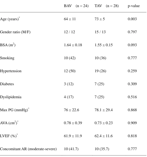
Figure 2: Region of interest (A) and 3D reconstruction of regions (B).
DICOM data from CT images were imported into Mimics software. Regions of interest on CT
images were selected and reconstructed in three dimensions.
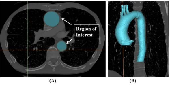
Figure 3: Assessment of aortic size.
The size of the thoracic aorta was evaluated by calculating the cross-sectional area of the
vertical cross-section against the 3D centerlines.
Figure 4: Assessment of symmetry of the ascending aorta.
This was evaluated by the ellipticity of maximum vertical section of the ascending aorta.
Figure 5: Symmetry of the ascending aorta.
The ellipticity was significantly larger in the bicuspid group, suggesting that patients with a
stenotic BAV had a more asymmetrical, dilated ascending aorta.
References
1. Cripe L, Andelfinger G, Martin LJ, et al. Bicuspid aortic valve is heritable. J Am Coll Cardiol
2004.7; 44: 138-43.
2. Abdulkareem N, Smelt J, Jahangiri M, et al. Bicuspid aortic valve aortopathy: Genetics,
pathophysiology and medical therapy. Interact Cardiovasc Thorac Surg 2013.9; 17: 554-9.
3. Pisano C, Maresi E, Balistreri CR, et al. Histological and genetic studies in patients with
bicuspid aortic valve and ascending aorta complications. Interact Cardiovasc Thorac Surg
2012.3; 14: 300-6.
4. Tadros TM, Klein MD, Shapira OM, et al. Ascending aortic dilatation associated with
bicuspid aortic valve. Pathophysiology, molecular biology, and clinical implications. Circulation
2009.2; 119: 880-90.
5. Warnes CA. Bicuspid aortic valve and coarctation: Two villains part of a diffuse problem.
Heart 2003.9; 89: 965-6.
6. Yasuda H, Nakatani S, Stugaard M, et al. Failure to prevent progressive dilation of
ascending aorta by aortic valve replacement in patients with bicuspid aortic valve:
Comparison with tricuspid aortic valve. Circulation 2003.9; 108 (Suppl.1):Ⅱ291-4.
7. Lu MT, Thadani SR, Hope MD, et al. Quantitative assessment of asymmetric aortic dilation
with valve-related aortic disease. Acad Radiol 2013.1; 20: 10-5.
8. Hope MD, Hope TA, Meadows AK, et al. Bicuspid aortic valve: Four-dimensional MR
evaluation of ascending aortic systolic flow patterns. Radiology 2010.4; 255:53-61.
9. Hope MD, Hope TA, Crook SES, et al. 4D Flow CMR in assessment of valve-related
ascending aortic disease. J Am Coll Cardiol Imaging 2011.7; 4: 781-7.
10. Doyle BJ, Morris LG, Callanan A, et al. 3D reconstruction and manufacture of real
abnormal aortic aneurysms: From CT scan to silicone model. J Biomech Eng 2008.6; 130:
034501-1-034501-5.
11. Doyle BJ, Gallanan A, Burke PE, et al. Vessel asymmetry as an additional diagnostic tool in
the assessment of abdominal aortic aneurysms. J Vasc Surg 2009.2; 49: 443-54.
12. Finol EA, Keyhani K, Amon CH, et al. The effect of asymmetry in abdominal aortic
aneurysms under physiologically realistic pulsatile flow Conditions. J Biomech Eng 2003.4;
125: 207-17.
13. Vorp DA, Raghavan ML, Webster MW, et al. Mechanical wall stress in abdominal aortic
aneurysm: Influence of diameter and asymmetry. J Vasc Surg 1998.4; 27: 632-9.
14. Raghavan ML, Kratzberg JA, Golzarian J, et al. Introduction to biomechanics related to
endovascular repair of abdominal aortic aneurysm. Tech Vasc Interv Radiol 2005.3; 8: 50-5.
15. Bissell MM, Hess AT, Biasiolli L, et al. Aortic dilation in bicuspid aortic valve Disease.
Flow pattern is a major contributor and differs with valve fusion type. Circ Cardiovasc Imaging
2013.7; 6: 499-507.
16. Girdauskas E, Borger MA, Secknus M, et al. Is aortopathy in bicuspid aortic valve disease a
congenital defect or a result of abnormal hemodynamics? A critical reappraisal of a one-sided
argument. Eur J Cardiothorac Surg 2011.6; 39: 809-14.
17. de Sa M, Moshkovitz Y, Butany J, et al. Histologic abnormalities of the ascending aorta and
pulmonary trunk in patients with bicuspid aortic valve disease: Clinical relevance to the Ross
procedure. J Thorac Cardiovasc Surg 1999.10; 118: 588-94.
18. Huntington K, Hunter AGW, Chan K, et al. A prospective study to assess the frequency of
familial clustering of congenital bicuspid aortic Valve. J Am Coll Cardiol 1997.12; 30: 1809-12.
19. Biner S, Rafique AM, Ray I, et al. Aortopathy is prevalent in relatives of bicuspid aortic
valve patients. J Am Coll Cardiol 2009.6 ; 53 : 2288-95.
20. Sorrell VL, Panczyk E, Alpert JS, et al. A new disease: Bicuspid aortic valve aortopathy
syndrome. Am J Med 2012.4; 125: 322-3.
Reproduced with permission of The Journal of Heart Valve Disease and ICR
Publisher Limited “http://icr-heart.com”.