Analysis of Medialization of the Femoral Head in Periacetabular Osteotomy Using Three-Dimensional Computed Tomography
Yoshitsugu TANAKA1), Shigeaki MORIYAMA2), Yoshinari NAKAMURA1), Masatoshi NAITO1)
1) Department of Orthopaedic Surgery, Faculty of Medicine, Fukuoka University
2) Department of Mechanical Engineering, Faculty of Engineering, Fukuoka University
Abstract
Background: Periacetabular osteotomy has been established as an effective treatment for osteoarthritis caused by developmental dysplasia of the hip. Medialization of the femoral head during periacetabular osteotomy is crucial to providing significant reduction in joint contact stress. The purpose of this study was to develop a novel method for a special analysis of reoriented acetabulum movement and to evaluate effective procedures for medialization of the femoral head during periacetabular osteotomy using three-dimensional computed tomography (3DCT).
Methods: Thirty hips in 30 patients with hip dysplasia underwent both radiographic evaluations including lateral center-edge (CE) angle and acetabular retroversion, and 3DCT scans with a slice thickness of 0.5 mm before and after surgery. CT study consisted of the following procedures: 1. 3D acetabular positions and center of the femoral head were measured; 2. the 3D shape data were registered automatically and the coordinate systems were normalized; 3. the rotation matrix and translation vector were calculated. We evaluated direction of the rotation of the acetabular fragment and movement of the femoral head to analyze which parameters contributed to the medialization of the femoral head using differential volume ratio and six degrees of freedom search method whereby all six variables were varied independently.
Results: The average lateral CE angle, acetabular roof obliquity, and acetabulum-head index improved in all patients. All hips which had had acetabular retroversion preoperatively had an anteverted acetabulum postoperatively. Anterior coverage of the acetabular fragment and internal rotation in the axial plane contributed to both medialization of the femoral head and anteversion of the retroverted acetabulum, while posterior rotation of the acetabular fragment caused lateralization of the femoral head.
Conclusions: This study demonstrated that reorientation of the acetabulum was spatially confirmed by our method using 3DCT images. This method may be a valuable addition to preoperative planning and intraoperative assessment of joint realignment in periacetabular osteotomy.
Key words:Medialization of the femoral head, Periacetabular osteotomy, 3DCT, Dysplastic hip, Acetabular retroversionn
Correspondence to : Yoshitsugu Tanaka, Department of Orthopaedic Surgery, Faculty of Medicine, Fukuoka University, 7-45-1, Nanakuma, Jonan-ku, Fukuoka, 814-0180, Japan
Tel:+81-92-801-1011 (3465) Fax:+81-92-864-9055 E-mail:[email protected]
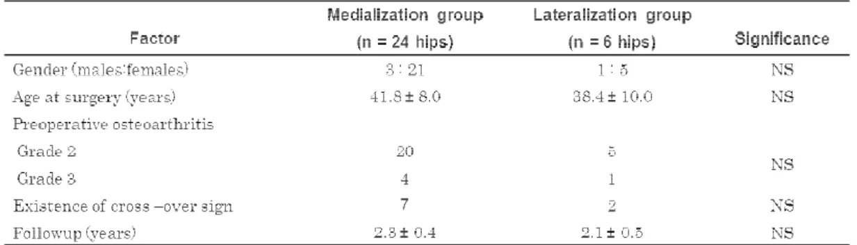
Table I Patient Data in the Medialization and Lateralization groups
Data are mean ± standard deviation NS = not significant
Introduction
Acetabular dysplasia is the most common cause of secondary hip osteoarthritis1,2. To improve acetabular coverage in patients with dysplastic hips, various osteotomies have been developed3–9. Among them, curved periacetabular osteotomy (CPO) is a modification of the Bernese periacetabular osteotomy technique developed by Ganz6 and is a method we have performed in our facility since 1995 to treat acetabular dysplasia from the prearthrosis stage to the advanced stage in patients less than sixty-five years of age8,10. CPO provides good coverage of the femoral head by the acetabular fragment, preserving the vascular supply of the fragment and allows medialization of the hip center. Medialization of the femoral head reduces the peak contact stress even if the CE angle is the same7, so we have recently put emphasis on medializing the femoral head11. During the surgery, we used an image intensifier while rotating the acetabulum, with the main indices being femoral head coverage, acetabular roof obliquity (ARO), medialization of the femoral head and avoidance of acetabular retroversion and impingement. However, the key parameter for medialization of the femoral head remains unclear. The purpose of this study was to investigate a novel method for the three-dimensional (3D) analysis of acetabular movement and to determine effective procedures for medialization of the femoral head during CPO.
Patients and Methods
Patients
From the records of CPO procedures performed by two of the authors (M. N. and Y. N.) between 2008 and 2010, 30 patients involving 30 right hip joints were randomly
selected for this retrospective study. There were 4 male and 26 female patients. According to the postoperative position of the femoral head as described later, patients were divided into two groups of the medialization group and the lateralization group (Table I). Surgical indications for CPO were based on a CE angle of less than 20 degrees on the anteroposterior radiographs and a congruent hip joint with hip abduction. The severity of secondary osteoarthritis was classified into four grades using the Tönnis12 classification system. Specifically, hips with subchondral sclerosis were classified as grade 1, hips with subchondral cyst formation and partial cartilage interval narrowing were classified as grade 2, hips with severe or complete but localized cartilage interval narrowing were classified as grade 3, and hips with extensive and severe or complete non-localized cartilage interval loss were classified as grade 4. No hips were classified as grade 1 osteoarthrosis; 25 hips, grade 2; and 5 hips, grade 3. The average age of the patients at the time of surgery was 40 years (range, 20 to 64 years).
All 30 patients were evaluated preoperatively and at the last follow-up using the Harris hip score. The median clinical follow-up was 2.2 years (range, 1.3 to 2.8 years).
This study was approved by the Institutional Review Board. All patients gave informed consent prior to their participation.
Surgical Procedure
This operation was performed with patients in the supine position, through a direct anterior approach, with an incision of approximately 9 cm for surgical exposure.
The osteotomy was performed in the same manner as a procedure described previously in detail13. Briefly, the osteotomy was begun from the innominate groove of the ischial bone, using a curved osteotome, and the
Fig. 1 Radiographic indices measured from AP radiographs, namely Kohler’s ilioischial line (b-c), distance between the bilateral Kohler ilioischial lines (t) and distance from each Kohler ilioischial line to the femoral head center (a). Acetabular head lateralization index was calculated using the following formula: acetabular head lateralization index = (a)/(t)/2.
pubic bone cut at an angle of 30 degrees to the horizontal surface of the pelvis. The iliac bone was cut beginning under the anterior inferior iliac spine, maintaining a distance of approximately 15 mm from the surface of the hip joint. An image intensifier was used after the acetabular fragment was redirected and fixed temporarily with Kirschner wires to ensure that there was: adequate coverage of the femoral head by the reoriented acetabular fragment, a horizontal position of ARO, that the center of the femoral head was medialized. To confirm a lack of femoroacetabular impingement, the anterior part of the hip joint capsule was palpated when the hip was placed in flexion and internal rotation. If abutment of the anterior femoral head and neck on the anterior rim of the acetublum was detected, the reoriented acetabular fragment was rotated posteriorly. Three poly-L-lactic acid cortical screws were used to fix the acetabular fragment.
Radiographic Evaluations
Anteroposterior radiographs of the pelvis were taken with the patient in a supine position. The tube-to-film distance was 120 cm, and the tube was perpendicular to the table. The center beam was directed toward the midpoint between the upper border of the symphysis and a horizontal line connecting both anterior superior iliac spines. The rotation of the lower limbs was standardized by positioning both patellae in the exact frontal position.
The method described by Siebenrock et al.14 was used to judge the extent of pelvic inclination. No corrections were made for radiographic magnification. Radiographic measurements included the CE angle, ARO15, acetabulum- head index (AHI)16, and acetabular head lateralization index (HLI)17. The ratio of femoral head lateralization was calculated using the HLI and modified Ninomiya measurements18(Fig. 1). The distance between the two Kohler ilioischial lines (t) was referred to as the inter-hip distance. The acetabular HLI was calculated using (a)
/(t)/2, and the ratio of femoral head lateralization was calculated as postoperative acetabular HLI /preoperative HLI. Based on the HLI, the hips were divided into two groups: 1. a medialization group, in which the ratio of the HLI was decreased postoperatively and 2. a lateralization group, in which the ratio of the HLI was increased postoperatively(Table I). Acetabular retroversion was evaluated using the criteria proposed by Reynolds et al.19 who defined it as a cross-over sign created by the anterior aspect of the acetabular rim being more lateral than the posterior aspect in the proximal part of the
acetabulum. Radiographic measurements were taken using a digital caliper (Mitutoyo, Tokyo, Japan) with an accuracy of ±0.02 mm. Measurements of the radiographs were carried out by two authors (Y.T. and Y.N.) who completed repeated training sessions and were blinded to the clinical results. In addition, the radiographs were reviewed three times on different days by the same observers, and the average values were calculated. The data from the radiographic measurements were analyzed
(intraclass correlation coefficient) for intraobserver and interobserver reliability, and these data were reasonably reproducible.
Computed Tomography Evaluations
Before conducting the measurements described here, the validity of the evaluation using differential volume ratio was assessed. Differential volume ratio was calculated using six degrees of freedom search method whereby all six variables were varied independently of each other with regard to postoperative 3D images, and superimposition on preoperative images was performed.
If the state of superimposition was poor, the number of voxels in the superimposed image was greater than the original number of voxels. That is, a search was performed for the position and posture was minimized.
Based on this information, we chose a slice thickness of 0.5 mm.
Pelvic computed tomography was performed on all 30 patients with developmental dysplasia of the hip. With the patients in a supine position, images were obtained at 0.5 mm intervals from the anterior superior iliac
Fig. 2 Three-dimensional evaluation using the left and right symphyseal surfaces of the anterior superior iliac spine and pubis as datum points.
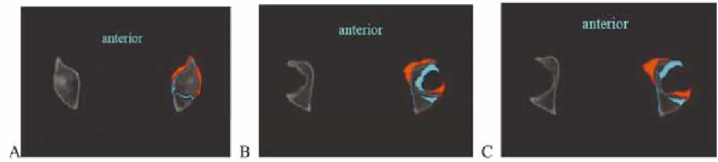
Fig. 3 (A,B and C) Images are a series of axial slices. Areas indicated in blue are preoperative sections, while the reoriented acetabular fragments are indicated in red.
spines to the inferior rim of the pelvis before and after surgery. After downloading of data from these scans in Digital Imaging and Communications in Medicine format
(DICOM; NEMA [National Electrical Manufacturers Association], Rosslyn, Virginia) onto a personal computer, we performed multiplanar reconstruction imaging using image-processing and analysis software (ZedView ver.
6.0 LEXI Co., Ltd., Tokyo, Japan). Centers of gravity of the left and right anterior superior iliac spine and the pubic symphysis were calculated, and the Y axis defined as the line joining these centers of gravity and the central point. The Z axis was oriented vertically downward to the Y axis in the calculated planar state, with the X axis perpendicular to the Y and Z axes (Fig. 2). Directions of rotation were all clockwise with respect to each of these axes. After the hip region of interest for reconstructing 3D images had been identified, bone CT values were binarized. In the two images, positions of bones in three directions and three rotation angles were required to
express the three dimensional state. The principle of 3D image superimposition used in the present study was a six degrees of freedom search method whereby all six variables for postoperative 3D images were varied independently of each other and superimposition on preoperative images was performed (Figs. 3-A through 3-C). Movement of the acetabulum was calculated as
(x, y, z), rotation direction as (φ, θ, ψ) and change in position of the center of femoral head as (X, Y, Z) using the least-squares method. Especially, X in plus means the medializaition of the femoral head, and minus shows the lateralization. Distance and direction of movement between the centers of femoral heads, and direction of rotation were measured on pre- and postoperative CT, and relationships were evaluated.
Statistical Evaluations
Multiple regression analysis was used to clarify the relationships in the resulting data. Data analysis was done using the amount of movement X (mm) of the femoral head as the dependent variable and the acetabular fragment rotation φ, acetabular fragment rotation ψ, acetabular fragment rotation θ, preoperative CE angle and presence of retroversion as the independent variables. The Shapiro-Wilk test was used for the data normalization assay, and the stepwise method was used to select the variables for multiple regression analysis.
The significance level of these analyses was 5%.
Results
In the radiographic measurements, the mean values of all the postoperative parameters (CE angle, acetabular head index, A RO and head lateralization index)
showed satisfactory improvements compared with
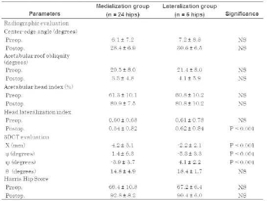
Table II Radiographic and 3DCT Evaluations, Clinical Outcomes
Data are mean ± standard deviation NS = not significant
Fig. 4 Differential volume ratio decreased sharply from a pitch of 4 mm, 4° to 0.5 mm, 0.5°, but little change was seen from a pitch of 0.125 mm, 0.125° to 0.0625°.
those of the preoperative parameters (Table II). The mean CE angle significantly increased from 7.0 ± 6.8°
preoperatively to 29.2 ± 5.1° postoperatively (p < 0.001). Medialization of the femoral head was observed in 24 of the 30 patients, and the mean ratios of femoral head lateralization were 0.92 ± 0.1. Among 30 hips, 9 (30%)
had acetabular retroversion preoperatively. All of these hips had an anteverted acetabulum postoperatively. The clinical scores between the medialization group and the lateralization group were similar. The average Harris hip score in the medialization group improved from 66.4 points (range, 48–82 points) preoperatively, to 92.8 points
(range, 88–100 points) at the last follow-up, and from 67.2 points (range, 44–88 points) to 90.4 points (range, 78–
100 points) in the lateralization group.
Figure 4 shows differential volume ratios when the search pitch for position and posture was varied during superimposition. As can be seen from the table, the differential volume ratio decreased sharply from a pitch of 4 mm, 4° to 0.5 mm, 0.5°, but little change was seen from a pitch of 0.125 mm, 0.125° to 0.0625°. To demonstrate the validity of using differential volume ratio at a pitch of 0.125 mm, 0.125° for evaluation, Figure 5 shows changes in differential volume ratio when images of an identical
phantom were displaced at the specified pitch. As can be seen from this table, pitch when the differential volume ratio was 4% was approximately 0.5 mm, 0.5°. As this is almost equivalent to the size of voxels in CT images, the automatic processing used for superimposition in this study was regarded as sufficiently accurate for evaluation.
Referring to the overall mean rotation direction
Fig. 5 Pitch when the differential volume ratio was 4%, was approximately 0.5 mm, 0.5°.
Fig. 6 (A, B, C and D): The relationships of rotation directions and CE angle to medialization of the femoral head (X mm). Rotation direction of acetabular fragment φ (r = 0.42, p = 0.02) and ψ (r = 0.43, p = 0.02) significantly contribute to medialization of the femoral head. There were no significant relationships in θ (r = 0.01, p = 0.95) and CE (r = 0.04, p = 0.81).
measurements of the acetabular fragment, φ was -2.1° ± 6.0°, ψ was -2.3° ± 4.7°, and θ was 15.5° ± 4.6°. The mean medialized distance, which was measured on pre- and postoperative CT, was 3.1 ± 3.8 mm (p < 0.001). There was a high correlation between medialization of the femoral
head and the rotation direction of the acetabular fragment of φ (p < 0.001) and ψ (p < 0.001). In addition, dividing all the results into the medialization group and lateralization group, we can see significant findings especially in the rotation direction of φ and ψ (TABLE. II). The mean degrees of each rotation direction were as follows: φ was 1.4 ± 6.3 in the medialization group, and -5.3 ± 3.3 in the lateralization group; ψ was -3.9 ± 3.7 in the medialization group, and 4.1 ± 2.2 in the lateralization group. These results suggest that medialization of the femoral head depends on whether the rotation direction of φ and ψ are plus or minus.
Neither CE angle preoperatively, nor rotation direction θ had any relationship to the medialization of the femoral head (Figs. 6-A through 6-D).
Discussion
The use of CT to assess reoriented bone fragments is occasionally seen in the fields of orthopaedic surgery and dental and oral surgery20,21, where extremely detailed
calculations are required, but a thorough review of the literature did not produce any reports pertaining to both periacetabular osteotomy and medialization of the femoral head. Until now, the authors have used plain radiographs for evaluation, but because the procedure includes tracing, which is a human-based process using pre- and postoperative orientation and a tester, a certain amount of error was involved. From the viewpoint that analysis of the reoriented acetabular fragment is crucial to precise evaluation of the joint and to efficient surgical technique, 3DCT was used and an actual evaluation was conducted.
In this study, patients age at surgery, severity of osteoarthritis and pre- and postoperative CE angle were not significantly different from the conventional report6,17,18. Radiographic evaluations revealed significant improvements in CE angle, AHI, ARO, and the mean ratios of femoral head lateralization were comparable to values found in the literature11. The prevalence of preoperative acetabular retroversion was 30% of 30 hips with developmental dysplasia and all acetabular retroversion was corrected postoperatively.
In various conventional approaches for acetabular reorientation, the primary emphasis has been on appropriate coverage of the femoral head with the acetabular fragment. However, a number of problems such as lateralization of the femoral head, acetabular retroversion and FA I were of ten seen follow ing surgery22,23. Postoperative acetabular retroversion and FAI are reported to cause degenerative hip disease24. At our facility, as described earlier, various means have been utilized to achieve medialization along with covering of the femoral head, without causing problems such as these11. In the present cases of CPO performed at our facility, sufficient covering of the femoral head has been achieved using the reoriented acetabular fragment.
Moreover, this is also advantageous in that it enables stabilization of the hip joint25.
On pre- and postoperative radiographic evaluation, there were no statistically significant relationships in CE angle, ARO, and AHI between the medialization group and the lateralization group. Two-dimensional analysis, alone, could not provide enough information regarding the key factors related to medialization of the femoral head. The shape of the acetabular fragment and pelvis are complex and precise movement is not easily demonstrated by plain radiographs.
The position and orientation of the reoriented acetabular
fragment were evaluated using all 6 variables, with the differential volume ratio, using 3D imaging. We demonstrated 3D evaluation was possible without being affected by the posture of the subject being studied, or by intra-tester variability. CT using slices with a thickness of 0.5 mm was found to be effective in accurately evaluating the reoriented acetabular fragment both before and after surgery. In CT evaluation, factors with significant effects on medialization of the femoral head were rotation direction of φ and ψ.
In our 3DCT analysis, the mean degrees of each rotation direction of the acetabular fragment were as follows: φ was 1.4 ± 6.3 in the medialization group, and -5.3 ± 3.3 in the lateralization group; ψ was -3.9 ± 3.7 in the medialization group, and 4.1 ± 2.2 in the lateralization group; θ was 14.8 ± 4.9 in the medialization group, and 18.4 ± 1.7 in the lateralization group (Table II). The mean medialization distance was 4.4 ± 3.1 mm in the medialization group, and -2.2 ± 2.1 mm in the lateralization group. These data suggest that preservation of anterior coverage of the acetabular fragment and internal rotation in the axial plane contribute to medialization of the femoral head. In other words, posterior rotation of the acetabular fragment may cause lateralization of the femoral head during CPO. However, if abutment of the anterior femoral head and neck on the anterior rim of the acetabulum was detected, the reoriented acetabular fragment was rotated posteriorly which may lead to the lateralization of the femoral head. These results indicate that 3D analysis of the hip joint provides precise information about the pelvis and improves preoperative planning and determination of the correct orientation for the acetabular fragment during surgery.
In cases where sufficient rotation of the acetabular fragment in the anterior direction was not achieved, the following are considered to be causes: 1. Patients with flattening of the femoral head and deformity of the weight-bearing area of the acetabular fragment. 2.
Insufficient rotation resulting from surgical techniques aimed at improving acetabular retroversion and avoiding FAI. When sufficient pre- and postoperative assessment is done using CT, CPO may affords a surgical method for adequately covering the femoral head using the acetabular fragment, and for alleviating peak contact stress caused by medialization, while avoiding FAI and acetabular retroversion.
Our study had several limitations. It included a relatively small number of patients (30 hips), which
limited the statistical power. A second limitation is the selection of the femoral head center, which was determined using the least-squares method. Patients with non-spherical femoral heads may introduce additional errors. There is also concern about the dose of radiation to the patients, especially since most patients with adult acetabular dysplasia are women of child-bearing age.
In summary, this retrospective study demonstrated that reorientation of the acetabulum was spatially confirmed by our method using 3DCT images. Anterior coverage of the acetabular fragment and internal rotation in the axial plane contributed to both medialization of the femoral head and anteversion of the retroverted acetabulum, while posterior rotation of the acetabular fragment caused lateralization of the femoral head. Our method may be a valuable addition to preoperative planning and intraoperative assessment of various orthopaedic surgeries.
Acknowledgements
No benefits in any form have been received or will be received from a commercial party related directly or indirectly to the subject of this article.
References
1. Harris W: Etiology of osteoarthritis of the hip. Clin Orthop Relat Res 213: 20-33, 1986.
2. Solomon L: Patterns of osteoarthritis of the hip. J Bone Joint Surg Br 58: 176-183, 1976.
3. Salter RB: Innominate osteotomy in the treatment of congenital dislocation and subluxation of the hip. J Bone Joint Surg Am 43B: 518-539, 1961.
4. Steel HH: Triple osteotomy of the innominate bone. J Bone Joint Surg Am 55A: 343-350. 1973.
5. Tönnis D, Aming A, Bloch M et al: Triple pelvic osteotomy. J Pediatr Orthop 3:54-67, 1994.
6. Ganz R , K laue K , V inh T S, Mast J W.: A new periacetabular osteotomy for the treatment of hip dysplasias. Technique and preliminary results. Clin orthop Relat Res 232: 26-36, 1988.
7. Kralj M, Mavcic B, Antolic V, Iglic A, Kralj-Iglic V.:
The Bernese periacetabular osteotomy: clinical, radiographic and mechanical 7-15-year follow-up of 26 hips. Acta Orthop 76: 833-40, 2005.
8. Na ito M, Shirami z u K , A k iyoshi Y, Ezoe M, Nakamura Y: Curved periacetabular osteotomy for
treatment of dysplastic hip. Clin Orthop Relat Res 433: 129-135, 2005.
9. Steppacher SD, Tannast M, Ganz R, Siebenrock KA.:
Mean 20-year followup of Bernese periacetabular osteotomy. Clin Orthop Relat Res 466: 1633-44, 2008.
10. Teratani T, Naito M, K iyama T, Maeyama A .:
Periacetabular osteotomy in patients fifty years of age or older. J Bone Joint Surg Am 92: 31-41, 2010.
11. Teratani T, Naito M, Shiramizu K, Nakamura Y, Moriyama S.: Modified pubic osteotomy for femoral head medialization in periacetabular osteotomy: A retrospective study of 144 hips. Acta Orthopaedica 79
(4): 474-82, 2008.
12. Tönnis D.: Congenital dysplasia and dislocation of the hip in children and adults. Springer-Verlag, New York 1987. pp. 167-71.
13. Teratani T, Naito M, K iyama T, Maeyama A .:
Periacetabular osteotomy in patients fifty years of age or older. Surgical Technique. J Bone Joint Surg Am 93(Supplement 1): 30-39, 2011.
14. Siebenrock K A, Schoeniger R, Ganz A.: Anterior femoro-acetabular impingement due to acetabular retroversion. J Bone Joint Surg Am 85: 278-86, 2003.
15. Massie WK, Howorth MB.: Congenital dislocation of the hip. J Bone Joint Surg Am 32: 519-31, 1950.
16. Heyman CH, Herndon CH.: Legg-Perthes disease: a method for the measurement of the roentgenographic results. J Bone Joint Surg Am 32: 767-78, 1950.
17. Yasunaga Y, Takahashi K, Ochi M, Ikuta Y, Hisatome T, Nakashiro J, Yamamoto S.: Rotational acetabular osteotomy in patients forty-six years of age or older:
Comparison with younger patients. J Bone Joint Surg Am 85: 266-72, 2003.
18. Ninomiya S.: Rotational acetabular osteotomy for the severely dysplastic hip in the adolescent and adult.
Clin Orthop Relat Res 247: 127-37,1989.
19. Reynolds D, Lucas J, Klaue K. Retroversion of the acetabulum. A cause of hip pain. J Bone Joint Surg Br 81: 281-8, 1999.
20. Karami M, Gouran Savadkoohi D, Ghadirpoor A A computer model for evaluating the osteotomy parameters of Chiari pelvic osteotomy. Int Orthop 34
(3): 329-33, 2010.
21. Uechi J, Okayama M, Shibata T, Muguruma T, A novel method for the 3-dimensional simulation of orthognathic surgery by using a multimodal image- fusion technique. Am J Orthod Dentofacial Orthop 130(6): 786-98, 2006.
22. Myers SR, Eijer H, Ganz R. Anterior femoroacetabular impingement after periacetabular osteotomy. Clin Orthop Relat Res 363: 93-99, 1999.
23. Xie J,Naito M, Maeyama A Evaluation of acetabular versions after a curved periacetabular osteotomy for dysplastic hips Int Orthop 34(4): 473–477, 2010.
24. K iyama T, Naito M, Shiramizu K , Shinoda T.
Postoperative acetabular retroversion causes posterior osteoarthritis of the hip. Int Orthop 33(3): 625-631, 2009.
25. Maeyama A, Naito M, Moriyama S, Yoshimura I:
Evaluation of dynamic instability of dysplastic hip with use of triaxial accelerometry. J Bone Joint Surg Am 90: 85-92, 2008.
(平成25.11.1受付,平成26.1.20受理)