• 検索結果がありません。

Implantation of Autologous Bone Marrow-derived Cells Improves Erectile Dysfunction in Spontaneously Hypertensive Rats

N/A
N/A
Protected

Academic year: 2021

シェア "Implantation of Autologous Bone Marrow-derived Cells Improves Erectile Dysfunction in Spontaneously Hypertensive Rats"

Copied!
8
0
0

読み込み中.... (全文を見る)

全文

(1)

Introduction

Erectile dysfunction (ED) has a negative impact on the quality of life

1)2)

over 50 % of men beyond 40 years of age are affected by ED

3)-6)

. Administration of phosphodiesterase-5 (PDE-5) inhibitors improves cyclic guanosine monophosphate (cGMP)-dependent smooth muscle and arteriole relaxation. This widely accepted treatment for ED acts by amplification of the nitric oxide (NO) signaling level within the cor- pus cavernosum penis. While approximately 70 % of

patients benefit from PDE-5 inhibition

7)

, many oth- ers do not

1)6)

. For those refractory patients, implan- tation of autologous bone marrow-derived cells, which are capable of differentiating into nerve, mus- cle, and other tissues, may be an effective alternative treatment

8)9)

.

Spontaneously hypertensive rats (SHRs) are affect- ed with ED

10)-13)

. While the mechanism of the hyper- tensive-related ED remains to be clarified

14)-16)

, these animals may serve as a suitable model to test the ability of implanted autologous bone marrow-de- rived cells to relieve ED. These cells, when implant- ed into freeze-injured urinary bladders or urethras, differentiate into smooth and skeletal muscle cells and restore physiological functions to these damaged

Implantation of Autologous Bone Marrow-derived Cells Improves Erectile Dysfunction in Spontaneously Hypertensive Rats

Masakuni I shikawa 1)* , Tetsuya I mamura 1) , Osamu N ishizawa 2) and Osamu I shizuka 1)

1)Department of Urology, Shinshu University School of Medicine 2)Department of Urology, North Alps Medical Center, Azumi Hospital

Purpose : Erectile dysfunction (ED) decreases the quality of life. However, some patients are refractory to phos- phodiesterase-5 (PDE-5) inhibitors. This preliminary study investigated the possibility that autologous bone marrow-derived cells could improve the ED of spontaneously hypertensive rats (SHRs).

Materials and Methods : Erectile responses were induced by apomorphine. Bone marrow cells were harvested from femurs of SHRs, and following culturing and labeling, they were implanted autologously into the corpus cavernosum penis. Control SHRs received cell-free injections. At 7 days after the implantation, apomorphine-in- duced erectile responses were estimated. The presence of implanted bone marrow-derived cells in the corpora cavernosa and the expression of neuronal nitric oxide synthase (nNOS) were determined by microscopy and re- verse transcription polymerase chain reaction.

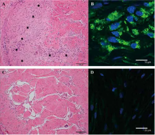
Results : The number of the negative reactions for apomorphine in the SHRs was significantly higher compared to the Wister Kyoto (WKY) rats (P=0.045). Ten of 15 cell-free control SHRs did not respond to apomorphine, while 8 of 9 cell-implanted SHRs responded. Significantly more cell-implanted SHRs responded to apomorphine than did cell-free SHRs (P<0.013). The implanted cells formed clusters within the corpora cavernosa, and some expressed nNOS mRNA and protein.

Conclusions : Bone marrow-derived cells autologously implanted into the corpus cavernosum penis significantly increased the number of SHRs having erectile responses to apomorphine. Shinshu Med J 65 : 37―44, 2017

(Received for publication July 22, 2016 ; accepted in revised form September 14, 2016) Key words : apomorphine, autologous bone marrow-derived cells, erectile dysfunction,

neuronal nitric oxide synthase, spontaneously hypertensive rats

Corresponding author : Masakuni Ishikawa

Department of Urology, Shinshu University School of Medi-

cine, 3-1-1 Asahi, Matsumoto, Nagano 390-8621, Japan

E-mail : [email protected]

(2)

organs

17)18)

. Furthermore, considering future clinical applications, autologous cells are superior to allogen- ic cells because the use of autologous cells is not burdened with immunological rejection or ethical problems. Therefore in this preliminary study, we investigated the ability of implanted autologous bone marrow-derived cells to remediate hypertensive-re- lated erectile dysfunction in SHRs.

Materials and Methods A Animals

Male Wister Kyoto (WKY) rats and SHRs at post- natal week 25 (Japan SLC Inc., Shizuoka, Japan) were used to assess the erectile responses. In sepa- rate experiments, SHRs were used to assess the ef- fects of implanted autologous bone marrow-derived cells. All animals were treated in accordance with National Institutes of Health Animal Care Guidelines and the guidelines approved by the Animal Ethics Committee of Shinshu University School of Medicine.

B Drug

This study used apomorphine (Sigma-Aldrich, Inc., St. Louis, MO, USA), which is the central dopa- mine D1/D2-recepter agonist in the central nervous system to induce erectile responses

19)-21)

. The apo- morphine was dissolved to the desired concentration with 0.9 % saline.

C Measurements of intracavernous pressure The apomorphine-induced erectile responses of untreated SHRs (n=15) and WKY rats (n=16) were assessed by intracavernous pressure measurements.

As previously described

22)

, a 22-gauge needle at- tached to a polyethylene catheter (PE-50) was in- serted into the corpus cavernosum penis of animals anesthetized by inhalation of sevoflurane (Sevof- rane®, Abbot Japan Co., Ltd., Tokyo, Japan). The catheter was sealed and brought out subcutaneously to the back of the neck. For measurements of blood pressure, another single catheter was inserted into the right carotid artery and then brought subcuta- neously to the back of the neck. The skin incision was closed. After the surgery, each animal was placed without any restraint in a cage to recover for over 2 hours.

After confirming that each rat was alert, each catheter was attached to a pressure transducer (DX-100, Nihon Kohden, Tokyo, Japan). Intracavern- ous pressure signals were passed through a carrier amplifier (AP-601G, Nihon Kohden) and recorded by a multichannel pen-recorder (Nihon Kohden). After the pressures demonstrated stable traces, 0.1 mg/kg apomorphine was administered subcutaneously just below the back of the neck. Our previous studies confirmed that the dose was able to induce erectile responses in healthy rats

22)

. We quantified the fol- lowing parameters : blood pressure (cmH

2

O), time to first response (TFR ; minutes), number of peak pres- sures per 30 minutes (PP30), tonic peak pressure (TPP ; cmH

2

O), and burst peak pressure (BPP ; cm- H

2

O).

D Harvest and culture of autologous bone mar- row-derived cells

Twenty-four SHRs were anesthetized by inhala-

tion of sevoflurane (Abbot Japan Co., Ltd.). In each

animal, two pediatric bone marrow needles were in-

serted approximately 1 cm apart into a femur. The

bone marrow cells were flushed out with saline and

collected through the other needle

11)23)

. After centrif-

ugation, the cells were suspended with culture medi-

um composed of Dulbeccoʼs modified eagle medium

(Invitrogen Life Technologies, Carlsbad, CA, USA)

supplemented with 15 % fetal bovine serum (Biow-

est, Paris, France) and 0.1 % penicillin-streptomycin

(Invitrogen). The harvested bone marrow cells were

cultured on type I collagen-coated culture dishes

(Asahi Glass Co., Ltd., Tokyo, Japan). During the cul-

ture, the medium was completely replaced every

day, and non-attached cells were discarded. At 5

days, the Qtracker 525 Cell Labeling Kit (Invitrogen)

was used for 24 hours to label the cytoplasm with

nanocrystals. The cells taking the marker during the

culture period were easily detected within the recip-

ient tissues by a Leica DAS Microscope (Leica Mi-

crosystems GmbH, Wetzlar, Germany) at 488 nm ex-

citation. At two days after labeling, the adherent,

proliferating, labeled bone marrow-derived cells

were collected after brief incubation with a 0.25 %

trypsin solution

17)18)

and then suspended in culture

(3)

medium at 1.0×10

7

cells/ml (viability > 80 %).

E Implantation of autologous bone marrow-de- rived cells

Seven days after the culture, the SHRs from which the bone marrow cells were collected were randomly divided into cell implantation (n=9) and cell-free injection control (n=15) groups. After re-anesthetizing the SHRs, the corpus cavernosum penis was exposed.

For the cell implantation group, the suspended autologous bone marrow-derived cells (1.0×10

6

cells/100μl) were autologously implanted with a 29-gauge syringe into the corpus cavernosum penis at the 3- and 9-oʼclock positions. For the cell-free in- jection control group, 100 μl of cell-free culture me- dium was similarly injected at the same positions.

The implantation cell number and volume were cho- sen to avoid further damaging the host tissues or the implanted cells with shear stress

17)18)

. At each operation, the retention of small swellings that in- cluded the implanted cells or control media was vi- sually confirmed.

At 7 days after these operations, the apomor- phine-induced erectile responses of the cell-implant- ed and control injected rats were estimated by intra- cavernous pressure measurements (described above).

Following the measurement, the rats were eutha- nized, and the corpus cavernosum penis was re- moved for immunohistochemical investigations and real-time reverse transcription polymerase chain re- action (RT-PCR) as described below.

F Immunohistochemistry

Tissue samples were fixed in 4 % paraformalde- hyde and 4 % sucrose in 0.1 M phosphate buffer (PB), pH 7.4, for 12 hr at 4 ℃, and then embedded in par- affin. The sections (3μm) were cut on a microtome and then deparaffinized, rehydrated, rinsed three times with 0.01 M phosphate buffer saline (PBS), and immersed in 10 mM sodium citrate, pH 6.0. For anti- gen retrieval, they were then microwaved at 100 ℃ for 5 min. The specimens were treated with 1.5 % normal donkey serum (Chemicon International Inc., Temecula, CA, USA) and 1.5 % non-fat milk in PBS for 1 hr at 4 ℃. The sections were incubated for 12 hr at 4 ℃ with antibody for neuronal nitric oxide

synthase (nNOS, 1 : 300, goat polyclonal, Osenses Pty Ltd, Flagstaff Hill, SA, Australia). The sections were rinsed with PBS at 4 ℃, and then incubated with secondary antibody consisting of donkey anti-goat IgG conjugated with Alexa Fluor 594 (1 : 250, Molec- ular Probes, Eugene, OR, USA) for 1 hr at 4 ℃. Neg- ative controls were performed without the primary antibody, and positive controls were performed with rat lungs. Following rinsing, the nuclei were counter- stained with 4ʼ, 6-diamidino-2-phenylindole dihydro- chloride (DAPI, 5μg/ml, Molecular Probes), and then coated with Fluorescent Mounting Medium (Dako Cytomation, Carpinteria, CA, USA). The slides were observed and photographed with a Leica DAS Mi- croscope (Leica Microsystems GmbH). The other sections from each sample were stained with hema- toxylin and eosin (H&E).

G Real-time RT-PCR

For real-time RT-PCR, the corpus cavernosum of SHRs were homogenized, and total RNA was ex- tracted with the RNeasy Mini Kit (Qiagen Inc., Va- lencia, CA, USA). Complementary DNA (cDNA) was synthesized from 0.2μg of total RNA with the High-Capacity cDNA Archive Kit (Applied Biosyste- ms, Foster City, CA, USA). Real time RT-PCR of the cDNA was performed at 50 ℃ for 2 minutes fol- lowed by 95 ℃ for 10 minutes. These were followed by 40 cycles at 95 ℃ for 15 seconds and 60 ℃ for 1 minute. The primer of neuronal nitric oxide synthase 1 (Rn00583793_m1, Applied Biosystems) was used to measure the nNOS mRNA expression level. Gene activity was expressed as the ratio to the internal standard gene beta-actin (Rn00667869_m1).

H Statistical analysis

Results were expressed as means ± standard de- viation. We determined statistical differences with Fisherʼs exact probablility test. Non-repeated mea- sures ANOVA followed by the Tukey procedure was used among experimental groups having the apomorphine-induced erectile responses. Also, unpaired t-tests were used to compare between the cell-implantation and control injection group. P- values less than 0.05 were considered significant.

These tests were performed with GraphPad InStat®

(4)

(GraphPad Software, Inc. San Diego, CA, USA).

Results

The blood pressure in normal SHRs, 236.4±26.4 cmH

2

O, was significantly higher than that of the nor- mal WKY rats, 154.4±21.7 cmH

2

O (P<0.01).

A Erectile functions of normal WKY rats and SHRs Apomorphine induced an erectile response in 14 of 16 WKY rats (Table 1). In contrast, 7 of 15 of the untreated SHRs responded to the apomorphine, while 8 did not. Thus, the negative responses by the nor- mal SHRs were significantly greater than that by the normal WKY rats (P<0.05). For the 14 responsive WKY rats and the 7 responsive SHRs, there were no differences in TFR, PP30, TPP, or BPP (Table 2).

B Cultured autologous bone marrow-derived cells Immediately after seeding, the harvested bone marrow cells consisted of heterogeneous, spin- dle-shaped, round, and polygonal cells, along with red blood cells. After 7 days of culture, the adhered proliferating cells reached approximately 90 % con- fluence and were relatively homogenous in spin- dle-shaped appearance. The cultured autologous bone marrow-derived cells did not stain with anti- bodies for nNOS just prior to implantation. Thus, this finding suggested that the cells might not differ- entiate into nNOS-produced cells during the culture period.

C Effects of autologous bone marrow - derived cells

Blood pressure of the cell-implanted SHRs, 225.3±

16.9 cmH

2

O, was not significantly different from the cell-free injected control SHRs, 234.0±47.4 cmH

2

O (P

=0.623). Seven days after implantation, 8 of 9 cell- implanted SHRs developed apomorphine-induced erectile responses. In the cell-free control SHRs, only 5 of 15 rats (P=0.013) responded (Table 3). Neither TFR nor PP30 for the cell-implanted SHRs differed significantly from the cell-free controls. While TPP and BPP in the cell-implanted SHRs tended to be greater than in cell-free controls, the difference was not significant ( Table 4 ).

D Histological and gene expression evaluation of the corpus cavernosum penis

Seven days after implantation in the penile corpo- ra cavernosa of SHRs, the bone marrow-derived cells formed clusters (Fig. 1A) that contained numer- ous Qtracker 525 nanocrystal-labeled cells (Fig. 1B).

In contrast, the cell-free control injected SHRs did not have the cell clusters (Fig. 1C) or nanocrystal-la- beled cells (Fig. 1D). Some of the labeled implanted cells were positive for nNOS antibody (Fig. 2). The nNOS mRNA expression level of cell-implanted SHRs, 1.18±0.43, tended to be higher than the con- trols, 0.93±0.25, but the difference was not signifi- cant (P=0.296).

Table 1 Apomorphine-induced erectile responses in normal WKY rats and SHRs Apomorphine-induced Erectile Response Positive Response Negative Response

WKY rats (n=16) 14 2

SHRs (n=15) 7 9

Fisherʼs exact probability test. P=0.045, erectile response for WKY rats vs SHRs.

Table 2 Intracavernous pressure measurements of rats having apomorphine-induced erectile responses

TFR (minutes) PP30 TPP (cmH

2

O) BPP (cmH

2

O)

Untreated

WKY rats (n=14) 9.6±5.6 2.4±1.3 76.4±47.8 159.6±97.0

SHRs (n=7) 8.5±3.2 2.7±2.1 49.2±30.0 93.5±63.1

TFR ; time to first response, PP30 ; number of peak pressures per 30 minutes,

TPP ; tonic peak pressure, BPP ; burst peak pressure.

(5)

Table 3 Apomorphine-induced erectile responses in cell-implanted and  cell-free control SHRs

Erectile Response

Positive Response Negative Response

Cell-free control SHRs (n=15) 5 10

Cell-implanted SHRs (n=9) 8 1

Fisherʼs exact probability test. P=0.013, erectile response for cell-free control SHRs vs cell-implanted SHRs.

Table 4 Intracavernous pressure measurements of rats having apomorphine-induced erectile responses

TFR (minutes) PP30 TPP (cmH

2

O) BPP (cmH

2

O) Cell-treatment

Cell-free control

group (n=5) 8.7±7.5 2.2±1.3 85.3±46.3 206.8±125.4

Cell-implantation

group (n=8) 8.3±4.9 2.5±1.4 130.3±83.4 317.2±185.6

TFR ; time to first response, PP30 : number of peak pressures per 30 numites, TPP ; tonic peak pressure, BPP ; burst peak pressure.

Fig. 1 Histological evaluation of the penile corpus cavernosum. (A) The cell-implanted corpus cavernosum con-

tained clusters of cells (asterisks). (B) These cells contained the green fluorescent-labeled nanocrystals

(green) taken up while the cells were in culture before implantation. (C) The cell-free control injected cor-

pora cavernosa did not have any clusters or (D) nanocrystal-labeled cells. Blue, DAPI-labeled nuclei.

(6)

Discussion

Along with aging, hypertension, diabetes mellitus, and hyperlipemia are considered to be risk factors for ED

1)5)

. The SHRs are used as an animal model of hypertension as well as models for lower urinary tract symptoms such as C-fiber related detrusor overactivity

24)-26)

, benign prostatic hypertrophy

27)

, and hypertension-related ED

12)-15)

. In our study, the blood pressure of the sham-operated SHRs was sig- nificantly higher than that of the normal sham-oper- ated WKY rats. The frequency of erectile response to apomorphine in SHRs was significantly less than that in WKY rats. These results are consistent with damage to the nervous system, microcirculation, and/or smooth muscle of the corpus cavernosum in SHRs.

The percent of SHRs having apomorphine-induced erections was significantly greater in the cell-im- planted rats compared to the cell-free injected ones.

The erectile response is initiated by NO produced by nNOS and liberated from nonadrenergic and non- cholinergic neurons in the corpora cavernosa

28)

. We showed that the implanted bone marrow-derived cells formed clusters within the corpora cavernosa.

Prior to implantation, these cells did not express nNOS, but one week afterwards, some of them did.

The implanted cells differentiated into nerve-like cells that produced nNOS, while this study did not identify these cell types. However, the overall level of nNOS mRNA expression was similar between the cell-free injected tissues and the cell-implanted ones. Thus, our results suggest that the apomor-

phine-induced erectile responses of SHRs might be mediated by not only the nNOS-related NO-depen- dent mechanisms, but also by other neurogenic and/

or muscular systems to induce smooth muscle and arteriole relaxations.

Bone marrow-derived cells are capable of differ- entiating both in vitro and in vivo along multiple pathways that include nerve, muscle, bone, cartilage, adipose tissue, tendon, and connective tissue

8)9)

. These cells, when situated in areas of tissue damage, have paracrine effects that produce cytokines and growth factors based on available signals from the surrounding tissues

29)30)

. Thus, the paracrine effects of the implanted bone marrow-derived cells might contribute to the reconstruction of healthy peripher- al nervous system elements, microcirculation, and/or smooth muscle systems within the penile corpora cavernosa.

This study had some limitations. While our results showed that the implantation of bone marrow-de- rived cells improved the apomorphine-induced erec- tions, we have not thoroughly demonstrated the mechanisms of the recovery. In this study, we per- formed cell direct-injection methods to implant the cells. While we detected the implanted cells within the penile corpora cavernosa, the cell numbers were very small. Thus, we need to improve the survival rates of the implanted cells in the penile corpora cavernosa by using the novel techniques, such as cell sheet engineering

31)

. In our next study, we will test these effects and the safety over a long period.

In this study, we conducted autologous cell im- plantation by harvesting bone marrow cells from a Fig. 2 Immunohistochemistry for nNOS antibodies.

The Qtracker-labeled implanted cells (green)

differentiated into nNOS-expressing cells (red,

arrows) within the penile corpora cavernosa.

(7)

femur of each anesthetized animal. The cultured au- tologous cells were similar to the cells harvested from femurs of the euthanized SHRs according to our previous methods

10)

. The majority of patients with ED are elderly or have other diseases that may affect the developmental potential of bone marrow cells. However, our results suggest that bone mar- row-derived cells harvested from the SHRs behave with the same potential as those from healthy ani- mals. Therefore, the bone marrow-derived cells har- vested from aging and/or diseased patients also might retain their developmental potential and be available in treatments for ED.

Conclusions

The proportion of normal SHRs with an erectile response to apomorphine was significantly lower than that of normal WKY rats. SHRs implanted with autologous cultured bone marrow-derived cells had a higher frequency of response than did cell-free in- jected control SHRs. Therefore, the implantation of autologous bone marrow-derived cells has the po- tential for development into a treatment for refracto- ry ED.

Conflict of Interest There are no conflicts of interest to declare.

References

 1) Levine LA : Diagnosis and treatment of erectile dysfunction. Am J Med 109 : 3-12, 2000

 2) Sivalingam S, Hashim H, Schwaibold H : An overview of the diagnosis and treatment of erectile dysfunction. Drugs 66 : 2339-2355, 2006

 3) Ayta IA, McKinlay JB, Krane RJ : The likely worldwide increase in erectile dysfunction between 1995 and 2025 and some possible policy consequences. BJU Int 84 : 50-56, 1999

 4) Blumentals WA, Gomez-Caminero A, Joo S, Vannappagari V : Is erectile dysfunction predictive of peripheral vascu- lar disease? Aging Male 6 : 217-221, 2003

 5) Safarinejad MR, Hosseini S : Erectile dysfunction : clinical guidelines (1). Urol J 1 : 133-147, 2004  6) Safarinejad MR, Hosseini SY : Erectile dysfunction : clinical guidelines (2). Urol J 1 : 227-239, 2004

 7) Shirai M, Tsukamoto T, Kato S, Yachiku S, Fujioka T, Okada K, Takimoto Y, Koshido Y, Ishizu H : Randomized, pla- cebo-controlled, double-blind study of oral sildenafil in Japanese men with erectile dysfunction. Nishinihon J Urol 62 : 373-382, 2000

 8) Peister A, Mellad JA, Larson BL, Hall BM, Gibson LF, Prockop DJ : Adult stem cells from bone marrow (MSCs) iso- lated from different strains of inbred mice vary in surface epitopes, rates of proliferation, and differentiation poten- tial. Blood 103 : 1662-1668, 2004

 9) Pittenger MF, Mackay AM, Beck SC, Jaiswal RK, Douglas R, Mosca JD, Moorman MA, Simonetti DW, Craig S, Mar- shak DR : Multilineage potential of adult human mesenchymal stem cells. Science 284 : 143-147, 1999

10) Hannan JL, Smallegange C, Hale TM, Heaton JP, Adams MA : Impact of antihypertensive treatments on erectile re- sponses in aging spontaneously hypertensive rats. J Hypertens 24 : 159-168, 2006

11) Ushiyama M, Kuramochi T, Katayama S : Treatment with hypotensive agents affects the impaired relaxation of the penile corpus cavernosum in hypertensive rats. Hypertens Res 29 : 523-532, 2006

12) Toblli JE, Cao G, Lombrana A, Rivero M : Functional and morphological improvement in erectile tissue of hyperten- sive rats by long-term combined therapy with phosphodiesterase type 5 inhibitor and losartan. J Sex Med 4 : 1291- 1303, 2007

13) Ushiyama M, Kuramochi T, Yagi S, Katayama S : Antioxidant treatment with alpha-tocopherol improves erectile function in hypertensive rats. Hypertens Res 31 : 1007-1013, 2008

14) Behr-Roussel D, Chamiot-Clerc P, Bernabe J, Mevel K, Alexandre L, Safar ME, Giuliano F : Erectile dysfunction in

spontaneously hypertensive rats : pathophysiological mechanisms. Am J Physiol Regul Integr Comp Physiol

(8)

284 : R682-688, 2003

15) Toblli JE, Stella I, Inserra F, Ferder L, Zeller F, Mazza ON : Morphological changes in cavernous tissue in sponta- neously hypertensive rats. Am J Hypertens 13 : 686-692, 2000

16) Ushiyama M, Morita T, Kuramochi T, Yagi S, Katayama S : Erectile dysfunction in hypertensive rats results from impairment of the relaxation evoked by neurogenic carbon monoxide and nitric oxide. Hypertens Res 27 : 253-261, 2004

17) Imamura T, Kinebuchi Y, Ishizuka O, Seki S, Igawa Y, Nishizawa O : Implanted mouse bone marrow-derived cells reconstruct layered smooth muscle structures in injured urinary bladders. Cell Transplant 17 : 267-278, 2008 18) Imamura T, Ishizuka O, Kinebuchi Y, Kurizaki Y, Nakayama T, Ishikawa M, Nishizawa O : Implantation of Autolo-

gous Bone Marrow-derived Cells Reconstructs Functional Urethral Sphincters in Rabbits. Tissue Eng Part A 17 : 1069-1081,2011

19) Heaton JP, Morales A, Adams MA, Johnston B, el-Rashidy R : Recovery of erectile function by the oral administra- tion of apomorphine. Urology 45 : 200-206, 1995

20) Anden NE, Rubenson A, Fuxe K, Hokfelt T : Evidence for dopamine receptor stimulation by apomorphine. J Pharm Pharmacol 19 : 627-629, 1967

21) Roos BE : Decrease in homovanillic acid as evidence for dopamine receptor stimulation by apomorphine in the neostriatum of the rat. J Pharm Pharmacol 21 : 263-264, 1969

22) Aizawa N, Ishizuka O, Ogawa T, Mizusawa H, Igawa Y, Nishizawa O : Role of 5-HT receptor antagonists in mediat- ing intracavernous pressure response induced by fluvoxamine in conscious rats. J Sex Med 4 : 1108-1116, 2007 23) Kushida T, Inaba M, Ikebukuro K, Ngahama T, Oyaizu H, Lee S, Ito T, Ichioka N, Hisha H, Sugiura K, Miyashima S,

Ageyama N, Ono F, Iida H, Ogawa R, Ikehara S : A new method for bone marrow cell harvesting. Stem Cells 18 : 453-456, 2000

24) Kok ET, Schouten BW, Bohnen AM, Groeneveld FP, Thomas S, Bosch JL : Risk factors for lower urinary tract symptoms suggestive of benign prostatic hyperplasia in a community based population of healthy aging men : the Krimpen Study. J Urol 181 : 710-716, 2009

25) Jin LH, Andersson KE, Kwon YH, Park CS, Yoon SM, Lee T : Substantial detrusor overactivity in conscious sponta- neously hypertensive rats with hyperactive behaviour. Scand J Urol Nephrol 43 : 3-7, 2009

26) Imamura T, Ishizuka O, Zhong C, Ogawa T, Nakayama T, Kurizaki Y, Tanabe T, Nishizawa O, Andersson KE : An extract (THC-002) of Ba-Wei-Die-Huang-Wan inhibits expression of tachykinins, and P2X3 and TRPV1 receptors, and inhibits ATP-induced detrusor overactivity in spontaneously hypertensive rats. Neurourol Urodyn 28 : 529-534, 2009 27) Steers WD, Clemow DB, Persson K, Sherer TB, Andersson KE, Tuttle JB : The spontaneously hypertensive rat : in- sight into the pathogenesis of irritative symptoms in benign prostatic hyperplasia and young anxious males. Exp Physiol 84 : 137-147, 1999

28) Burnett AL : Novel nitric oxide signaling mechanisms regulate the erectile response. Int J Impot Res 16 : 15-19, 2004 29) Huang NF, Lam A, Fang Q, Sievers RE, Li S, Lee RJ : Bone marrow-derived mesenchymal stem cells in fibrin aug-

ment angiogenesis in the chronically infarcted myocardium. Regen Med 4 : 527-538, 2009

30) Matsushima A, Kotobuki N, Tadokoro M, Kawate K, Yajima H, Takakura Y, Ohgushi H : In vivo osteogenic capabili- ty of human mesenchymal cells cultured on hydroxyapatite and on beta-tricalcium phosphate. Artif Organs 33 : 474-481, 2009

31) Imamura T, Ogawa T, Minagawa T, Yokoyama H, Nakazawa M, Nishizawa O, Ishizuka O : Engineered bone mar- row-derived cell sheets restore structure and function of radiation-injured rat urinary bladder. Tissue Eng Part A 21 : 1600-1610, 2015

(2016. 7. 22 received;2016. 9. 14 accepted) 

Table 2 Intracavernous pressure measurements of rats having apomorphine-induced erectile responses
Fig. 1  Histological evaluation of the penile corpus cavernosum. (A) The cell-implanted corpus cavernosum con- con-tained clusters of cells (asterisks)

参照

関連したドキュメント

The findings obtained in this study can be summarized as follows: i) CD56(-)CD16(+) NK cells were increased around the small bile ducts and beneath the biliary epithelia of large

Character- ization and expression analysis of mesenchymal stem cells from human bone marrow and adipose tissue. IGFBP-4 is an inhibitor of canonical Wnt signalling

We herein report two cases of disseminated adenovirus infection that presented with nodular shadows on chest X-ray after allogeneic bone marrow transplantation from unrelated

During land plant evolution, stem cells diverged in the gametophyte generation to form different types of body parts, including the protonema and rhizoid filaments, leafy-shoot

These findings further suggest that CD45 + /ColI + may contribute to kidney fibrosis by producing MCP-1/CCL2 and TGF-beta, which may be responsible for chronic

8)Takahashi S, et al : Comparative single-institute analysis of cord blood transplantation from unrelated donors with bone marrow or peripheral blood stem-cell trans- plants

32) Braga TT, Correa-Costa M, Guise YF, Castoldi A, De Oliveira CD, Hyane MI, Cenedeze MA, Teixeira SA, Muscara MN, Perez KR, Cuccovia IM, Pacheco-Silva A, Gonçalves GM, Camara NO.

It is a new contribution to the Mathematical Theory of Contact Mechanics, MTCM, which has seen considerable progress, especially since the beginning of this century, in