九州大学学術情報リポジトリ
Kyushu University Institutional Repository
悪性末梢神経鞘腫瘍におけるH3K27me3の免疫組織化 学的評価
大塚, 洋
http://hdl.handle.net/2324/1937172
出版情報:Kyushu University, 2018, 博士(医学), 課程博士 バージョン:
権利関係:© 2018 Elsevier GmbH. All rights reserved.
Contents lists available atScienceDirect
Pathology - Research and Practice
journal homepage:www.elsevier.com/locate/prp
Immunohistochemical evaluation of H3K27 trimethylation in malignant peripheral nerve sheath tumors
Hiroshi Otsuka
a,b, Kenichi Kohashi
a, Masato Yoshimoto
a,b, Shin Ishihara
a,b, Yu Toda
a, Yuichi Yamada
a, Hidetaka Yamamoto
a, Yasuharu Nakashima
b, Yoshinao Oda
a,⁎aDepartment of Anatomic Pathology, Graduate School of Medical Sciences, Kyushu University, Fukuoka, Japan
bDepartment of Orthopaedic Surgery, Graduate School of Medical Sciences, Kyushu University, Fukuoka, Japan
A R T I C L E I N F O
Keywords:
Malignant peripheral nerve sheath tumor H3K27me3
Neurofibromatosis type 1 Polycomb repressive complex 2 Heterochronous
A B S T R A C T
The histological definitive diagnosis of malignant peripheral nerve sheath tumor (MPNST) is quite difficult because the morphological features are not specific and no useful immunohistochemical marker has been identified. Loss-of-function mutations in EED or SUZ12, which encode the core subunit of polycomb repressive complex 2 (PRC2), were reported in MPNSTs, and the mutations were shown to cause inactivation of PRC2, leading to loss of trimethylation of histone H3 at lysine 27 (H3K27me3). Immunohistochemistry of H3K27me3 is expected to be a specific marker for MPNSTs. We evaluated immunohistochemical expression of H3K27me3 in MPNSTs with heterologous components and metachronous cases of MPNSTs. Among 145 MPNST samples, 50 (34.5%) showed complete loss of staining, and 45 (31.0%) showed partial loss of staining. Regarding the backgrounds of MPNSTs, 43 patients of neurofibromatosis type 1 (NF-1)-associated MPNST demonstrated 19 (44.2%) complete and 12 (27.9%) partial loss of H3K27me3. Among MPNSTs with heterologous component, almost all of MPNSTs with epithelioid differentiation (8/9 samples, 88.9%) retained H3K27me3, and malignant Triton tumors without epithelioid component lacked H3K27me3 at high rate (91.7%). Five of 20 metachronous MPNST cases showed significantly reduced expression of H3K27me3 between primary and later-occurring tu- mors, but in some cases increased expression of H3K27me3 in the clinical course (such as complete loss to partial loss) was observed. If the tumors are recurrent or metastatic, H3K27me3 expression should be reduced or at least maintained because loss of H3K27me3 is due to genetic mutation ofEEDorSUZ12. MPNSTs, especially those associated with NF-1, can occur in heterochronous and multiple patterns, and the identification of increased expression of H3K27me3 during a patient’s clinical course can be helpful for determining whether the tumors are heterochronous, multiple or not. As heterochronous and multiple tumors may show lower malignancy compared to recurrent or metastatic tumors, favorable prognosis may be expected when H3K27me3 expression is in- creased.
1. Introduction
Malignant peripheral nerve sheath tumors (MPNSTs) are rare tu- mors, accounting for 4%–10% of soft tissue sarcomas [1]. MPNSTs can arise from benign peripheral nerve sheath tumors, and 50% of MPNSTs are associated with neurofibromatosis type 1 (NF-1). Ten percent of MPNSTs arise following radiation therapy, and others are regarded as sporadic [1–3]. MPNSTs are typically seen in middle-aged adults, but can occur in all age groups. These tumors are usually located in the extremities, the trunk, the retroperitoneum, and the head and the neck region [1]. Most MPNSTs are high-grade malignancy, with a 5-year survival rate of 35%–60% [1,4,5].
Histologically, MPNSTs are typically composed of spindle cells
showing a fascicular growth pattern with varying cellularity, the so- called‘conventional’type [1,3]. Heterologous differentiation is present in up to 10%–15% of MPNSTs. Malignant Triton tumors, which are MPSNTs with rhabdomyoblastic differentiation, are the most frequently observed variantof MPNST, and epithelioid MPNSTs account for <5%
of cases. Glandular formation is very rare [1].
Immunohistochemically, MPNSTs are positive for S-100 protein (40%–50%, focally), SOX10 (30%) and glialfibrillary acidic protein (GFAP) (30%) [1,6,7]. Because the sensitivity and specificity of these immunohistochemical markers are not high, the histological diagnosis of conventional MPNSTs is quite difficult, especially in sporadic cases.
Loss-of-function mutations inEEDorSUZ12, which encode the core subunit of polycomb repressive complex 2 (PRC2), were reported in
https://doi.org/10.1016/j.prp.2017.12.015
Received 17 November 2017; Received in revised form 11 December 2017; Accepted 31 December 2017
⁎Corresponding author at: Department of Anatomic Pathology, Graduate School of Medical Sciences, Kyushu University, 3-1-1 Maidashi, Higashi-ku, Fukuoka, 812-8582, Japan.
E-mail address:[email protected](Y. Oda).
0344-0338/ © 2018 Elsevier GmbH. All rights reserved.
T
MPNSTs, and these mutations were shown to cause inactivation of PRC2 [8–11]. PRC2 plays an important role in trimethylation of histone H3 at lysine 27 (H3K27), and PRC2 inactivation leads to loss of tri- methylated H3K27 (H3K27me3) [8–13]. Immunohistochemical loss of H3K27me3 expression is expected to be a specific marker for MPNST.
In this study, we evaluated H3K27me3 expression in MPNSTs with heterologous components and metachronous cases of MPNSTs, and we examined whether the immunohistochemicalfindings have any relation to these tumors’clinical features.
2. Materials and methods 2.1. Patients and tissue samples
We retrieved 145 MPNST samples from 97 patients from the soft tissue tumorfiles that were registered at the Department of Anatomic Pathology, Graduate School of Medical Sciences, Kyushu University, Fukuoka, Japan between 1975 and 2016. The average age of the pa- tients was 48.0 years (range 1–88 years).
The diagnosis of MPNST was confirmed by clinical and histo- pathologicalfindings according to 2013 WHO classification of tumors of soft tissue and bone [1] such as the typical morphological patterns (i.e., a fascicular growth pattern with“tapestry”appearance), location by neural tissue, a clinical history of NF-1, or the preexistence of benign peripheral nerve sheath tumor. In cases with heterologous components (such as epithelioid differentiation, rhabdomyoblastic differentiation and glandular differentiation), the existence of the typical morpholo- gical pattern was strictly required. We diagnosed epithelioid MPNST by histological features: i.e., clearly typical population of epithelioid cells arranged in lobular, nested or cord-like pattern surrounded by myxoid and/orfibrous stroma [14].
Among the 145 samples from 97 patients, MPNSTs associated with NF-1 were observed in 74 samples from 43 patients, and MPNSTs arising from schwannoma were identified infive samples from three patients. Epithelioid MPNSTs were revealed in nine samples from eight patients. MPNSTs with rhabdomyoblastic differentiation (malignant Triton tumors) were identified in 17 samples from 16 patients. A single sample showed MPNST with glandular differentiation. Five samples from four patients had both epithelioid differentiation and rhabdo- myoblastic differentiation. We confirmed that epithelioid components were immunohistochemically positive for S-100 protein.
We reviewed 68 samples from 20 patients as metachronous MPNSTs. Among these cases, 39 samples (from 11 patients) occurred at the same sites and 29 samples (from nine patients) occurred at the differing sites.
2.2. Immunohistochemistry
Immunohistochemical (IHC) staining was performed using the rabbit polyclonal antibody H3K27me3 (dilution 1:250; Millipore, Billerica, MA, USA) as the primary antibody. Formalin-fixed, paraffin- embedded tissue was sectioned at 3-μm thickness. Antigen retrieval was performed by autoclaving in citrate buffer (pH 6.0, 99 °C, 20 min).
The percentages of immunoreactive tumor cells were scored from 0 to 4 (IHC score 0: 0%–5%, 1: 6%–24%, 2: 25%–49%, 3: 50%–74%, 4:
75%–100%). We regarded IHC score 0 as complete loss of H3K27me3, and IHC score of 1, 2, or 3 as partial loss of H3K27me3. IHC score 4 was considered to represent intact H3K27me3 (Fig. 1).
At the staining evaluation, the presence of positive internal controls (endothelial cells or lymphocytes) was required.
The immunohistochemical results were independently evaluated by three investigators (H.O., M.Y., K.K.) who were blinded to the clinical
data of the patients, and a consensus judgement was adopted. We analyzed the results by Mann-Whitney’sUtest and proportional hazard analysis.
3. Results
3.1. Correlation between H3K27me3 immunohistochemical expression and clinicopathologicalfindings
The results of the immunohistochemical staining for H3K27me3 in MPNSTs and the clinicopathological findings are summarized in Table 1. Among 145 MPNST samples, 50 (34.5%) showed complete loss of staining, and 45 (31.0%) showed partial loss of staining. A total of 95 samples (65.5%) revealed lack of H3K27me3, whereas 50 samples (34.5%) retained H3K27me3.
Complete loss of staining was observed in 23.9% of FNCLCC grade 1 samples, 35.3% of grade 2 samples and 45.2% of grade 3 samples.
Regarding the clinical backgrounds of MPNSTs, among 43 patients of NF-1-associated MPNST, 19 (44.2%) demonstrated complete loss of H3K27me3 and 12 (27.9%) demonstrated partial loss. Among four patients of MPNST arising from schwannoma, lack of staining was ob- served in three (75.0%).
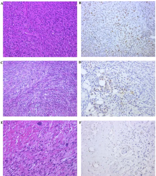
As for histological subtypes, all four samples of epithelioid MPNST showed intact H3K27me3 (Fig. 2a,b). Twelve (70.6%) of 17 malignant Triton tumors revealed lack of H3K27me3 staining. When they were classified according to the existence of epithelioid component, only one (20%) of the malignant Triton tumors with epithelioid component showed partial loss of staining, whereas 11 (91.7%) malignant Triton tumors without epithelioid component lacked staining (Fig. 2c,d).
These histological variants revealed significant differences in H3K27me3 expression. Compared to conventional MPNSTs, epithelioid MPNSTs showed preserved expression of H3K27me3 and malignant Triton tumors revealed reduced expression of H3K27me3 (Table 1).
One MPNST with glandular formation also showed complete loss of H3K27me3 (Fig. 2e,f).
3.1.1. H3K27me3 expression and prognosis
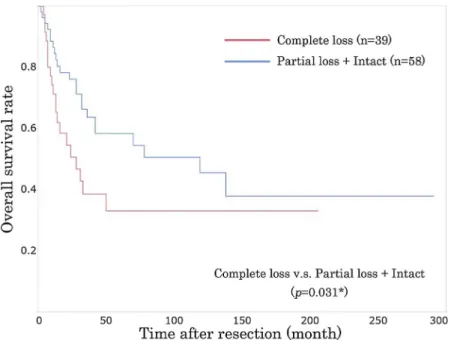
The patients with complete loss of H3K27me3 showed significantly poorer prognoses compared to the cases with partial loss or intact H3K27me3 (p = 0.031) (Fig. 3). In univariate analysis, significantly adverse prognoses were observed in the patients with morphological high-grade malignancy (FNCLCC grade 3), NF-1 associated MPNSTs, the tumors with heterologous components and the cases with complete loss of H3K27me3. However, in multivariate analysis (proportional hazard analysis), any factor did not show significant prognostic value (Table 2).
3.2. H3K27me3 expression in the metachronous cases (same sites)
We reviewed 39 samples (from 11 patients) of metachronous MPNSTs at the same sites (Table 3). Four of the 11 patients’cases (Cases 1, 2, 5, and 6) maintained the percentage of immunoreactive tumor cells (i.e., complete loss to complete loss, or partial loss to partial loss between the primary and later-occurring tumors). Two cases showed significantly reduced expression of H3K27me3 as the tumor recurred (partial loss to complete loss) (Cases 3 and 4) (Fig. 4), but three cases showed increased expression of H3K27me3 (complete loss to partial loss, partial loss to intact) (Cases 7–9). In the two multi-recurrent cases (Cases 10 and 11), the immunoreactivity did not show a stable pattern.
H. Otsuka et al. Pathology - Research and Practice 214 (2018) 417–425
418
Fig. 1.Immunohistochemical staining pattern of H3K27me3 in MPNST. The evaluation was performed with the presence of positive internal controls (endothelial cells or lymphocytes).
IHC score 0 (immunoreactive rate: 0–5%), complete loss of H3K27me3 (a). IHC score 1+ (6–24%) (b), 2+ (25–49%) (c) and 3+ (50–74%) (d) were regarded as partial loss of H3K27me3. IHC score 4+ (75–100%) (e) was considered as intact H3K27me3.
Table 1
Immunohistochemical H3K27me3 expression and clinicopathologicalfindings.
Complete loss, n (%) Partial loss, n (%) Intact, n (%) p−value N
Total MPNST cases 50 (34.5%) 45 (31.0%) 50 (34.5%) 145 samples
Clinical features
Sex Males 22 (43.1%) 11 (21.6%) 18 (35.3%) (p= 0.47) 51 patients
Females 17 (40.0%) 15 (32.6%) 14 (30.4%) 46 patients
Location Trunks 29 (30.2%) 34 (35.4%) 33 (34.4%) (p= 0.40) 96 samples
Extrimities 20 (40.8%) 13 (26.5%) 16 (32.7%) 49 samples
FNCLCC grade 1 11 (23.9%) 16 (34.8%) 19 (41.3%) (p= 0.15) 46 samples
2 24 (35.3%) 22 (32.4%) 22 (32.4%) 68 samples
3 14 (45.2%) 9 (29.0%) 8 (25.8%) 31 samples
Clinical backgrounds v.s.Sporadic
NF1-associated MPNST 19 (44.2%) 12 (27.9%) 12 (27.9%) (p= 0.57) 43 patients
MPNST arising from schwannoma 2 (50.0%) 1 (25.0%) 1 (25.0%) (p= 0.83) 4 patients
Sporadic MPNST cases 18 (45.0%) 13 (32.5%) 19 (47.5%) 40 patients
Histological subtypes v.s.Conventional
Epithelioid MPNST 0 0 4 (100%) p= 0.015* 4 samples
Malignant Triton tumor with epithelioid component 0 1 (20.0%) 4 (80%) p= 0.029* 5 samples
Malignant Triton tumor without epithelioid component 8 (66.7%) 3 (25.0%) 1 (8.3%) p= 0.019* 12 samples
MPNST with glandular formation 1 (100%) 0 0 1 sample
Conventional MPNST 41 (33.3%) 41 (33.3%) 41 (33.3%) 123 samples
Fig. 2.In epithelioid MPNST (a), both epithelioid cells and spindle-shaped cells retained H3K27me3 IHC (b).In malignant Triton tumor (c), rhabdomyoblastic cells showed lack of H3K27me3 (d). In MPNST with glandular formation (e), glandular component was negative for H3K27me3 (f).
Fig. 3.Kaplan-Meier survival curves of the relationship between H3K27me3 expression and overall survival.
H. Otsuka et al. Pathology - Research and Practice 214 (2018) 417–425
420
3.3. H3K27me3 expression in the metachronous cases (differing sites)
We reviewed 29 samples (from nine patients) of metachronous MPNSTs at the differing sites (Table 4). Three of these nine patients’ cases showed reduced expression of H3K27me3 in the later occurring tumor compared to the primary tumor (partial loss to complete loss, intact to partial loss) (Cases A, B, and C). In two of these cases, the later- occurring tumors were located in the lung, indicating lung metastasis.
The other cases showed no remarkable change or an unstable pattern of H3K27me3 expression, but in some cases with favorable prognosis, the later occurring tumors tended to show increased H3K27me3 expression.
3.4. H3K27me3 expression and morphological features
Some cases showed a correlation between the morphology of the tumors and H3K27me3 expression. Below, two cases with typical re- sults were presented. In the case of MPNST arising from plexiform neurofibroma, lack of H3K27me3 was correlated with the morpholo- gical malignant features (Fig. 5). In Case 3, as the recurrence was re- peated, the tumor showed higher malignancy and the percentage of H3K27me3-positive cells decreased (Table 3,Fig. 4).
4. Discussion
We focused this retrospective analysis on two issues: the relation between the immunohistochemical expression of H3K27me3 and clin- icopathologicalfindings, and the transition of H3K27me3 expression in metachronous MPNST cases.
Regarding all MPNST samples and NF-1-associated MPNSTs, our findings of complete or partial loss of H3K27me3 expression are similar to those of previous studies [15–20]. Three (75%) of MPNSTs arising from schwannoma, which were histologically conventional MPNSTs, in our study showed lack of H3K27me3 and this rate was higher than the total average for our series of cases.
In our study, epithelioid MPNSTs retained H3K27me3, and previous studies reported the same result [16,19]. We also found that a high
percentage of MPNSTs with only rhabdomyoblastic differentiation lacked H3K27me3 (91.7%), but among MPNSTs with both rhabdo- myoblastic and epithelioid differentiation only one sample showed a lack of staining. Almost all of MPNSTs with epithelioid differentiation (88.9%) retained H3K27me3. This suggests that a mechanism other than PRC2 inactivation may be involved in the malignant transforma- tion of MPNSTs with epithelioid differentiation.
On the other hand, it is known that malignant Triton tumors tend to show poorer prognoses than conventional MPNSTs, and loss of H3K27me3 is related to poorer survival in the MPNST patients.
[17,21,22] Together with our result, malignant Triton tumors without epithelioid component might show lack of staining more frequently compared to conventional MPNSTs. In our study, one sample of MPNST with glandular formation showed loss of H3K27me3. Several previous reports described three cases of MPNSTs with glandular formation that retained H3K27me3, but our presentfindings demonstrate that loss of H3K27me3 can occur in MPNST with glandular formation [16,18].
A correlation between H3K27me3 expression and prognosis was demonstrated in our study. The patients with complete loss of HeK27me3 expression revealed poorer prognoses than the patients with partial loss and the intact cases. This result is consistents with the report that there are relationships both between H3K27me3 expression and morphological malignancy, and between morphological malignancy and prognosis [15–19].
Regarding metachronous MPNST cases, the issue is whether loss of H3K27me3 can be reversible. Loss of H3K27me3 is caused by PRC2 inactivation due to a loss-of-function mutation inEEDorSUZ12, and the rate of H3K27me3 loss in MPNSTs with histological high-grade malignancy is higher than that in MPNSTs with low-grade malignancy [8–10,15–20]. Loss of H3K27me3 is also related to adverse prognosis in MPNST [17]. In light of thesefindings, it seems that H3K27me3 ex- pression should be reduced or at least maintained as the passage be- comes longer, and it could be expected to be irreversible. Two of our present cases, the case of arising from plexiform neurofibroma and Case 3, showed typical results that percentage of H3K27me3-positive cells was correlated with the morphological malignant featurs (Fig. 5, Table 3andFig. 4). However in some cases, later-occurring tumors showed increased expression of H3K27me3 compared to that of the primary tumors. Below, the examination for each case was shown.
Among the cases of metachronous MPNSTs at the same sites (Table 3), Cases 1, 2, 5 and 6 showed no change in H3K27me3 ex- pression. We speculate that these tumors showed the natural course.
Cases 3 and 4 showed reduced expression of H3K27me3 in the later- occurring tumors compared to the primary tumors, and these MPNSTs resulted in death within 2 years; we consider these to be recurrent and aggressive cases. In five cases (Cases 7–11), H3K27me3 expression showed an unstable pattern. These can be classified as the cases with poor prognosis and the cases with long-term passage.
As it is reported that H3K27me3 IHC sometimes shows a mosaic pattern of staining, our present cases with poor prognoses (Cases 8 and 9) may be recurrent. [15–20] However, in the cases with long-term passage, i.e., >10 years from the primary occurrence to the final oc- currence (Cases 7, 10, and 11), since the passage is too long to consider them as recurrent cases, they may be heterochronous and multiple tu- mors located in the same regions, rather than recurrent.
Among our cases of metachronous MPNSTs at different sites (Table 4), Cases A, B and C showed more frequent loss of staining in the later-occurring tumors. They could be considered metastasis from the primary lesion. Cases E and F had died within 1 year of the primary occurrence, and these aggressive cases may also be considered meta- static cases. However, the remaining cases (Cases D, G–I) revealed an Table 2
Univariate analysis (a) and multivariate analysis (b) for the prognosis.
(a)
Variables Analyzed groups p-value
Sex M/F p= 0.86
Location Trunks/Extrimities p= 0.28
FNCLCC grade 1 + 2/3 p= 0.030*
NF-1 NF-1/non-NF-1 p= 0.028*
Histological feature Epithelioid MPNST/
Malignant Triton tumor/
conventional MPNST
p= 0.0029*
H3K27me3 expression Complete loss/Partial loss + Intact
p= 0.031*
(b)
Variables Analyzed groups Hazard ratio p-value
FNCLCC grade 1 + 2/3 0.962 p= 0.11
NF-1 NF-1/non-NF-1 0.669 p= 0.21
Histological feature Epithelioid MPNST/
Malignant Triton tumor/
conventional MPNST
0.797 p= 0.16
H3K27me3 expression Complete loss/Partial loss + Intact
0.853 p= 0.14
unstable expression of H3K27me3 and showed favorable prognoses. In these four cases, the later-occurring tumors might not be metastatic;
rather, they may be heterochronous and multiple lesions located in different regions from the primary lesion. This hypothesis may be supported by the findings that Cases D and G were NF-1-associated MPNSTs and Cases H and I were suspected (but not genetically proved) to be associated with NF-1 because of their clinical features.
In conclusion, MPNSTs, especially those associated with NF-1, can occur in heterochronous and multiple patterns, but it is difficult to distinguish these cases from recurrent or metastatic cases. Because loss of H3K27me3 is due to a genetic mutation ofEEDorSUZ12, expression of H3K27me3 can be expected to be reduced or at least maintained as the disease progresses. It was reported that H3K27me3 sometimes shows a mosaic pattern of staining, and the possibility cannot be denied Table 3
Summary of H3K27me3 IHC in metachronous MPNSTs at the same sites.
Complete : Complete loss of H3K27me3.
Partial : Partial loss of H3K27me3.
Intact : Intact H3K27me3.
H. Otsuka et al. Pathology - Research and Practice 214 (2018) 417–425
422
that both complete loss and partial loss would be present in respective parts of the same tumor [15–20]. However, a case with increased ex- pression in the long term, such as that seen in complete loss to partial loss, should be considered a heterochronous and multiple-tumor case.
As heterochronous and multiple tumors may show lower malignancy compared to recurrent or metastatic tumors, a favorable prognosis may
be expected when H3K27me3 expression is increased.
Disclosure/Conflicts of interest
The authors declare no significant relationships with, or financial interest in, any commercial companies pertaining to this article.
Fig. 4.The recurrent case of MPNST (case 3,Table 3). As the recurrence was repeated, the tumor showed severer nuclear atypia, more pleomorphism and more frequent atypical mitosis, so obtained higher malignancy. In primary lesion (a), H3K27me3 IHC score (the rate of immunoreactive cell for H3K27me3) was 3+ (b). H3K27me3 IHC score of 1st recurrence was 2+
(c: HE, d: IHC) and that of 2nd recurrence was 0, complete loss (e: HE, f: IHC).
Table 4
Summary of H3K27me3 IHC in metachronous MPNSTs at the different sites.
Complete : Complete loss of H3K27me3.
Partial : Partial loss of H3K27me3.
Intact : Intact H3K27me3.
H. Otsuka et al. Pathology - Research and Practice 214 (2018) 417–425
424
References
[1] C.D.M. Fletcher, J.A. Bridge, P.C.W. Hogendoorn, et al. (Eds.), WHO Classification of Tumours of Soft Tissue and Bone, IARC Press, Lyon, 2013, pp. 187–189.
[2] B.S. Ducatman, B.W. Scheithauer, Postirradiation neurofibrosarcoma, Cancer 51 (1983) 1028–1033.
[3] J.R. Goldblum, A.L. Folpe, S.W. Weiss, et al. (Eds.), Malignant Peripheral Nerve Sheath Tumors. Enzinger & Weiss’s Soft Tissue Tumors, 6th ed., Elsevier, Philadelphia, PA, 2014, pp. 855–879.
[4] F.J. Rodriguez, A.L. Folpe, C. Giannini, et al., Pathology of peripheral nerve sheath tumors: diagnostic overview and update on selected diagnostic problems, Acta Neuropathol. 123 (2012) 295–319.
[5] C. Zou, K.D. Smith, J. Liu, et al., Clinical, pathological, and molecular variables predictive of malignant peripheral nerve sheath tumor outcome, Ann. Surg. 249 (2009) 1014–1022.
[6] Y. KoKang, M. Pekmezci, A.L. Folpe, et al., Diagnostic utility of SO×10 to distin- guish malignant peripheral nerve sheath tumor from synovial sarcoma, including intraneural synovial sarcoma, Mod. Pathol. 27 (2014) 55–61.
[7] J.R. Karamchandani, T.O. Nielsen, M. van de Rijn, et al., So×10 and S100 in the diagnosis of soft-tissue neoplasms, Appl. Immunohistochem. Mol. Morphol. 20 (2012) 445–450.
[8] W. Lee, S. Teckie, T. Wiesner, et al., PRC2 is recurrently inactivated through EED or SUZ12 loss in malignant peripheral nerve sheath tumors, Nat. Genet. 46 (2014) 1227–1232.
[9] M. Zhang, Y. Wang, S. Jones, et al., Somatic mutations of SUZ12 in malignant peripheral nerve sheath tumors, Nat. Genet. 46 (2014) 1170–1172.
[10] T. De Raedt, E. Beert, E. Pasmant, et al., PRC2 loss amplifies Ras-driven tran- scription and confers sensitivity to BRD4-based therapies, Nature 514 (2014) 247–251.
[11] J.S. Pyo, D.W. Kang, Prognostic role of EZH2 in gliomas: a meta-analysis, Int. J.
Biol. Markers 28 (2017) 0.
[12] R. Cao, L. Wang, H. Wang, et al., Role of histone H3 lysine 27 methylation in polycomb-group silencing, Science 298 (2002) 1039–1043.
[13] J.A. Simon, C.A. Lange, Roles of the EZH2 histone methyltransferase in cancer epigenetics, Mutat. Res. 647 (2008) 21–29.
[14] Y.J. Vickie, Fletcher CDM epithelioid malignant peripheral nerve sheath tumor clinicopathologic analysis of 63 cases, Am. J. Surg. Pathol. 39 (2015) 673–682.
[15] A.H. Cleven, G.A. Sannaa, I. Briaire-de Bruijn, et al., Loss of H3K27 tri-methylation is a diagnostic marker for malignant peripheral nerve sheath tumors and an in- dicator for an inferior survival, Mod. Pathol. 29 (2016) 582–590.
[16] I.M. Schaefer, C.D. Fletcher, J.L. Hornick, et al., Loss of H3K27 trimethylation distinguishes malignant peripheral nerve sheath tumors from histologic mimics, Mod. Pathol. 29 (2016) 4–13.
[17] N. Carlos, R. Prieto-Granada Cristina, et al., Loss of H3K27me3 expression is a highly sensitive marker for sporadic and radiation-induced MPNST, Am. J. Surg.
Pathol. 40 (2016) 479–489.
[18] N. Asano, A. Yoshida, N. Hiraoka, et al., Immunohistochemistry for trimethylated H3K27 in the diagnosis of malignant peripheral nerve sheath tumours, Histopathology 70 (2017) 385–393.
[19] C.N. Prieto-Granada, T. Wiesner, J.L. Messina, et al., Loss of H3K27me3 expression is a highly sensitive marker for sporadic and radiation-induced MPNST, Am. J. Surg.
Pathol. 40 (2016) 479–489.
[20] M. R€ohrich, C. Koelsche, D. Schrimpf, et al., Methylation-based classification of benign and malignant peripheral nerve sheath tumors, Acta Neuropathol. 131 (2016) 877–887.
[21] C.J. Stasik, O. Tawfik, Malignant peripheral nerve sheath tumor with rhabdomyo- sarcomatous differentiation (malignant triton tumor), Arch. Pathol. Lab. Med. 130 (2006) 1878–1881.
[22] S.C. Kamran, A.B. Shinagare, N.H. Ramaiya, et al., Malignant peripheral nerve sheath tumors: prognostic impact of rhabdomyoblastic differentiation (malignant triton tumors), neurofibromatosis 1 status and location, Eur. J. Surg. Oncol. 39 (2013) 46–52.
Fig. 5.The case of MPNST arising from plexiform neurofibroma. The component of low-grade malignancy (a,b) showed a proliferation of spindle-shaped cells arranged in whorl pattern, having the feature of plexiform neurofibroma. In this component (a), H3K27me3 IHC score (the rate of immunoreactive cell for H3K27me3) was 3+ (partial loss) (b). The component having higher cellularity and frequent atypical mitosis (c) also revealed partial loss of H3K27me3, but IHC score was 2+ (d), lower than that of the component of low-grade malignancy.
The component of histological high-grade malignancy, having pleomorphism of tumor cells and nuclear atypia (e), exhibited complete loss of H3K27me3 (IHC score: 0) (f).