Antibiotic Production by Marine Pigmented
Bacteria I : Antibacterial Effect of
Alteromonas luteo-violaceus
著者
SAKATA Taizo, SAKAGUCHI Kunio, KAKIMOTO
Daiichi
journal or
publication title
鹿児島大学水産学部紀要=Memoirs of Faculty of
Fisheries Kagoshima University
volume
31
page range
243-250
別言語のタイトル
海洋色素細菌による抗菌物質の産生 I :
Alteromonas luteo-violaceusの抗菌作用について
Vol. 31 pp. 243-250 (1982)
Antibiotic Production by Marine Pigmented Bacteria-1
Antibacterial Effect of Alteromonas luteo-violaceusTaizo Sakata, Kunio Sakaguchi and Daiichi Kakimoto*
Abstract
Marine purple bacteria belonging to the species Alteromonas luteo-violaceus were isolated from coastal water of Kinko Bay in southern Japan and examined for antibacterial action.
These isolates produce antibacterial substance(s) which inhibits the growth of various kinds of test organisms including marine purple bacteria themselves.
The antibacterial activity was found in the crude extract obtained from the cells by sonication, which contained heat-labile macromolecular substance(s).
On addition of the crude extract to natural sea water samples, colony forming abilities in sea water
were inhibited completely.
Some chromogenic marine bacteria have recently reported to produce different types of
antibiotic substances. For example, Andersen et all) showed that a marine Chromobacterium sp produces a series of highly brominated metabolites which are responsible for antibiotic action as well as autoinhibition. Gauthier et al2,3,4,5) also described that four species of marine Alteromonas synthesize polyanionic polysaccharide antibiotics which act on bacterial respiration and that some strains of A. luteo-violaceus concomitantly produced brominated antibiotics which are small
molecules but not diffusible due to association with cells.
As described in previous paper67', we isolated Alteromonas luteo-violaceus strains from coastal water of Kinko Bay in Japan, which showed antibacterial effects against various kinds of bacteria. These isolates usually die out within one week unless subcultured in fresh media, suggesting their
autotoxication.
In this paper, we examined the inhibitory effect of antibacterial substance(s) produced by marine isolates belonging to the species Alteromonas luteo-violaceus against various kinds of bacteria
including A. luteo-violaceus.
Materials and Methods
Organisms Ten strains of marine purple bacteria, belonging to A. luteo-violaceus, isolated from Kinko Bay, Kagoshima, Japan in 1979 as shown in Fig.5. One strain 9K-V9 among ten isolates, which formed the strongest purple color colony, was used exclusively in this study.
* Laboratory of Microbiology, Faculty of Fisheries, Kagoshima University, 4-50-20 Shimoarata, Kagoshima 890, Japan.
244 Mem. Fac. Fish., Kagoshima Univ. Vol. 31 (1982)
All strains were grown on modified ZoBell 2216E slants (pH 7.6) at 25 C and subcultured every
four days.
The following organisms were used for growth inhibition test: 1) Alteromonas luteo-violaceus
9K-V9, 2) Vibrio parahaemolyticus Vp H-K15 (serotype 05:K15 isolated from patient by Dr. K.
Takagi at Hokkaido University), 3) V. alginolyticus Va F-l (NIH 154-78), 4) A. haloplanktis
1055-1 (by Dr.M.SAKAl), 5) Pseudomonas I~6 (isolated from sea water), 6) Micrococcus lysodeikticus
(laboratory strain), 7) Staphylococcus citreus, 8) Bacillus subtillis Marburg strain 6051, 9) Pseudomonas
aeruginosa, 10) Escherichia coli B strain, 11) Aeromonas hydrophila AHH (IFO 12981), 12) A.
salmonicida ASS (IFO 12718).
Extraction of antibacterial substances from the cells Purple bacterium 9K-V9 was
incubated statically in 4 flasks containing 250 ml modified ZoBell broth at 25 C for 3 days.
Bacterial cells were harvested with centrifugation at 8,000 g for 20 min and then the bacterial
suspension in deionized water (30 ml) was submitted to sonic disintegration for 3 min at 2A
(Kaijo Denki type 4280). The supernatant solution after centrifuged at 17,000 g for 20 min was
dialyzed against deionized water for one day and filtrated with Millipore filter (HAWP 0.45 ^m)
to sterilize contaminated bacterial cells.
Antibacterial activity Growth inhibition oftest organisms was examined by two methods.
One is the double agar method, in which test organisms were seeded in ZoBell agar (pH 6.3 for
marine bacteria) and TSA (pH 6.3 for terrestrial bacteria and Vibrio strains) plates. The paper
disks impregnated with the culture filtrate orcell extract ora loopful ofbacterial cells were placed
on the bacterial lawns of test organisms.
Inoculated plates were incubated at 25 C for 48 hr,
after which time zones of growth inhibition were measured.
The other is the broth culture
method, in which 0.1 ml culture of test organisms were inoculated into L-from test tubes
containing 9.5 ml ZoBell broth (pH 7.6 for marine bacteria) or Z~A (pH 7.2 for terrestrial
bacteria) and the cell extract (0.3 ml), and then incubated at 30C for 72 hr on a shaking
incubator. The turbidity ofculture was determined by using a spectrophotometer (Fuji Kogyo,
660 nm filter).
Effect of the cell extract on colony forming ability of natural sea water Soft agar
containing half volume of the cell extract and 0.1 ml natural sea water was overlayered on basal
agar plate and incubated at 25C for 72 hr. After incubation period, bacterial colonies formed
were counted, and compared with those of control plate without the addition of the cell extract.
Results
Changes with cultivation time in antibacterial activity
Figs. 1 and 2 show the zone sizes
of growth inhibition against test organisms caused by the cellsor culture filtrate of purple bacteria
during culture period.
Antibacterial activity appeared in the cells but not released into culture medium at one day
days Zones of inhibition (mm) 9K-V5 5 10 9K-V9 5 10
Fig. 1. Growth inhibition of Vibrio parahaemolyticus H-K15 by strains of Alteromonas luteo-violaceus. The cells (C) or paper disks impregnated whth the supernatant (S) from Alteromonas strains were placed on the surface of a double layer agar (TSA pH 6.3) seeded with Vibrio parahaemoly ticus H-K15 and incubated for one day at 25C. Blank columns indicate diameters of the col
onies or paper disks
filtrate was occasionally detected at 5 or 7 days (Fig.l).
If large amount of active substances
would be released from the cells into medium due to autolysis, a paper disk impregnated with the
filtrate has activity enough to inhibit the growth of test organisms on agar plates.
The production of antibacterial substances by a strain 9K-V9 varied depending on culture
methods (Fig.2).
Growth inhibition against Vp H-K15 was detected at one day with static
£ 3
Incubation time (day)
Fig. 2. Time course of growth and antibiotic production of Alteromonas 9K-V9.
Staticcultureina flask
Incubationtime (day)
3 4
Incubation time (day)
Plate culture
^ ^ VP.H-K15
246
Mem. Fac. Fish., Kagoshima Univ. Vol. 31 (1982)
culture in a flask, shaking culture in a test tube and on an agar plate cultureof 9K-V9 but no or
slight inhibition occurred with shaking culture in a flask (100 st/min) during early period of
culture.
On the other hand, selfinhibition against 9K-V9 appeared at 2 days with static culture
in a flask and at some days later with plate culture and test tube culture.
In thecase ofshaking
culture in a flask (100 st/min), the appearance of antibacterial activity was retarded after 5"6
days, just before which time bacterial viability had decreased rapidly.
The greater and earlier
production of antibacterial substance(s) and longer viability occurred concomitantly with static
culture than with shaking culture of 9K-V9.
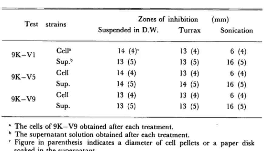
Treatment for the release of antibacterial substance(s) from the cells
As shown in Table
1, antibacterial substance(s) was associated with the cells not so tightlyand considerable amount
of the substance(s) was released from the cells into suspending solution (deionized water).
Furthermore, sonictreatment (5 min at 2A) attained the release of the substance(s) from the cells
almost completely.
Turrax treatment (Ultra-Turrax, Ikawerk TP 18" 10 type, 2 min) gave no or
slight effect on the release of activity compared with sonication.
Table 1. Comparison among treatments for the release of antibacterial substances from the cells.
Test strains Zones of inhibition Suspended in D.W. Turrax (mm) Sonication 9K-V1 Cella Sup.b 14 (4)c 13 (5) 13 (4) 13 (5) 6 (4) 16 (5) 9K-V5 Cell Sup. 14 (4) 14 (5) 13 (4) 14 (5) 6 (4) 16 (5) 9K-V9 Cell Sup. 13 (4) 13 (5) 13 (4) 13 (5) 6 (4) 16 (5)
a The cells of 9K—V9 obtained after each treatment. b The supernatant solution obtained after each treatment.
c Figure in parenthesis indicates a diameter of cell pellets or a paper disk
soaked in the supernatant.
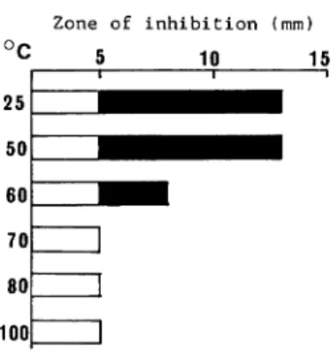
Heat treatment of antibacterial substance(s) The crude extract obtained after sonic
treatment was heated at each temperature for 10 min as shown in Fig.3. The inhibition activity
against VP H-K15 was lost at 60°C by half and at 70°C completely.
Precipitation of antibacterial substance(s) Table 2 demonstrates that antibacterial substance(s) was precipitated with 80% saturated (NHi^SCh, ethanol (50% v/v) and acetone (50% v/v). The substance(s) was not dialyzable and not inactivated in deionized water. These results indicate that the antibacterial substance(s) has high molecular weight.
Growth inhibition of various kinds of bacteria Growth inhibition of various test
organisms by the crude extract obtained from the cells of 9K-V9 is demonstrated in Fig.4. On
Zone of inhibition (mm)
°c
5
Fig. 3. Effect of heat treatment on antibacterial activity of the cell extract from 9K-V9 against V.
parahaemolyticus H - K15.
The cell extract was heated at each temperature for 10 min.
Table 2. Effect of precipitation and dialysis of the cell extract from Alteromo nas 9K—V9 on its antibacterial activity.
Treatment Dialysis3 (NH4)2S04 ppt.1 Ethanol ppt. Acetone ppt. Zones of inhibition (mm) 9K-V9 Vp H-K15 17 (5)' 14 (5) 12 (5) 14 (5) 10 (5) 8 (5) 7 (5) 10 (5)
a The crude extract from 9K—V9 dialyzed directly without precipitation. b The crude extract dialyzed after precipitation.
c Figure in parenthesis indicates a diameter of a paper disk.
organisms were inhibited, even if to different extent. Most of test organisms were not able to grow at all but some terrestrial strains grew after long lag period under existence of the crude
extract.
Antibacterial activity of the crude extract of 9K-V9 was examined against natural sea water by
using double agar method (Fig.6).
It is very interesting that the crude extract inhibited colony
forming ability of natural sea water completely, in contrast with 600~ 1,000 c.f.u. per plate for
natural sea water samples without the crude extract. Same results were obtained with the
culture filtrate, which was separated from the cells after 3 days culture was treated directly with
sonication.Discussion
Marine purple bacteria, Chromobacterium spp and Alteromonas spp, have been reported to
248 Mem. Fac. Fish., Kagoshima Univ. Vol. 31 (1982)
500 r 9K-V9 Va F-l r 1055-1
Fig. 4. Effect of the addition of crude extract from the cells of 9K-V9 on the growth of test organ
isms.: without crude extract. •-•••: with crude extract.
Fig. 5. Plate cultures of 11 strains of Alteromonas luteo-violaceus. isolated from Kinko Bay.
1: 9K-V1, 2: 9K-V2, 3: 9K-V3, 4: 9K-V4, 5: 9K-V5, 6: 9K-V6, 7: 9K-V7, 8: 9K-V8, 9: 9K-V9, 10: 9K-V10, 11: 8K-V2.
Fig. 6. Antibacterial activity of the crude extract from 9K-V9 against natural sea water,
left: without the crude extract, right: with the crude extract.
to be brominated pyrrole or benzene derivatives" or polyanionic polysaccharide compounds".
They are also responsible for autoinbition as well as for antibiotic action against other kinds of
bacteria. Especially, GAUGHIER" presented that an antibacterial polysaccharide could act at the level of the respiratory chain by setting up a flavinic respiration.
The crude extract obtained from the cells of our strain A. luteo-violaceus 9K.-V9, which isolated from sea water of Kinko Bny, was demonstrated to include heat-labile, non-dialyzable macromolecular subatance(s). Antibacterial substance(s) was shown to comprise different kinds of activities, for example, against VP H~K15 and 9K~V9 itself, judging from difference in production time of the substance(s). The characteristics of antibacterial substance(s) extracted
from the cells of 9K~V9 suggest the possibility that it is composed of proteinous compound(s),
such as bacteriolytic or autolytic enzyme(s).
Time course experiments on production of antibacterial substance(s) indicate that static
culture gave rise to earlier and greater production than shaking culture did. The cells of purple
bacteria grew well in surface layer of broth medium with static culture and sank into bottom layer, followed with newly growing cells in surface layer. In surface layer of static culture or on agar plate, Os supply is considered to be sufficient for bacterial cells to synthesize antibacterial culture to be homogenous and supply On in lower concentration as a whole culture.
Further experiments will be studied with purified substance to elucidate the structure and mechanism of antibacterial substance(s) produced by Alteromonas luteo-violaceus.
250
Mem. Fac. Fish., Kagoshima Univ. Vol. 31 (1982)
Apknowledgment
This work was supported by a scientific research fund granted by the Ministry of Education,
Science and Culture (No. 511916).
References
1) ANDERSEN, R.J., M.S. WOLFE and D.J. Faulkner (1974): Autotoxic antibiotic production by a marine
Chromobacterium. Marine Biology, 27, 281-285.
2) Gauthier, MJ. (1975): Morphological, physiological, and biochemical characteristics of some violet-pigmented bacteria isolated from seawater. Can. J. Microbiol., 22, 138-149.
3) GAUTHIER, M.J. and G.N. FLATAU (1976): Antibacterial activity of marine violet-pigmented Alteromonas with specisl reference to the production of brominated compounds. Can. J. Microbiol., 22, 1612-1619. 4) GAUTHIER, M.J. (1976): Modification of bacterial respiration by a macromoleculer polyanionic
antibiotic production by a marine Alteromonas. Antimicrob. Agents Chemother., 9, 361-366.
5) Gauthier, M.J. and V.A. Breittmayer (1979): A new antibiotic-producing bacterium from seawater:
Alteromnas aurantia sp. nov. /./. J. Syst. Bacteriol., 29, 366-372.
6) Kakimoto, D., H. Maeda, T. S akata, W. Sharp, R.M. Johnson(1980): Study on marine pigmented
bacteria-I: Distribution and characteristics of pigmented bacteria. Mem. Fac Fish., Kagoshima Univ., 29,
339-347.
7) JOHNSON, R.M., H. Maeda and D. Kakimoto (1981): Factors effecting viabillity and pigment of