Title
[症例報告]Nonfunctioning islet cell tumor associated with
concurrent renal cell carcinoma : A case report
Author(s)
Shimoji, Hideaki; Shiraishi, Masayuki; Oshiro, Takashi; Soda,
Noriko; Tomori, Takehiko; Muto, Yoshihiro; Ogawa,
Yoshihide; Sawada, Satoshi
Citation
琉球医学会誌 = Ryukyu Medical Journal, 18(1-2): 33-36
Issue Date
1998
URL
http://hdl.handle.net/20.500.12001/3340
Nonfunctioning islet cell tumor associated with concurrent renal cell carcinoma: A case report
Hideaki Shimojill, Masayuki Shiraishill, Takashi Oshiro", Noriko Sodal), Takehiko Tomorill, Yoshihiro Muto", Yoshihide Ogawa21 and Satoshi Sawada31
'First Department of Surgery, 2)Department of Urology, and 3) Department of Radiology, Faculty of Medicine,
University of the Ryukyus, Okinawa, Japan
(Received on December 22, 1997, accepted on June 30, 1998)
ABSTRACT
A case of concurrent nonfunctioning pancreatic islet cell tumor and renal cell carci-noma of the right kidney in a 62-year-old woman is reported. She initially presented with a complaint of right flank pain at a hospital where she was suspected to have a tumor in the right kidney. Subsequently, the patient was referred to our University Hospital for
fur-ther examination. A tumor in the head of the pancreas was incidβntally detected during the
examination of the renal tumor. On sophisticated imaging studies (US, CT, and MRI), both pancreatic and renal tumors showed similar findings. Only proton density weighted MRI allowed for a differentiation of the two tumors, but the tumor of the pancreatic head could not be definitely diagnosed. An exploratory laparotomy revealed a renal tumor in the right kidney and a well encapsulated tumor in the uncinate process of the pancreatic head.
A right radical nephrectomy and an enucleation of the pancreatic tumor were formed. The right kidney showed renal cell carcinoma (clear cell type), while atic tumor was determined to be an islet cell tumor. Both tumors showed similar appearance on a cut surface, that was attributed to the similar imaging Since this concurrence of these two tumors is extremely rare, in our opinion, able to report this case so that more information can be gathered for assessing sis and providing best treatment. RyukyuMed. J., 18(1, 2)33-36, 1998 Key words: nonfunctioning islet cell tumor, renal cell carcinoma
INTRODUCTION
The simultaneous occurrence of two distinct neoplasms of different origin occasionally presents in all organs, however, the combination of this phenomenon is very rare between the pancreas and kidney. Nonfunctioning islet cell tumor1 21 and renal cell carcinoma3-61 are rarely concurrent or coincident. TO our knowledge, such concur-rent two tumors have never been previously reported.
We report a rare instance of a concurrent nonfunctioning islet cell tumor of the pancreatic head and renal cell carci-noma of the right kidney. Since each primary tumor exhib-ited similar diagnostic images except for the findings of
proton density weighted magnetic resonance imaging (M RI), we could not make a precise diagnosis before opera-tion. The rarity and diagnostic difficulty of this case prompted us to describe our findings.
easily per-the pancre-a grossly features. it is valu-the diagno-CASE REPORT
A 62-year-old woman presented at Ryukyu University Hospital for thorough examination in October 1996. She had been receiving medical treatment for hypertension in a local clinic. A dull pain in the right flank developed in August 1995 which was diagnosed as a tumor of the right kidney. Her past medical history revealed that she had un-dergone a hysterectomy for uterine myoma in 1974 and a subtotal thyroidectomy for goiter in 1989.
After a thorough examination at the University Hospital, two distinct tumors were identified: one was in the right kidney while the other was found. Each tumor revealed the same findings, that is, a hypoechoic mass on ultrasonography, a hyperdense mass on enhanced CT, and a hypervascular mass on angiography. These similar findings thus made it difficult to differentiate one from the other. Subsequently MRI demonstrated the same image in each tumor. However, only the proton
34 The Concurrence of islet cell tumor with renal cell carcinoma
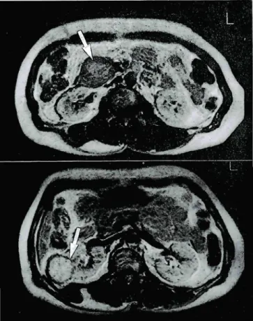
Fig. 1 Proton density weighted MRI demonstrating low intensity of pancreatic tumor (top, arrow) compared with high intensity of renal cell carcinoma (bottom, arrow).
density weighted MRI findings demonstrated a tumor of
low intensity in the pancreatic head and a tumor of high
intensity in the right kidney (Fig. 1). Based on these fin
dings, the tumor of the right kidney was diagnosed as
renal cell carcinoma, while that in the pancreatic head
could not be definitely diagnosed. Both the laboratory
data and tumor markers (CEA, CA19-9, elastase I, and
DUPAN) were within the reference ranges. In addition,
the patient did not present with any clinical
manifesta-tion of functioning islet cell tumors (insulinoma,
glucagonoma, gastrinoma, somatostatinoma, vipoma).
The patient underwent an exploratory laparotomy
on January 10, 1997. Both tumors bulged out and were
confined to each organ without any spread to the
sur-rounding or adjacent tissue. A radical nephrectomy and
enucleation of the pancreatic tumor in the head (uncinate
process) were thus performed with no difficulty.
GROSS PATHOLOGY
The enucleated tumor from the uncinate process of
the pancreatic head was encapsulated, and measured 5 X
4 cm in size. Its cut surface was pink-purple with areas
of hemorrhage and necrosis (Fig. 2, top).
The tumor of the kidney bulged out and measured 4 X
Fig. 2 Macrophotographs of the cut surface of islet cell carcinoma (top) and renal cell carcinoma (bottom). Both tumors show hemorrhaging, necrosis and cystic change.
3cm in size. On a cut section, it was found to be a
well-circumscribed, spherical, orange tumor with areas of
hemor-rhage, necrosis, and cystic change (Fig. 2, bottom). In
addi-tion, no thrombus was found in the renal veins.
MICROSCOPIC PATHOLOGY
The pancreatic tumor cells were arranged in a
rib-bon pattern mimicking an acinar or tubular structure
(Fig. 3, top; HE, X 25). The cells were small, round
and uniform with small nuclei. The tumor cells were
posi-tive for Grimelius's silver impregnation and
chromo-granin-A (Fig. 3, bottom; HE, X 50).
The renal tumor cells were large with clear
cyto-plasm in the tubular structures and stained slightly for
eosin. They were uniform in size and shape with small
dense nuclei (Fig. 4, HE, X50).
The patient was uneventful postoperatively, and
re-mains disease free at the time of this writing. DISCUSSION
We report the findings of a patient with two
concur-rent common neoplasms. Renal cell carcinoma2' arises
r .蝣 丁 *-【 pf - て
壁諒針骨鵬
H, , ヽ`. ・ {- ' .<'・ 〆.- _I 一,」* -..A* .Tuも-ゥ f *蝣 、 ごⅠ. ㌢- 一 iJ I- ′r i A--M J ' 1 1」IA蓑童震恵
ノ I.ヽ ∴ !蝣...ふ∴.上l_I. Fig. 3 Microphotographs of the islet cell tumor showingthe typical histology (top; HE,X 25), with both posi-tive argyrophil cells (bottom, left; Silver impregnation, X 50) and positive chromogranin cells (bottom, right;
X50).
originates from the islet cells of the pancreas. However,
this concurrence of two distinct tumors is extremely
rarely reported. In this patient, renal cell carcinoma of
the right kidney was initially diagnosed. On the other
hand, nonfunctioning islet cell tumor was also
inciden-tally detected during further imaging examinations of
the kidney tumor.
The concurrence of the two tumors may be an
inci-dental occurrence because there is no definite evidence of
a known genetic predisposition to these tumors and both
tumors do not tend to occur in familiar forms.
Moreo-ver, they may not be part of any recognized neoplasia
syn-drome hitherto described5'7'.
In clinical practice, we were confronted with a
diag-nostic problem in this case. Renal cell carcinoma could
easily be diagnosed by the characteristic findings using
imaging studies5'8'"". However, the tumor in the head
(uncinate process) of the pancreas demonstrated
identi-cal features to those of renal cell carcinoma using
sophis-ticated imaging studies" 12'. Consequently, the pancreatic
tumor could not be clearly diagnosed before surgery. Only
la ヽ BE I ・声車 一、、 l
葬
>aCl , -^A-`Il サ V *fォ」-* .+. こil !・要 >・!Fig. 4 Microphotograph of the renal cell carcinoma show-ing a clear cell type (HE, X 50).
proton density weighted MRI findings enabled us to differen-tiate these two tumors without any definite diagnosis of the pancreatic tumor. This differentiation was largely at-tributed to the difference in the water content5'.
Regarding the spread of renal cell carcinoma, the di-rect extension of renal cell carcinoma into the renal vein al-lows this tumor access to the blood stream which is unique among carcinomas. Thus, the early metastasis of renal cell carcinoma to the lung is a characteristic finding. Other com-mon sites of metastasis are the lymph nodes, bones, liver and adrenal glands. Regarding the lymph nodes, those in the para-aortic chain are the most common sites of metastasis3'. As a result, we were inclined to believe that the pancreatic tumor was a metastatic lesion of the renal cell carcinoma. Other differential diagnoses in-cluded nonfunctioning pancreatic islet cell tumor and pan-creatic mucinous cystadenocarcinoma.
The rarity of pancreatic islet cell tumors and cystadenocarcinoma reported in the literature means that no pathologist has yet had the opportunity to study any great number of these tumors. Therefore, the isolated case reports that have so far been published do not pro-vide reasonable data from which to extract percentages of metastasis to the kidney. To the best of our knowledge, no previous case with concurrent pancreatic islet cell tumor and renal cell carcinoma has yet been reported in the literature (Medline 1990-1997). Although an analysis of the pancreatic islet cell tumors previously reported in the literature does not represent the whole experience, this may indeed be the first case with these two tumors concurrently.
In conclusion, since concurrent pancreatic islet cell tumor and renal cell carcinoma is an uncommon phenomenon, more information on such cases should be gathered in order to better assess and diagnose such cases and thus de-velop an optimal treatment strategy.
36 The Concurrence of islet cell tumor with renal cell carcinoma
REFERENCES
1 ) Frantz V.K.: Tumor of the pancreas. Atlas of tumor pathology, Section VII - Fascicles 27 and 28, pp. 79-134, AFIP, Washington DC, 1959.
2 ) Kissane J. M.: Anderson's pathology, pp.1252-1254, The C.V. Mosby Company, St. Louis, 1985.
3) Luke B. and Schlumberger H.G.: Tumor of the kidney, renal pelvis and ureter. Atlas of tumor pa-thology, Section VIII - Fascicle 30, pp.42-78, AFIP, Washington DC, 1957.
4 ) Kissane J. M.: Anderson's pathology, pp.768-769, The C.V. Mosby Company, St. Louis, 1985. 5) Krane R.J., Siroky M.B. and Fitzpatrick J.M.:
Clinical urology. pp.359-373, J. B. Lippincott Company, Philadelphia, 1994.
6) Goto M., Nakano I., Sumi K., Yamaguchi H., Kimura T., Sako Y., Nawata H., Tanaka M. and Nagai E. : Cystic insulinoma and nonfunctioning islet cell tumor in multiple endocrine neoplasia type I. Pancreas. 9: 393-395, 1994.
7 ) NgadimanS., Horenstein M.G. and Campbell W.G. Jr.: The concurrence of duodeneal epitheliod stromal sarcoma, pulmonary chondromatous hamatoma, and nonfunctioning pancreatic islet cell tumor. A
possi-ble analogue of Carney's triad? Arch. Pathol. Lab. Med. 118: 840-843, 1994.
8 ) Takahashi S., Ueda J., Furukawa T. and Higashino K., Tsujihata M., Itatani H., Narumi Y. and Nakamura H.: Renal cell carcinoma: preoperative as-sessment for enucleative surgery with angiography, CT, and MRI. J. Compute Assist Tomogr.20: 863-8 70, 1996.
9) SoyerP., DufresneA., Klein I., Barbagelatta M.,
Herve J.M. and Scherrer A.: Renal cell carcinoma of
clear type: correlation of CT features with tumor
size, architectural patterns, and pathologic staging.
Eur. Radiol. 7: 224-229, 1997.
10) Bono A.V. and Lovisolo J.A. : Renal cell carcinoma-diagnosis and treatment: state of the art. Eur. Urol. 31 Suppl 1: 47-55, 1997.
LI) Brambs H.J. and Claussen CD. : Pancreatic and ampullary carcinoma. Ultrasound, computed tomogra phy, magnetic resonance imaging and angiography. Endoscopy 25: 58-68, 1993.
L2) Takeshita K., Furui S., Makita K., Yamauchi T., Irie T., Tsuchiya K., Kusano S. and Ohtomo K.: Cys-tic islet cell tumors: radiologic findings in three cases. Abdom. Imaging 19: 225-228, 1994.