Diagnostic Value of Serum Amylase Levels
Indicating Computed Tomography-Defined
Post-Endoscopic Retrograde
Cholangiopancreatography Pancreatitis: A
Prospective Multicenter Observational Study.
著者
INATOMI Osamu, BAMBA Shigeki, NAKAI Yoshitaka,
KUSUMOTO Kiyonori, KAWAKAMI Takumi, SUZUKI
Takahiro, SUZUKI Azumi, ENDOH Bunji, OHTA
Shinichi, INOUE Akitoshi, ITOKAWA Yoshio,
CHIKUGO Koki, ANDOH Akira, MIZUMOTO Yoshinori,
TANAKA Kiyohito
journal or
publication title
Pancreas
volume
49
number
7
page range
955-959
year
2020-08
URL
http://hdl.handle.net/10422/00012772
doi: 10.1097/MPA.0000000000001606(https://doi.org/10.1097/mpa.0000000000001606)Diagnostic Value of Serum Amylase Levels Indicating Computed
Tomography-defined Post-Endoscopic Retrograde
Cholangiopancreatography Pancreatitis: A Prospective Multicenter
Observational Study
Running title: Amylase Cut-off Value for Post-ERCP Pancreatitis
Osamu Inatomi, MD, PhD
Division of Gastroenterology, Department of Medicine, Shiga University of Medical Science, Otsu, Japan
Shigeki Bamba, MD, PhD
Division of Clinical Nutrition, Shiga University of Medical Science, Otsu, Japan
Yoshitaka Nakai, MD
Department of Gastroenterology and Hepatology, Kyoto Katsura Hospital, Kyoto, Japan
Kiyonori Kusumoto, MD
Department of Gastroenterology and Hepatology, Kyoto Katsura Hospital, Kyoto, Japan
Takumi Kawakami, MD
Takahiro Suzuki, MD, PhD
Department of Gastroenterology, Kyoto First Red Cross Hospital, Kyoto, Japan
Azumi Suzuki, MD
Department of Gastroenterology, Kyoto Second Red Cross Hospital, Kyoto, Japan
Bunji Endoh, MD
Department of Gastroenterology, National Hospital Organization Kyoto Medical Center, Kyoto, Japan
Shinichi Ota, MD, PhD
Department of Radiology, Shiga University of Medical Science, Otsu, Japan
Akitoshi Inoue, MD, PhD
Department of Radiology, Shiga University of Medical Science, Otsu, Japan
Yoshio Itokawa, MD
Department of Gastroenterology and Hepatology, Kyoto Katsura Hospital, Kyoto, Japan
Department of Gastroenterology, National Hospital Organization Kyoto Medical Center, Kyoto, Japan
Akira Andoh, MD, PhD
Division of Gastroenterology, Department of Medicine, Shiga University of Medical Science, Otsu, Japan
Yoshinori Mizumoto, MD
Department of Gastroenterology, National Hospital Organization Kyoto Medical Center, Kyoto, Japan
Kiyohito Tanaka, MD
Department of Gastroenterology, Kyoto Second Red Cross Hospital, Kyoto, Japan
Keywords: ERCP, pancreatitis, amylase
Correspondence to:
Osamu Inatomi, M.D., Ph.D.
Division of Gastroenterology, Department of Medicine, Shiga University of Medical Science Seta Tsukinowa-cho, Otsu, Shiga 520-2192, Japan
FAX: +81 77 548 2219
E-MAIL: [email protected]
Abstract Objectives:
Post-endoscopic retrograde cholangiopancreatography (ERCP) pancreatitis involves persistent serum amylase levels of ≥three times the standard upper limit. However, these criteria were mostly based on retrospective studies and not necessarily supported by
diagnostic imaging. Our prospective study aimed to investigate cut-off serum amylase levels suggesting post-ERCP pancreatitis using computed tomography as the gold standard.
Methods:
We prospectively followed 2078 cases. Computed tomography was performed in patients whose serum amylase levels exceeded the institutional upper limit 12–24 h after ERCP. Two expert radiological diagnosticians blindly assessed the images and judged the presence or absence of pancreatitis. Correlations between serum amylase levels with pancreatitis were investigated using receiver operating characteristic analysis.
Results:
Amylase levels increased in 416 (23.2%) of 1789 cases included, and 350 cases were analysed using computed tomography. Post-ERCP pancreatitis was diagnosed in 12.0% (214/1789). The cut-off amylase levels for judging pancreatitis after 12–24 h was 2.75 times higher than the institutional upper limit, with an area under the curve of 0.77.
The appropriate cut-off serum amylase level for judging post-ERCP pancreatitis at 12–24 h after ERCP was 2.75 times higher than the institutional upper limit. These results may clarify the definition of post-ERCP pancreatitis.
Introduction
Severe post-endoscopic retrograde cholangiopancreatography (ERCP) pancreatitis is potentially fatal. It is reported that the incidence was 2.6–9.7% and the mortality rate was 0.3– 0.7%.1,2 Therefore, it is important to diagnose pancreatitis rapidly and accurately in routine practice. In general, acute pancreatitis is diagnosed on the basis of the presence of two of the following three features: (1) abdominal pain and tenderness, (2) elevated pancreatic enzyme blood levels and (3) abnormal diagnostic imaging findings in the pancreas associated with acute pancreatitis.3 Abdominal pain and serum amylase (AMY) levels have high sensitivity and specificity for the diagnosis of acute pancreatitis.4-6 Recent systematic review reported that the sensitivity was 0.72, and the specificity was 0.93.7 Accordingly, it is often easy to diagnose acute pancreatitis without detailed image inspection.
On the other hand, after ERCP, hyperamylasemia can occur because of non-specific elevation of salivary amylase. It can be difficult to evaluate abdominal pain because of sedation or distinguish it from abdominal pain secondary to endoscopic procedures, such as insufflation of the intestinal tract. Therefore, a definitive diagnosis of post-ERCP pancreatitis can be elusive.8,9
Diagnosis of post-ERCP pancreatitis is defined as serum AMY levels at least three times higher than the upper limit of the standard for 24 h after the procedure, requiring admission or prolongation of planned admission to more than 2 days, as reported by Cotton et al.10 Abdominal pain and hyperamylasemia over three times normal upper limit were important findings; however, cases with pancreatitis and no abdominal pain or with serum AMY <three times the normal value upper limit after ERCP exist.10-12 In addition, it was unclear as to when post-ERCP pancreatitis is highly suspected.
On the basis of these previous studies, definitive criteria including cut-off AMY values for post-ERCP pancreatitis are insufficient. Furthermore, many studies were retrospective and included small samples. In this study, we used computed tomography (CT) findings as the gold standard indicator of ERCP-induced pancreatitis. We aimed to clarify the cut-off serum AMY levels that suggest post-ERCP pancreatitis in a prospective multicenter observational study.
Patients and Methods Study design
A multicenter, prospective observational study was performed in patients who underwent ERCP. In total, five high-volume centres in Japan were included in this study. The recruitment period was April 2015–May 2016.
We prospectively enrolled 2078 patients who underwent ERCP examinations. We excluded patients on the basis of predetermined conditions (unreachable papilla of Vater, biliary tract reconstruction, pancreatitis observed before the examination, patients with increased serum AMY levels before the examination, ascites retention or massive tumour invasion that rendered investigation of pancreatitis by images difficult) (Figure 1). The protocol of this study was registered in the UMIN clinical trial registration system (UMIN000024814). The study protocol conforms to the ethical guidelines of the 1975 Declaration of Helsinki as reflected in a priori approval by the institution's human research committee and received approval from the ethics committee of Shiga University of Medical Sciences (# 26-191 on 3th, February 2015) and conformed to its guidelines. Each patient provided written informed consent to participate in all study-related procedures. The examination items were patients' backgrounds, procedures, the presence or absence of abdominal pain within 4 h of examination or that persisted for 12 to 24 h (the day after the examination) and serum AMY levels at 2 h after the examination and at 12 to 24 h. As for the volume of infusion during the examination and the type of medication administered, we followed each facility's policy. CT studies were promptly performed on patients whose serum AMY levels exceeded the institutional upper limit at 12 to 24 h after ERCP to investigate the presence or absence of pancreatitis-related findings. The decision to use contrast medium for CT was at the discretion of the attending doctors.
Two expert radiological diagnosticians (SO and AI) who were blinded to clinical information independently assessed the CT images and judged the presence or absence of pancreatitis on the basis of the Balthazar score and CT severity index (CTSI).13-18 All patients who participated in this study had CT scans within one month before ERCP. They compared CT image from before and after ERCP to precisely diagnose the pancreatitis. In the case that the diagnosis in the primary interpretation was different, they reassessed both images and reached a conclusion by consensus to minimize the influence of inter-reader variability.
Statistical analysis
All statistical analyses were performed using Prism version 6.05 (GraphPad, San Diego, Calif).
P < 0.05 was considered statistically significant. Receiver operating characteristic analyses
was performed to calculate the cut-off values of the serum AMY levels by identifying the point closest to the 1.0 angle. Abdominal pain under 4 h and pain that persisted longer than 12 h, with the presence or absence of pancreatitis, were investigated using the chi-square test.
Results
Among the 2078 ERCP cases enrolled, 1789 cases were analysed after exclusion. Of the 416 patients (23.2%) with high level of serum AMY on the following day, 350 underwent CT imaging (Figure 1). Of the 350 cases analysed by CT, there were no significant differences in age, sex, American Society of Anesthesiologists physical status and treatment contents for both the pancreatitis and non-pancreatitis diagnosis groups. The prevalence of biliary
pancreatic cancer was significantly higher in the pancreatitis group, and the prevalence of choledocholithiasis was lower in the non-pancreatitis group (Table 1). Contrast-enhanced CT was performed in 13 of 250 (3.7%) who underwent CT interpretation. One of them had necrotizing pancreatitis with less than 30% necrotic area.
The cut-off AMY level for judging the presence or absence of pancreatitis on the following day was 2.75 times higher than the institutional upper limit (sensitivity: 70.1%, specificity: 75.7%, positive likelihood ratio: 2.88, negative likelihood ratio: 0.39) with an area under the curve (AUC) of 0.77. The cut-off level after 2 h was 2.73 times higher than the institutional upper limit (sensitivity: 45.1%, specificity: 79.7%, positive likelihood ratio: 2.19, negative likelihood ratio: 0.69) with an AUC of 0.63 (Figure 2, Table 2).
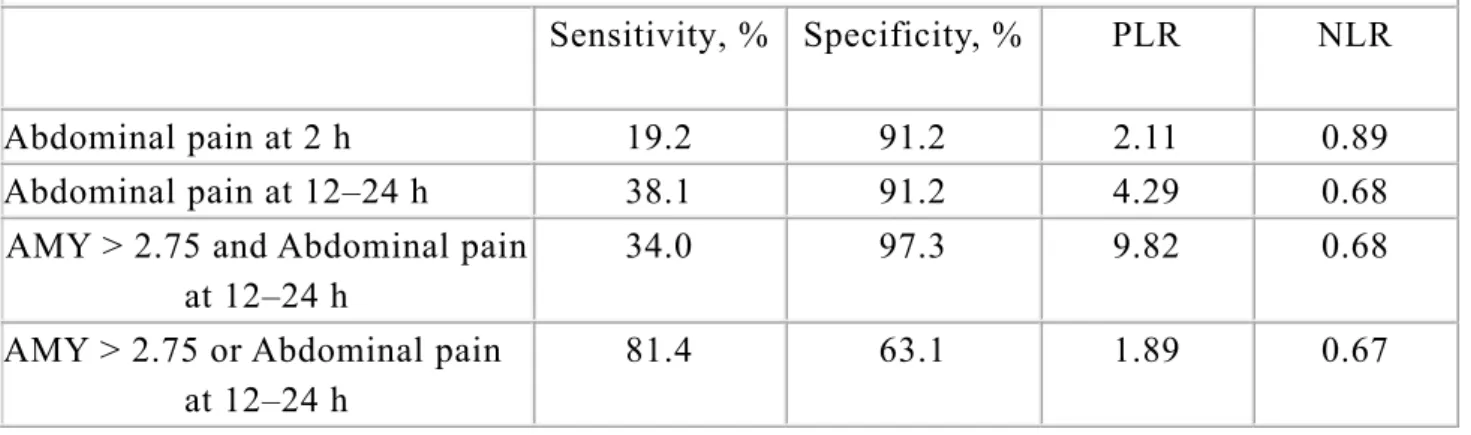
Abdominal pain under 4 h was noted in 40 of the 214 patients in the pancreatitis group (18.7%) and 12 of 136 patients in the non-pancreatitis group (8.8%) with a sensitivity of 18.7%; specificity, 91.2%; positive likelihood ratio, 2.11; and negative likelihood ratio, 0.89. Abdominal pain that persisted longer than 12 h occurred in 81 patients in the
pancreatitis group and 12 in the non-pancreatitis group with a sensitivity of 37.9%,
specificity of 91.2%, positive likelihood ratio of 3.97 and negative likelihood ratio of 0.71 (Table 3).
When we set the cut-off value of the serum AMY value on the next day judged as pancreatitis to 2.75 times at 1 day after ERCP, the sensitivity and specificity of pancreatitis in combination with AMY >2.75 and abdominal pain and AMY >2.75 or abdominal pain were 34.1%, 96.5%, 80.8% and 62.6%, respectively (Table 3).
Discussion
In this study, we analysed the correlation between serum AMY levels after ERCP and CT images using professional interpretation in a multicenter prospective study. We established a cut-off serum AMY level for the diagnosis of post-ERCP pancreatitis. With respect to abdominal pain, it was clear that the sensitivity was not sufficient although the specificity was high.
The cutoff value of serum amylase defining post-ERCP pancreatitis have remained controversial for many years. Artifon et alet reported that, based on ERCP, 41.9% of pancreatitis cases were overlooked and 30% of pancreatitis cases had AMY values that were less than three times the normal upper limit, as per Cotton’s definition alone, when lipase and CT findings were taken into consideration.11 Wang et al reported that asymptomatic hyperamylasemia (over three times the normal upper limit) was observed in 14.7% after ERCP.19 In addition, Uchino et al reported that post-ERCP pancreatitis was confirmed by detailed CT examination in 37% of patients with hyperamylasemia without abdominal pain at 18 h after ERCP.12
In this study, we set the cut-off value by performing CT for all cases where serum AMY exceeded the upper limit of the facility reference value, picking up as many cases of minimal pancreatitis as possible. The appropriate cut-off serum AMY level for judging ERCP-induced pancreatitis at 12–24 h after ERCP was 2.75 times higher than the institutional upper limit. Our data support the criteria based on the previous studies regarding serum AMY level
on the day following ERCP.
When the cut-off value is set to 2.75 times, it becomes possible to diagnose about 70% of post-ERCP pancreatitis by serum AMY alone. The severity of all pancreatitis cases overlooked by this cut-off value setting (30%) was mild; therefore, this threshold appears acceptable as a new clinical diagnostic criterion.
According to the previous reports, the frequency of post-ERCP pancreatitis is 2.6%– 9.7%.1,2,20-22 The frequency of post-ERCP pancreatitis in this study was 12.0%, which is slightly high; however, this is considered to be the result of picking up many more cases of mild pancreatitis using professional interpretation and correction prospectively.
The role of abdominal pain for the diagnosis of post-ERCP pancreatitis has also been ambiguous for many years. Our data showed that although its specificity is high, its sensitivity is low, especially for abdominal pain within 4 h. Patients may not complain of abdominal pain because of the prolonged effects of sedatives after examination or the fact that very mild pancreatitis may not be picked up in detail. Indeed, in non-ERCP-related pancreatitis, some patients do not complain of abdominal pain.23 Therefore, from the results of this study, if post-ERCP pancreatitis was diagnosed with weight on abdominal pain, many cases would be overlooked. Therefore, we should start treatment for the pancreatitis in some patients, even without abdominal pain. To increase the diagnostic accuracy of post-ERCP pancreatitis, a combination of elevated serum AMY value after 12–24 h and abdominal pain sustained for 12–24 h after examination was useful (Table 3).
The infusion volume at each facility was approximately 1500–2000 mL on the day of ERCP, and the volume after the following day depended on the physician’s decision. This suggested that the cut-off value of serum amylase defining post-ERCP pancreatitis is universal and is not affected by infusion volume.
This study had several limitations. Firstly, CT was not taken, for various reasons, in 63 (15.1%) of the 416 cases where pancreatic enzyme elevations were observed 12 to 24 h after ERCP. However, in 70% of cases, levels remained at the upper limit of the facility reference value of two times or less, and no cases reported abdominal pain. Therefore, the influence on detecting cases of pancreatitis was considered to be minimal. Secondly, the purpose of this study was to set a cut-off serum AMY value to indicate post-ERCP pancreatitis, not early detection or identification of predictive factors. The serum AMY value after 2 h had low sensitivity and specificity for pancreatitis diagnosis and is an unsuitable early predictive marker as reported in the previous reports.11,24 One of the reasons is that elevated serum AMY immediately after endoscopic examination is likely to be derived from salivary glands.25 Therefore, other pancreas-specific markers (pancreas-specific AMY, lipase, etc.) will be necessary as a predictive marker for early prediction of post-ERCP pancreatitis. We did not select pancreas-specific AMY or lipase for this study point because these were not measured at all facilities at nights or on holidays and, therefore, not considered suitable for identification of post-ERCP pancreatitis.
Serum AMY levels on the day following ERCP were highly correlated with CT-defined post-ERCP pancreatitis. The appropriate cut-off serum AMY level for judging post-ERCP-related pancreatitis is 2.75 times higher than the institutional upper limit. This newly set cut-off value of serum AMY can help diagnose ERCP-induced pancreatitis, independent of CT imaging. These results may clarify the definition of post-ERCP pancreatitis.
Conflict of interest
All authors disclosed no financial relationships relevant to this study, and there are no conflicts of interest in this study.
References
1. Kochar B, Akshintala VS, Afghani E, et al. Incidence, severity, and mortality of post-ERCP pancreatitis: a systematic review by using randomized, controlled trials.
Gastrointest Endosc. 2015;81:143–149.e9.
2. Cotton PB, Garrow DA, Gallagher J, et al. Risk factors for complications after ERCP: a multivariate analysis of 11,497 procedures over 12 years. Gastrointest Endosc.2009; 70:80–88.
3. Banks PA, Bollen TL, Dervenis C, et al. Classification of acute pancreatitis--2012: revision of the Atlanta classification and definitions by international consensus. Gut. 2013;62:102–111.
4. Malfertheiner P, Kemmer TP. Clinical picture and diagnosis of acute pancreatitis.
Hepatogastroenterology. 1991;38:97–100.
5. Dawra R, Sharif R, Phillips P, et al. Development of a new mouse model of acute pancreatitis induced by administration of L-arginine. Am J Physiol Gastrointest Liver
Physiol. 2007;292:G1009–18.
6. Agarwal N, Pitchumoni CS, Sivaprasad AV. Evaluating tests for acute pancreatitis.
Am J Gastroenterol. 1990;85:356–366.
7. Rompianesi G, Hann A, Komolafe O, et al. Serum amylase and lipase and urinary trypsinogen and amylase for diagnosis of acute pancreatitis. Cochrane Database Syst
Rev. 2017;4:CD012010.
8. Abid GH, Siriwardana HP, Holt A, et al. Mild ERCP-induced and non-ERCP-related acute pancreatitis: two distinct clinical entities? J Gastroenterol. 2007;42:146–151. 9. Testoni PA, Vailati C, Giussani A, et al. ERCP-induced and non-ERCP-induced acute
pancreatitis: Two distinct clinical entities with different outcomes in mild and severe form? Dig Liver Dis. 2010;42:567–570.
10. Cotton PB, Lehman G, Vennes J, et al. Endoscopic sphincterotomy complications and their management: an attempt at consensus. Gastrointest Endosc. 1991;37:383–393. 11. Artifon ELA, Chu A, Freeman M, et al. A comparison of the consensus and clinical
definitions of pancreatitis with a proposal to redefine post-endoscopic retrograde cholangiopancreatography pancreatitis. Pancreas. 2010;39:530–535.
computed tomography in patients with post-endoscopic retrograde
cholangiopancreatography hyperamylasemia. Pancreatology. 2014;14:17–20. 13. Balthazar EJ, Robinson DL, Megibow AJ, et al. Acute pancreatitis: value of CT in
establishing prognosis. Radiology. 1990;174:331–336.
14. Luiten EJ, Hop WC, Lange JF, et al. Controlled clinical trial of selective
decontamination for the treatment of severe acute pancreatitis. Ann Surg. 1995;222: 57–65.
15. Bollen TL, Singh VK, Maurer R, et al. A comparative evaluation of radiologic and clinical scoring systems in the early prediction of severity in acute pancreatitis. Am J
Gastroenterol. 2012;107:612–619.
16. Testoni PA, Mariani A, Giussani A, et al. Risk factors for post-ERCP pancreatitis in high- and low-volume centers and among expert and non-expert operators: a
prospective multicenter study. Am J Gastroenterol. 2010;105:1753–1761.
17. Lankisch PG, Weber-Dany B, Maisonneuve P, et al. Frequency and severity of acute pancreatitis in chronic dialysis patients. Nephrol Dial Transplant. 2008;23:1401– 1405.
18. Harshit Kumar A, Singh Griwan M. A comparison of APACHE II, BISAP, Ranson’s score and modified CTSI in predicting the severity of acute pancreatitis based on the 2012 revised Atlanta Classification. Gastroenterology Report. 2017;27:749.
19. Wang P, Li Z-S, Liu F, et al. Risk Factors for ERCP-Related Complications: A Prospective Multicenter Study. Am J Gastroenterol. 2009;104:31–40.
20. Freeman ML, Nelson DB, Sherman S, et al. Complications of endoscopic biliary sphincterotomy. N Engl J Med. 1996;335:909–918.
21. Andriulli A, Loperfido S, Napolitano G, et al. Incidence rates of post-ERCP
complications: a systematic survey of prospective studies. Am J Gastroenterol. 2007; 102:1781–1788.
22. Katsinelos P, Paroutoglou G, Kountouras J, et al. A comparative study of standard ERCP catheter and hydrophilic guide wire in the selective cannulation of the common bile duct. Endoscopy. 2008;40:302–307.
23. Read G, Braganza JM, Howat HT. Pancreatitis--a retrospective study. Gut. 1976;17: 945–952.
24. Ito K. Relationship between post-ERCP pancreatitis and the change of serum amylase level after the procedure. WJG. 2007;13:3855–3860.
25. Tsujikawa T, Bamba S, Inatomi O, et al. Factors affecting pancreatic
hyperamylasemia in patients undergoing peroral single-balloon enteroscopy. Dig
Figure legends
Figure 1. Flow diagram. There were 2078 patients who received ERCP. We excluded 289
cases (not reached the papilla in nine cases, post-biliary reconstruction in 63 cases, developed pancreatitis before ERCP in 74 cases, elevated amylase before ERCP in 120 cases and ascites or fluid collection due to biliary pancreatic cancer in 23 cases). Serum amylase level elevated in 416 of 1789 cases enrolled. Sixty-six cases were excluded (CT is not performed CT at the discretion of the physician in 63 cases, the patients refused CT examination in two cases and bile duct perforation in one case). We analysed 350 cases using CT diagnosis of two expert radiologists.
Figure 2. Receiver operating characteristic curves of serum amylase level at 2 h (A) and 12–
24 h (B) after ERCP for diagnosing post-ERCP pancreatitis. AUC: area under the curve
TABLE 1. The Clinical Characteristics of the Patients Received CT Analysis Pancreatitis (n = 214) No Pancreatitis (n = 136) P Male/female, n 101/113 66/70 0.81
Age, median (range), y 72.8 (33–106) 73.1 (41–96) 0.78 ASA, mean (SD) 1.56 (0.67) 1.68 (0.69) 0.10 Disease, n (%) CBD stone 77 (36.0) 72 (52.9) Neoplasm 80 (37.4) 27 (19.9) Others 57 (26.6) 37 (27.2) Procedure, n (%) Diagnostic 44 (20.6) 18 (13.2) Therapeutic 170 (79.4) 118 (86.8) CT grade *, n (%) A 0 136 (100) B 33 (15.4) 0 (0) C 123 (57.5) 0 (0) D 28 (13.1) 0 (0) E 30 (14.0) 0 (0)
*CT grade according to CT severity index Grade A; Normal pancreas
Grade B; Focal or diffuse enlargement of the pancreas
Grade C; Intrinsic pancreatic abnormalities associated with haziness and streaky densities representing inflammatory changes in the peripancreatic fat
Grade E; Two or more poorly defined fluid collections or presence of gas adjacent to the pancreas
TABLE 2. Sensitivity and Specificity of serum amylase at 2 and 12–24 h after ERCP for
post-ERCP pancreatitis Serum
amylase
AUC Sensitivity, % Specificity, % PLR NLR Cut-off value (times the upper limit of normal)
2 h 0.63 45.2 79.3 2.19 0.69 2.73
12–24 h 0.77 70.1 75.7 2.88 0.39 2.75
AUC: Area under the curve; PLR: Positive likelihood ratio; NLR: Negative likelihood ratio
TABLE 3. Sensitivity and Specificity of Abdominal Pain and Serum Amylase Level at 2
and 12–24 h After the Examination for Post-ERCP Pancreatitis
Sensitivity, % Specificity, % PLR NLR Abdominal pain at 2 h 19.2 91.2 2.11 0.89 Abdominal pain at 12–24 h 38.1 91.2 4.29 0.68 AMY > 2.75 and Abdominal pain
at 12–24 h
34.0 97.3 9.82 0.68
AMY > 2.75 or Abdominal pain at 12–24 h
81.4 63.1 1.89 0.67