patient‑derived ovarian cancer
organoids capture the genomic
profiles of primary tumours
applicable for drug sensitivity
and resistance testing
Yoshiko nanki
1,2, Tatsuyuki Chiyoda
1,2*, Akira Hirasawa
1,2,3*, Aki Ookubo
4, Manabu Itoh
4,
Masaru Ueno
4, Tomoko Akahane
2,5, Kaori Kameyama
6,7, Wataru Yamagami
1,
Fumio Kataoka
1,8& Daisuke Aoki
1The use of primary patient-derived organoids for drug sensitivity and resistance testing could play an important role in precision cancer medicine. We developed expandable ovarian cancer organoids in < 3 weeks; these organoids captured the characteristics of histological cancer subtypes and replicated the mutational landscape of the primary tumours. Seven pairs of organoids (3 high-grade serous, 1 clear cell, 3 endometrioid) and original tumours shared 59.5% (36.1–73.1%) of the variants identified. Copy number variations were also similar among organoids and primary tumours. The organoid that harboured the BRCA1 pathogenic variant (p.L63*) showed a higher sensitivity to PARP inhibitor, olaparib, as well as to platinum drugs compared to the other organoids, whereas an organoid derived from clear cell ovarian cancer was resistant to conventional drugs for ovarian cancer, namely platinum drugs, paclitaxel, and olaparib. The overall success rate of primary organoid culture, including those of various histological subtypes, was 80% (28/35). Our data show that patient-derived organoids are suitable physiological ex vivo cancer models that can be used to screen effective personalised ovarian cancer drugs.
Patient-derived tumour organoids have become important preclinical model systems in both cancer research and clinical settings1. In contrast to patient-derived xenograft (PDX) mouse models that need a large amount of
surgical specimen and 4–8 months for development2, organoids can be cultured from patient materials and can be
expanded with high efficiency in a relatively short period (typically < 1 month). Organoids from mouse intestine, as well as from various other mouse and human tissues, including the colon, stomach, liver, lung, prostate, and pancreas, have been established3,4. Patient-derived tumour organoids have also been generated from the colon,
pancreas, prostate, breast, gastric, lung, oesophageal, bladder, ovarian, kidney, and liver tumour tissues1.
Orga-noids maintain the key genetic and phenotypic features of primary tumours, thereby, enabling their use in a broad range of applications, such drug development and identification of the best therapeutic regimen for each patient.
Ovarian cancer is a devastating disease, with 295,000 new patients and 185,000 deaths each year, worldwide5.
The relative 5-year survival rate is 47% and has not apparently increased in the last 40 years. Debulking surgery with platinum-combination chemotherapy is usually administered to patients, irrespective of the histological
open
1Department of Obstetrics and Gynecology, Keio University School of Medicine, Tokyo, Japan. 2JSR-Keio University Medical and Chemical Innovation Center (JKiC), Keio University School of Medicine, Tokyo, Japan. 3Department of Clinical Genomic Medicine, Graduate School of Medicine, Dentistry and Pharmaceutical Sciences, Okayama University, Okayama, Japan. 4JSR-Keio University Medical and Chemical Innovation Center (JKiC), JSR Corp., Tokyo, Japan. 5Genomics Unit, Keio Cancer Center, Keio University School of Medicine, Tokyo, Japan. 6Department of Pathology, Keio University School of Medicine, Tokyo, Japan. 7Present address: Department of Pathology, Showa University Northern Yokohama Hospital, Yokohama, Japan. 8Present address: Department of Obstetrics and Gynecology, International University of Health and Welfare School of Medicine, Narita, Japan. *email: chiyoda@keio.jp; hir-aki45@umin.org
subtypes, namely high-grade serous (HGSC), endometrioid (EM), clear cell (CCC), and mucinous carcinoma. HGSC comprises 70–80% of all ovarian cancer cases and is characterised by TP53 mutation, with many chro-mosomal aberrations6. EM and CCC are endometriosis-associated ovarian cancers that frequently harbour the ARID1A mutation. Mucinous ovarian cancer is a rare tumour that accounts for 3% of all ovarian cancers and har-bours KRAS mutation, ERBB2 amplification, or TP53 mutation7. Anti-VEGF antibody, bevacizumab, and PARP
inhibitors, olaparib, rucaparib, and niraparib, are the molecular targeted drugs used in the clinical treatment of ovarian cancer. PARP inhibitors are effective against tumours with homologous recombination deficiency (HRD) and are mainly used against HGSC. Mismatch repair-deficient tumours make up less than 2% of the epithelial ovarian cancer8, and the overall response rate of single-agent immune checkpoint blockade by pembrolizumab
was reported to be 4.1% in ovarian cancer9. Therefore, there are still emergent needs in ovarian cancer
treat-ment for novel molecular targeted drugs and biomarkers for selecting the most effective therapeutic regimens. Recently, established ovarian cancer organoids that capture the genomic features of primary tumours have been reported10–12. Here, we have effectively established ovarian cancer organoids from several histologic
sub-types of ovarian cancer that can be utilised for biomedical applications, including drug sensitivity and resistance testing (DSRT).
Results
Establishment of primary ovarian cancer organoids.
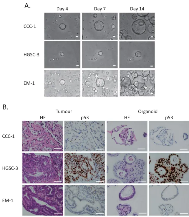
First, we established the protocol to culture and expand single cells dissociated from primary ovarian cancers. Culturing dissociated single tumour cells in Matrigel with a cocktail medium of niche factors (WNT-3A, R-Spondin, etc.) enabled us to develop ovarian can-cer organoids from different histologic subtypes (HGSC, EM, CCC) of stage I–III ovarian cancan-cer patients within 1–3 weeks (Fig. 1A, Table 1). We studied the growth of ovarian cancer organoids using over 20 combinations of various niche factor culture cocktails. Here, we present the organoids cultured with the cocktail medium most effective for multi-tissue type culture. The overall success rate of the primary organoid culture was 80% (28/35) (Table 2). The established organoids captured the histological characteristics and p53 positivity of the primary tumours (Fig. 1B).Capture of primary tumour genomic characteristics by organoids.
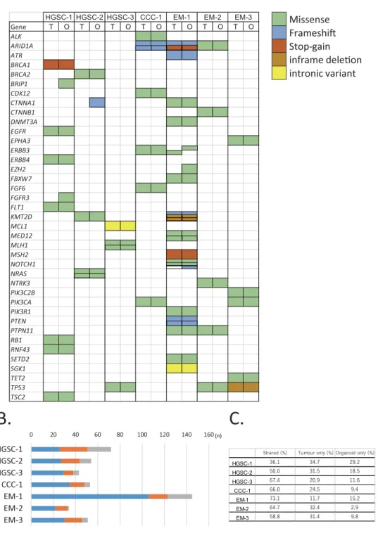
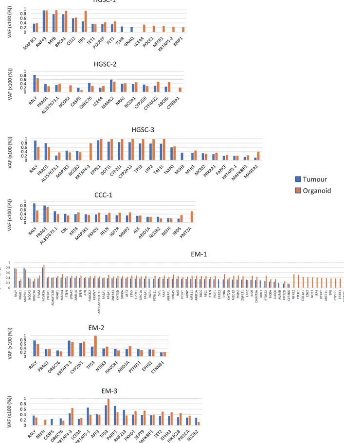
To compare the genomic characteristics of the parental tumours and derived organoids, we performed targeted capture sequencing of 1,053 cancer-related genes in the seven pairs (3 HGSC, 1 CCC, 3 EM) of organoids and primary tumours. The median passage number of organoids for analysis was 4 (range: 2–5). The analysis revealed that the pairs shared pivotal DNA variants, such as BRCA1, BRCA2, MLH1, PIK3CA, and TP53 (Fig. 2A, Supplementary Table 1). HGSC-1 harboured a stop-gain mutation in BRCA1 (p.L63*, pathogenic); CCC-1 had a frameshift mutation in ARID1A (p.P1995Lfs*22, p.Q1098Rfs*16); EM-1 had a frameshift mutation (p.K1072Nfs*21) and a stop-gain mutation (p.R1276*) in ARID1A; and EM-2 had a missense mutation in ARID1A (p.P251A) in both organoids and tumours. EM-1 also possessed a stop-gain mutation of the MSH2 gene (p.G220*). In total, 59.5% (range: 36.1–73.1%) of the variants were shared among organoids and primary tumours. A total of 26.7% (range: 11.7– 34.7%) of the variants were seen only in the tumour and 13.8% (range: 9.4–29.2%) were identified only in the organoid (Fig. 2B,C). EM-1 displayed a hypermutation pattern (145 gene variants). The variant allele frequency (VAF) was similar among organoids and primary tumours (Fig. 3, Supplementary Table 1). The VAF of 77.8% for BRCA1 variant (p.L63*) seen in the HGSC-1 tumour indicates a loss of heterozygosity (LOH) in the tumour or the presence of uniparental disomy with a germline mutation. The VAF of 92.4% for BRCA1 p.L63* in the orga-noid indicates that epithelial cells were concentrated in the orgaorga-noids. Following the results of BRCA1 variant analysis in the organoids, we performed genetic counselling of the patient. Genetic test revealed that the patient had a germline BRCA1 variant. The VAF of RB1 variant (p.E672Q) was 47.3% in HGSC-1 tumour and 92.2% in HGSC-1 organoid, which indicates that the RB1 wild type allele was lost during organoid development. HGSC-3 organoid and parental tumour both showed a ClinVar pathogenic TP53 variant (p.R248Q) with LOH. During development of the HGSC-3 organoid, a KRTAP4-3 variant was acquired with LOH in the main clone. In CCC-1, the VAF of ARID1A was 26.8% in the tumour and 27% in the organoid, which indicates that a subclone was maintained in the organoid culture. EM-1 displayed a hypermutation pattern, most of which was maintained in the organoid. EM-2 organoid gained a LOH of the TP53 variant (p.L130V), whereas the wild type allele of TP53 was identified in the tumour.Copy number variations of tumours and organoids.
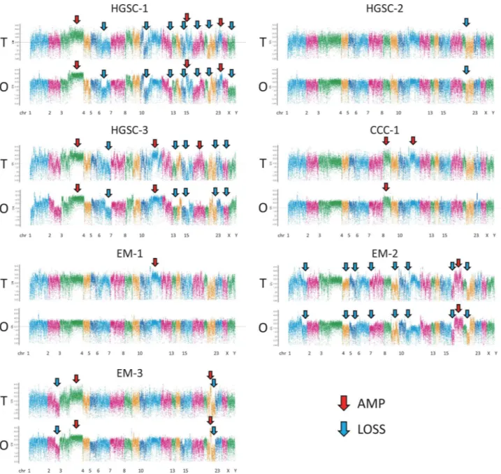
Copy number variations (CNVs) of the seven pairs of ovarian organoids and primary tumours showed a similar pattern of amplifications and losses through-out the chromosomes (Fig. 4). HGSC-1 and HGSC-3 had many amplifications and losses that may reflect HRD (HRD-like). HGSC-2 showed scarce CNVs (non-HRD like). Chromosome 8 amplification was seen in both the parental tumours and organoids of CCC-1, whereas amplification in the region of chromosome 11 was only seen in the primary tumour. Most of the changes in the copy number of chromosomes in the tumours were inherited in the organoids, and the organoids did not acquire major novel chromosomal aberrations.Organoid usability for personalised DSRT.
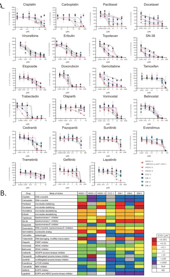
Finally, we performed DSRT using 23 FDA-approved drugs. HGSC-1 and HGSC-3 (HRD-like) displayed a similar drug sensitivity pattern that was different from that of HGSC-2 (non-HRD like) (Fig. 5A,B, Supplementary Table 2). HGSC-2 showed resistance to most of the drugs, except trabectedin. CCC-1 was resistant to platinum drugs (cisplatin and carboplatin) and paclitaxel, the key drugs in ovarian cancer therapeutics (Fig. 5A,B, Supplementary Table 2). HGSC-1 that harbours the deleterious BRCA1 variant and loss of wild type allele showed higher sensitivity to the PARP inhibitor, olaparib (p < 0.01), compared to other organoids (Fig. 5A,B). HGSC-1 also showed a tendency of higher sensitivity tocisplatin, although it was not significant (p = 0.14) (Fig. 5A,B). HGSC-1 was also sensitive to paclitaxel, docetaxel, topotecan, SN-38, gemcitabine, and trabectedin. Time interval to recurrence after completion of first-line plati-num regimen was 18 months in HGSC-1, which was longer than that in HGSC-2 (9 months), showing concord-ance with the results of DSRT (Table 1, Fig. 5A,B).
Discussion
Ovarian cancer is a heterogeneous disease that needs an appropriate tumour model for the development of novel therapeutics. We have developed patient-derived ovarian cancer organoids that capture the in vivo architecture, genetic signature, and heterogeneity of tumours. In accordance with studies carried out on ovarian cancer and other tumours, such as those of the colon, breast, and bladder, our ovarian cancer organoids retained both the histological and genetic features and intra-tumoural heterogeneity of the original tumours10,13–15. Our method
Day 4
Day 7
Day 14
CCC-1
EM-1
HGSC-3
CCC-1
d
i
o
n
a
g
r
O
r
u
o
m
u
T
HE
p53
HE
p53
HGSC-3
EM-1
A.
B.
Figure 1. Patient-derived primary ovarian cancer organoids maintain the histological architecture and
p53 positivity of the tumour subtype from which they are derived. (A) Brightfield microscopy images of the organoid lines. Scale bars 100 µm. (B) Haematoxylin and eosin (H&E) staining and p53 immunohistochemistry of primary ovarian tumours and derived organoids. It is noteworthy that organoids recapture the histologic features of the primary tumours (clear cell ovarian cancer, high-grade serous ovarian cancer, and endometrioid ovarian cancer) and p53 staining pattern. Scale bar = 100 µm.
enables the establishment of organoids from several histological subtypes of ovarian cancer, efficiently from a single cell, within 3 weeks. The established organoids recapitulated the CNVs of primary tumours. An organoid with BRCA1 variant (HGSC-1) displayed many chromosomal aberrations, which are characteristic of HGSC. A total of 59.1% of gene variants were shared among organoids and corresponding tumours in our study, which was lower than the percentage (98%) of shared variants reported previous12. The difference may be attributed
to the fact that we performed DNA sequencing at median passage number of 4 (> 4 weeks) but in the previous study, it was done after a short period (7–10 days) of culture. The pivotal DNA variants for tumourigenesis were shared among organoids and tumours in our study and the shared variants were reported to be maintained even after prolonged culture10.
In the DSRT, organoid from clear cell ovarian cancer (CCC-1) showed resistance to paclitaxel, cisplatin, and carboplatin compared to the other organoids; this was consistent with the fact that clear cell ovarian can-cer is resistant to platinum-based chemotherapy (response rate: clear cell 11.1%, serous 72.5%)16. CCC-1 has
mutations in the SWItch/Sucrose Non-Fermentable (SWI/SNF) related genes, PBRM1 (p.P1460L) and ARID1A (p.P1995Lfs*22, p.Q1098Rfs*16), indicating that immune checkpoint blockade might be an effective treatment strategy for this tumour17,18. HGSC-1 and HGSC-3 patients have HRD like CNVs, whereas HGSC-2 has limited
CNVs (Fig. 4). HGSC-1 and HGSC-3 showed sensitivity towards paclitaxel treatment; however, HGSC-2 was resistant to paclitaxel (Fig. 5A,B, Supplementary Table 2). HGSC-1 harbours a pathogenic BRCA1 variant and is sensitive to the PARP inhibitor, olaparib, and cisplatin compared to other organoids, which indicates that using organoid-based models is a reliable strategy for DSRT. In fact, HSGC1 and HGSC-2 were both from FIGO stage IIIC tumours. The disease-free period after platinum therapy was longer in 1 compared to that in HGSC-2, which can be considered as a reflection of DSRT (Table 1, Fig. 5A,B). Kopper et al. also reported that in vitro drug sensitivity was recapitulated in vivo using xenotransplantation of ovarian cancer organoids10.
As drug responses are more diverse and correlate better with genomic alterations in the 3D culture than in the 2D culture19, organoids can be considered as an appropriate culture format for drug sensitivity assays in
translational research and precision medicine. PDX of ovarian cancer also recapitulated the diversity of genomic alterations in tumours and can be used for drug testing20,21; however, organoids are considered a better 3D
culture system than PDX in terms of (1) amount of tumour needed, (2) engraftment time, and (3) engraftment success rate2.
The limitations of organoid models include the absence of cancer stroma, such as fibroblasts, blood vessels, and immune cells. Recently, however, the air–liquid interface method that retains tumour immune microen-vironment was reported22. A PDX mouse model can be efficiently created via engraftment of organoids, which Table 1. Characteristics of organoid cases. NAC neoadjuvant chemotherapy, ddTC dose-dense paclitaxel
carboplatin, Bev bevacizumab, M month, NED no evidence of disease, NA not applicable.
Case Age at diagnosis Stage NAC Debulking status Observation period Recurrence Time to recurrence after platinum therapy Status
HGSC-1 47 IIIC None Optimal 26 M Yes 18 M Alive with disease
HGSC-2 74 IIIC ddTC + Bev Optimal 26 M Yes 9 M Alive with disease
HGSC-3 77 IA None Complete 25 M No NA NED
CCC-1 50 IA None Complete 26 M No NA NED
EM-1 46 IC1 None Complete 22 M No NA NED
EM-2 42 IC2 None Complete 23 M No NA NED
EM-3 41 IIIB None Complete 22 M No NA NED
Table 2. Success rate of organoid culture and derived organoid lines from each histologic subtype of ovarian
tumour. HGSC high-grade serous, EM endometrioid, CCC clear cell, MC mucinous, MBT mucinous borderline tumour Others include dysgerminoma, thecoma, serous cystadenofibroma, carcinosarcoma, and fibroma. Organoid line was defined as an organoid that could be made from a single cell culture and for which a serial passage was successful for four times.
Number of cases Number of successful primary organoid culture Success rate of primary organoid culture (%) Number of derived organoid lines
HGSC 10 9 90 3 EM 5 3 60 3 CCC 10 10 100 9 MC 0 0 0 0 MBT 3 3 100 2 Others 7 3 43 1 Total 35 28 80 18
also enables the assessment of cancer stroma interactions. Organoid methodology allowing for cancer stroma integration is a vital next step in this field.
A.
B.
Gene T O T O T O T O T O T O T O ALK ARID1A ATR BRCA1 BRCA2 BRIP1 CDK12 CTNNA1 CTNNB1 DNMT3A EGFR EPHA3 ERBB3 ERBB4 EZH2 FBXW7 FGF6 FGFR3 FLT1 KMT2D MCL1 MED12 MLH1 MSH2 NOTCH1 NRAS NTRK3 PIK3C2B PIK3CA PIK3R1 PTEN PTPN11 RB1 RNF43 SETD2 SGK1 TET2 TP53 TSC2HGSC-1 HGSC-2 HGSC-3 CCC-1 EM-1 EM-2 EM-3
0 20 40 60 80 100 120 140 160 HGSC-1 HGSC-2 HGSC-3 CCC-1 EM-1 EM-2 EM-3
Shared Tumour only Organoid only
Missense
Stop-gain
intronic variant
C.
HGSC-1 HGSC-2 HGSC-3 CCC-1 EM-1 EM-2 EM-3 (n)Figure 2. Organoids preserve the genetic alterations of the original tumour. (A) A cancer-related set of variants
found in organoids and primary tumours (full list is shown in Supplementary Table 1). The type of mutation is indicated in the legend. Corresponding gene variant of tumour and organoid side by side in a same patient indicates a same variant. T tumour, O organoid. (B) The stacked bar graphs showing the total number of mutations per patient sample identified in both tumour and derived organoid, tumour only, and organoid only. (C) The percentage of shared, tumour only, and organoid only variants are indicated. Primary tumours and organoids share 59.5% (36.1–73.1%) of the variants.
CCC-1
HGSC-1
HGSC-3
EM-1
EM-2
HGSC-2
EM-3
Tumour
Organoid
)) %( 00 1x( FA V 0 0.2 0.4 0.6 0.81 0 0.2 0.4 0.6 0.81 0 0.2 0.4 0.6 0.81 0 0.2 0.4 0.6 0.81 0 0.2 0.4 0.6 0.81 0 0.2 0.4 0.6 0.81 )) %( 00 1x( FA V )) %( 00 1x( FA V )) %( 00 1x( FA V )) %( 00 1x( FA V )) %( 00 1x( FA V )) %( 00 1x( FA V 0 0.2 0.4 0.6 0.8 1 RA LY PR AG 1 MA P3 K1 NCOR 2 OR 6C 76 TF AM ACVR2A PIK3 R1 ADAMTS2 0 PK HD 1 MA RK 4 PT EN EP HA 2 ARID1A SPEN AT R FANCD2 FB XW 7 ARHGA P1 9-SL IT 1 RASA 1 RASA 1 ZN F8 83 KM T2 D SRPR A AFF3 ETV5 EPPK 1 SM C1 A RA SA 1 IK ZF 1 PT PN 11 SVIL PA X7 ARFRP1 EP HA 5 BA X PT PR T ADNP MRE1 1 ARID1A SB DS ABL1 PTEN PSIP 1 ER BB 3 CY BA KM T2 D MED1 2 MSH2 ZNF5 21 LR P2 IN SR DN MT 3A BR D3 PT DS S2 BI CR A CL OC K AU RK B KM T2 D CY P2 D6 NFKB 2 PT CH 1 COL1 A1 EZ H2 NOS3 IRF4 PTPR T ABCC11 TP R CY P2 F1 ER BB 3 NOTCH1 RA D5 0Figure 3. Prevalence of subclonal populations as revealed by the examination of variant allele frequency (VAF).
Genes with a VAF of 40–60% identified both in tumour and organoid were excluded from the graph as these may be germline variants. Genes with a VAF of < 20% both in tumour and organoid were also excluded.
The difference between our method and a previously reported one10 is that we did not use heregulinβ-1,
nicotinamide, forskolin, hydrocortisone, and estradiol, but used gastrin and insulin-like growth factor. Fibroblast growth factor, WNT, noggin, and R-spondin1 were the common niche factors. The primary organoid could be efficiently established with our method with a success rate of 80% (Table 2), which is similar to the success rate reported previously11.
In conclusion, the ovarian cancer organoids that we describe here recaptured the histological and genomic features of primary tumours, and thereby, present a useful platform for drug screening. By making it possible to progress from establishment to drug testing in a short time, this ovarian cancer organoid platform may serve as a pivotal experimental model, making it possible to predict drug responses before its administration to patients.
Figure 4. Copy number variation (CNV) profiles with correlations (Pearson’s r) of tumour tissue and
organoid samples in the seven cases. CNV profiles of the tumour tissue and organoids were created using a comprehensive capture-based cancer panel of 1053 genes. The pileup file was generated from processed BAM file that were used for variant call using SAMtools v. 1.2 (https ://samto ols.sourc eforg e.net/). The log-base-2 of the ratio of depth to average depth was calculated. CN was computed using the log-base-2 ratio and plotted. R script for these processes ran on R 3.4 (https ://www.R-proje ct.org). Red allow, chromosomal region with amplification; Blue allow, chromosomal region with loss. T: tumour, O: organoid.
A.
B.
(µM) (µM) (µM) (µM) Tamoxifen (µM) (µM) (µM) (µM) (µM) (µM) (µM) (µM) (µM) (µM) (µM) (µM) (µM) (µM) (µM) (µM) (µM) (µM) (µM) Paclitaxel Docetaxel n it a l p o b r a C Doxorubicin Topotecan GemcitabinePazopanib Sunitinib Everolimus SN-38
Etoposide
Belinostat Vinorelbine Eribulin
Trabectedin
Trametinib Gefitinib Lapatinib Cediranib
Vorinostat Cisplatin
Olaparib
Drug Mode of Action HGSC-1 HGSC-2 HGSC-3 CCC-1 EM-1 EM-2 EM-3 Cisplatin DNA crosslink
Carboplatin DNA crosslink Paclitaxel microtuble-stabilizing Docetaxel microtuble-stabilizing Vinorelbine microtuble-destabilizing Eribulin microtuble-destabilizing Topotecan topoisomerase inhibitor SN-38 topoisomerase inhibitor Etoposide topoisomerase inhibitor
Doxorubicin DNA crosslink, topoisomerase inhibition Gemcitabine nucleoside analog
Tamoxifen antiestrogen
Trabectedin DNA damaging, modifies transcription Olaparib PARP inhibitor
Vorinostat HDAC inhibitor Belinostat HDAC inhibitor
Cediranib VEGFR tyrosine kinases inhibitor Pazopanib multitargeted tyrosine kinase inhibitor Sunitinib multitargeted tyrosine kinase inhibitor Everolimus mTOR inhibitor
Trametinib MEK inhibitor Gefitinib EGFR inhibitor
Lapatinib EGFR and HER2 tyrosine kinase inhibitor
Figure 5. Ovarian cancer organoids as a platform for drug screening. (A) Dose–response curves of the organoid lines treated with 23
FDA-approved compounds. Dots represent the mean of the technical duplicates. Error bars represent the SEM of technical duplicates. ** < p = 0.01, N.S.: not significant (one-way ANOVA). Data analyses were performed using the GraphPad Prism 7.0b software. (B) Summary of the 23 FDA-approved compounds used in the drug sensitivity and resistance testing (DSRT) and the results. The corresponding colours for IC50 are depicted in the legend. HGSC-1 (BRCA1 pathogenic variant) showed higher sensitivity to cisplatin and olaparib compared to others. CCC-1 showed resistance to commonly used drugs for ovarian cancer; paclitaxel, carboplatin, and olaparib compared to other organoids. N = 3 distinct organoid lines. ND not determined.
Methods
Sample collection and tissue processing.
The collection of patient data and ovarian cancer tissues was performed at the Keio University Hospital with the approval of the institutional ethics committee (Approval No. 20070081). This study was performed in accordance with all relevant guidelines and regulations. All patients participating in this study signed informed consent forms. Ovarian cancer tissue was collected during surgery, and samples were stored in a sample storage solution at 4 °C during transportation to the laboratory. The sample storage solution consisted of Advanced DMEM/F12 (Thermo Fisher Scientific), 2 mM HEPES (Thermo Fisher Scientific), 1 × GlutaMAX-I (Thermo Fisher Scientific), and 200 U/mL penicillin/streptomycin (Thermo Fisher Scientific). The sample delivery time was 15–90 min. On arrival in the laboratory, the tissue samples were set on a sterile petri dish on crushed ice. Identifiable necrotic tissue and fat tissue were removed as much as possible and their size and weight were measured. An incision was made in the middle of the tissue to obtain a 5-mm slice for paraffin embedding of the primary tumour. Subsequently, the tumour was dissected to a 5 mm square under sterile conditions. A few pieces were stored at − 80 °C in 2 mL tubes with Recovery Cell culture freezing medium (Thermo Fisher Scientific) for later use. For organoid preparation, tissue samples were collected in 50 mL tubes containing HBSS 1 × (Thermo Fisher Scientific) and incubated on ice for 5 min. Thereafter, the supernatant was discarded. This wash step was repeated three times using HBSS 1 × . The cleaned tissue sample was placed on ice in a new petri dish, 100 µL of HBSS 1 × was added, and the sample was then minced into a paste. The minced tissue was collected in a new 50 mL tube and centrifuged at 300× g for 5 min at room temperature. The super-natant was discarded, pellet was gently loosened, and an enzymatic degradation solution containing HBSS 1 × , collagenase I (FUJIFILM Wako Pure Chemical Corporation), dispase II (FUJIFILM Wako Pure Chemical Cor-poration), Rock Inhibitor (FUJIFILM Wako Pure Chemical Corporation, CultureSure Y-27632), and DNase I (Roche) were added. The mixture was placed in a 50 mL tube in a water bath at 37 °C and shaken at 180–200 rpm for 30 min or up to 90 min. After the first 30 min, the dispersion status and live cells were checked every 15 min. The reaction was stopped when most of the cells had dispersed into single cells. The mixture was collected and dripped through a cell strainer on a new 50 mL tube to remove any residual tissue. The suspension was centri-fuged at 300× g for 5 min at room temperature, the supernatant was removed, and the pellet was loosened. In case of a visible red pellet, erythrocytes were lysed in ACK Lysis buffer (Thermo Fisher Scientific) for 5–10 min at room temperature followed by two wash steps with 45 mL of HBSS 1X and centrifugation at 300× g for 5 min.Organoid culture and passaging.
The concentration of the cell suspension was normalised to 1.0 × 10⁴ cells/drop and the suspension was centrifuged at 300× g for 5 min at room temperature. The cell pellet was then suspended in Matrigel (Corning), and 25 μL drops of matrix cell suspension were allowed to solidify on a pre-warmed 48-well plate at 37 °C for 15 min. We then calculated the volume to be transferred from the cell suspension into a new tube. On stabilisation of the Matrigel, we added the organoid medium cocktail. Advanced DMEM/F12 (Thermo Fisher Scientific) was supplemented with 2 mM HEPES (Thermo Fisher Scientific), 1 × GlutaMAX-I (Thermo Fisher Scientific), 1X B27 supplement (Thermo Fisher Scientific), 10 nM Leu15-Gas-trin I (Sigma-Aldrich), 1 mM N-acetylcystein (Sigma-Aldrich), 100 ng/mL recombinant human IGF-1 (R&D Systems), 50 ng/mL recombinant human FGF-2 (PeproTech), 20% Afamin/Wnt3a CM (JSR Life Sciences), 1 µg/ mL humanR-spondin (R&D Systems), 100 ng/mL Noggin (PeproTech), 500 nM A-83-01 (Tocris Bioscience), 200 U/mL penicillin/streptomycin (Thermo Fisher Scientific), and 10 µM Y-27632 (FUJIFILM Wako Pure Chemical Corporation). The medium was changed every 3–4 days, and the organoids were passaged at a 1:2–3 dilution every 1–4 weeks. Images of the organoids were taken every 3–4 days using a microscope. Organoids were mechanically and enzymatically dissociated into small clusters for passaging and collected in a 10 mL tube and sheared with a 1000 µL pipet tip without a filter. Thereafter, 100 µL of TrypLE Express (Invitrogen) was added and the mixture was incubated for 5 min at 37 °C. Subsequently, the organoids were centrifuged at 400× g for 3 min, washed with PBS, and centrifuged again briefly. All the seven organoids were successfully passaged and grown into organoids even after being isolated into single cells. For preservation of a stock, organoids in a Matrigel drop were suspended with Recovery Cell Culture Freezing Medium (Thermo Fisher Scientific), col-lected in a 2 mL tube, and then gradually frozen at − 80 °C using BICELL (Nihon Freezer).HE staining and immunohistochemistry of original tumours and organoids.
Tissues and orga-noids were processed for paraffin sectioning using standard protocols. Matrigel embedded orgaorga-noids were sus-pended in Cell Recovery Solution (Corning, 500 µL/well) and collected in a 15 mL tube. The organoid suspen-sion was occasuspen-sionally mixed with gentle pipetting for 30 min on ice to completely solubilise the Matrigel. The tube was then placed on ice to precipitate the organoids. The supernatant was removed, and organoids were washed with a small amount of cold PBS. Organoids were solidified using iPgell (NIPPON Genetics), and orga-noid blocks were fixed with 4% paraformaldehyde (PFA) for 20 min at room temperature, before embedding in paraffin. Fresh cancer tissue was embedded in paraffin after formalin fixation. After deparaffinisation, 5 μm sec-tions were stained with haematoxylin–eosin (H&E) and tumour protein p53 (DO-7, Dako). The organoids and primary tumour sections were evaluated for morphological and immunostaining similarity by the pathologist.DNA targeted analysis of original tumour and organoids.
DNA was isolated from 6–8 wells of cul-tured organoids with the QIAamp DNA Mini Kit (QIAGEN) according to the manufacturer’s instructions. Orig-inal tumour DNA was isolated from formalin-fixed, paraffin-embedded samples with the QIAamp DNA Mini Kit (QIAGEN). DNA purity and concentration were examined using the NanoDrop2000 spectrophotometer and Qubit 2.0 Fluorometer, respectively. The qualified genomic DNA sample was randomly fragmented with a Covaris ultrasonicator and adapters were ligated to both ends of the resulting fragments. The extracted DNA was then amplified via ligation-mediated PCR (LM-PCR), purified, and hybridised to the Roche NimbleGen SeqCapEZ Exome probe. Non-hybridised fragments were then washed off. Both the non-captured and captured LM-PCR products were subjected to quantitative LM-PCR to estimate the magnitude of enrichment. Target enrichment was performed with a cancer panel that targeted 1,053 cancer-related genes (Beijing Genomics Institute). Each captured library was then loaded on an Illumina Hiseq sequencing platform, and high-throughput sequencing was performed independently for each captured library to ensure that each sample met the desired average fold-coverage. Raw image files were processed with Illumina base calling Software 1.7 with default parameters and the sequences of each individual were generated as 90/100 bp paired-end reads23,24.
Genomic analysis.
After removing the adaptor reads, the clean reads were mapped to the reference genome (hg19) using Burrows–Wheeler Alignment with maximal exact matches (BWA-MEM), v. 0.7.1225. Readmap-ping was followed by indel-realignment by the assembly based realigner (ABRA), v. 0.9726. Duplicate reads were
marked for removal. SNVs and indels were called using VarScan, v. 2.4.227. The functional effect of the SNVs
and indels were predicted using SnpEff v. 4.228. CNV was detected using an in-house algorithm of Mitsubishi
Space Software. To determine the clinical actionability, all the variants were mapped to three clinical anno-tation databases, ClinVar (downloaded on 2017–02)29, COSMIC v. 8130, and CIViC (downloaded on 2016–
02)31. Common variants were detected using the following criteria: allele frequency of more than 0.01 in any
of the public database, Exome Aggregation Consortium database (ExAC) (https ://exac.broa-dinst itute .org/)32,
Human Genetic Variation Database (HGVD) (https ://www.genom e.med.kyoto -u.ac.jp/)33, and Tohoku Medical
Megabank Organization 2KJPN data (ToMMo 2KJPN) (https ://jmorp .megab ank.tohok u.ac.jp/)34. HGVD and
ToMMo 2KJPN were used as a reference for the Japanese controls.
DSRT on organoids.
Organoids were collected 4–5 days after passage and filtered through a 100 μm cell strainer to remove any large clumps. Values for each drug concentration point were averages of the values for the triplicate wells. Drugs were added 2 days after embedding. We selected 23 FDA-approved drugs including those covered by health insurance in Japan for ovarian cancer and endometrial cancer. Depending on the prop-erties of the individual drugs, the concentrations ranged from 10 μmol/L to 128 pmol/L or from 100 μmol/L to 1.28 nmol. Cell viability was assayed using CellTiter-Glo 3D (Promega) on day 6. Data analyses were performed using the GraphPad Prism 7.0b software to calculate IC5023.Data availability
The datasets generated during and/or analysed during the current study are available from the corresponding author on reasonable request.
Received: 26 March 2020; Accepted: 13 July 2020
References
1. Bleijs, M., van de Wetering, M., Clevers, H. & Drost, J. Xenograft and organoid model systems in cancer research. EMBO J. 38, e101654 (2019).
2. Hidalgo, M. et al. Patient-derived xenograft models: an emerging platform for translational cancer research. Cancer Discov. 4, 998–1013 (2014).
3. Sato, T. et al. Paneth cells constitute the niche for Lgr5 stem cells in intestinal crypts. Nature 469, 415–418 (2011).
4. Dutta, D., Heo, I. & Clevers, H. Disease modeling in stem cell-derived 3D organoid systems. Trends Mol. Med. 23, 393–410 (2017). 5. Bray, F. et al. Global cancer statistics 2018: GLOBOCAN estimates of incidence and mortality worldwide for 36 cancers in 185
countries. CA Cancer J Clin. 68, 394–424 (2018).
6. Chiyoda, T., Dozen, A., Saotome, K., Nanki, Y. & Aoki, D. Biomarkers of gynecological cancers. In Biomarkers in Cancer Therapy.
Liquid Biopsy Comes of Age (ed. Shimada, H.) (Springer, Berlin, 2019).
7. Morice, P., Gouy, S. & Leary, A. Mucinous ovarian carcinoma. N. Engl. J. Med. 380, 1256–1266 (2019).
8. Le, D. T. et al. Mismatch repair deficiency predicts response of solid tumors to PD-1 blockade. Science 357, 409–413 (2017). 9. Matulonis, U. A. et al. Antitumor activity and safety of pembrolizumab in patients with advanced recurrent ovarian cancer: results
from the phase II KEYNOTE-100 study. Ann. Oncol. 30, 1080–1087 (2019).
10. Kopper, O. et al. An organoid platform for ovarian cancer captures intra- and interpatient heterogeneity. Nat. Med. 25, 838–849 (2019).
11. Maru, Y., Tanaka, N., Itami, M. & Hippo, Y. Efficient use of patient-derived organoids as a preclinical model for gynecologic tumors.
Gynecol. Oncol. 154, 189–198 (2019).
12. Hill, S. J. et al. Prediction of DNA repair inhibitor response in short-term patient-derived ovarian cancer organoids. Cancer Discov.
11, 1404–1421 (2018).
13. van de Wetering, M. et al. Prospective derivation of a living organoid biobank of colorectal cancer patients. Cell 161, 933–945 (2015).
14. Sachs, N. et al. A living biobank of breast cancer organoids captures disease heterogeneity. Cell 172, 373-386.e310 (2018). 15. Lee, S. H. et al. Tumor evolution and drug response in patient-derived organoid models of bladder cancer. Cell 173, 515-528.e517
(2018).
16. Sugiyama, T. et al. Clinical characteristics of clear cell carcinoma of the ovary: a distinct histologic type with poor prognosis and resistance to platinum-based chemotherapy. Cancer 88, 2584–2589 (2000).
17. Miao, D. et al. Genomic correlates of response to immune checkpoint therapies in clear cell renal cell carcinoma. Science 359, 801–806 (2018).
18. Shen, J. et al. ARID1A deficiency promotes mutability and potentiates therapeutic antitumor immunity unleashed by immune checkpoint blockade. Nat. Med. 24, 556–562 (2018).
19. Jabs, J. et al. Screening drug effects in patient-derived cancer cells links organoid responses to genome alterations. Mol. Syst. Biol.
13, 955 (2017).
20. Cybulska, P. et al. A genomically characterized collection of high grade serous ovarian cancer xenografts for preclinical testing.
novel therapeutics. Clin. Cancer Res. 23, 1263–1273 (2017).
22. Neal, J. T. et al. Organoid Modeling of the Tumor Immune Microenvironment. Cell 175, 1972-1988.e1916 (2018).
23. Hou, H. et al. Discovery of targetable genetic alterations in advanced non-small cell lung cancer using a next-generation sequenc-ing-based circulating tumor DNA assay. Sci. Rep. 7, 14605 (2017).
24. Wu, X. et al. The first nationwide multicenter prevalence study of germline BRCA1 and BRCA2 mutations in chinese ovarian cancer patients. Int. J. Gynecol. Cancer. 27, 1650–1657 (2017).
25. Li, H. & Durbin, R. Fast and accurate short read alignment with Burrows-Wheeler transform. Bioinformatics 25, 1754–1760 (2009). 26. Mose, L. E., Wilkerson, M. D., Hayes, D. N., Perou, C. M. & Parker, J. S. ABRA: improved coding indel detection via assembly-based
realignment. Bioinformatics 30, 2813–2815 (2014).
27. Koboldt, D. C. et al. VarScan 2: somatic mutation and copy number alteration discovery in cancer by exome sequencing. Genome
Res. 22, 568–576 (2012).
28. Cingolani, P. et al. A program for annotating and predicting the effects of single nucleotide polymorphisms, SnpEff: SNPs in the genome of Drosophila melanogaster strain w1118; iso-2; iso-3. Fly 6, 80–92 (2012).
29. Landrum, M. J. et al. ClinVar: public archive of interpretations of clinically relevant variants. Nucleic Acids Res. 44, D862-868 (2016).
30. Tate, J. G. et al. COSMIC: the catalogue of somatic mutations in cancer. Nucleic Acids Res. 47, D941-d947 (2019).
31. Griffith, M. et al. CIViC is a community knowledgebase for expert crowdsourcing the clinical interpretation of variants in cancer.
Nat. Genet. 49, 170–174 (2017).
32. Lek, M. et al. Analysis of protein-coding genetic variation in 60,706 humans. Nature 536, 285–291 (2016).
33. Higasa, K. et al. Human genetic variation database, a reference database of genetic variations in the Japanese population. J. Hum.
Genet. 61, 547–553 (2016).
34. Tadaka, S. et al. jMorp: Japanese Multi Omics Reference Panel. Nucleic Acids Res. 46, D551-d557 (2018).
Acknowledgements
This research was supported by JSPS Bilateral program, JSPS KAKENHI Grant Numbers: 17K19611, 17K19613, 18K09298, and 19K18651. This work was also supported by JSR Corporation as a JKiC Strategic Project. The authors thank Ms. Tomomi Noda, Ms. Atsuko Fukushima for their biobank and data management, and Ms. Keiko Abe and Ms. Shihomi Yamaguchi for secretarial assistance. The authors also thank colleagues at the Department of Obstetrics and Gynecology Keio University School of Medicine for sample collection.
Author contributions
Y.N., T.C., A.H., A.O., M.I., and M.U. designed the project. Y.N., T.C., A.H., A.O., and M.I. wrote the manuscript. Y.N., T.C., W.Y., F.K., and D.A. collected the samples and clinical data. Y.N., A.O., and T.A. conducted experi-ments. K.K. performed pathological review. T.C., A.O., and M.I. did the data analysis. All authors reviewed the manuscript.
competing interests
A.O., M.I., and M.U. are employees of JSR Corporation. Y.N., T.C., A.H., and T.A. report receiving a research grant from JSR Corporation. Other authors declare no conflict of interest regarding this manuscript.
Additional information
Supplementary information is available for this paper at https ://doi.org/10.1038/s4159 8-020-69488 -9.
Correspondence and requests for materials should be addressed to T.C. or A.H. Reprints and permissions information is available at www.nature.com/reprints.
Publisher’s note Springer Nature remains neutral with regard to jurisdictional claims in published maps and
institutional affiliations.
Open Access This article is licensed under a Creative Commons Attribution 4.0 International
License, which permits use, sharing, adaptation, distribution and reproduction in any medium or format, as long as you give appropriate credit to the original author(s) and the source, provide a link to the Creative Commons license, and indicate if changes were made. The images or other third party material in this article are included in the article’s Creative Commons license, unless indicated otherwise in a credit line to the material. If material is not included in the article’s Creative Commons license and your intended use is not permitted by statutory regulation or exceeds the permitted use, you will need to obtain permission directly from the copyright holder. To view a copy of this license, visit http://creat iveco mmons .org/licen ses/by/4.0/.