Vol. 4, No. 1-4, 1971 5
Additional
Report
on the
Dichotomous
Snakes
with 20 text-figures
Toshi Niimi
Biological Laboratory, Chukyo Woman's University Recieved September 20, 1971
There are many reports on the double monster of reptiles, but there is a few reports on the dicho-tomous snake observed with X-ray, except the report by the present author, in 1965. Therefore, the present author added three more samples of the double monster snake.
I. Dichotomous Elaphe climacophora (Fig. 1-8)
The male specimen had been dead, when he was collected in Misakubo-Cho, Iwata-Gun, Shizuoka-Ken, in June 3, 1967. He was one year old.
The body length is 30.5cm, the right head 18.0mm and the left head 18.0mm. The scale counts of the both heads is shown in table 1. Ventrals 231 and subcaudals 117 pairs.
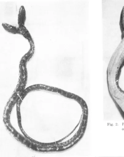
Fig. 1. Photograph of total dorsal view.
Fig. 2. Photograph of ventral view of the head and neck part.
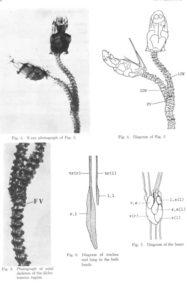
Fig. 3. X-ray photograph of Fig. 2. Fig. 4. Diagram of Fig. 3
Fig. 5. Photograph of axial skeleton of the tomous region.
Fig. 6. Diagram of trachea and lung in the both
heads.
Vol. 4, No. 1-4, 1971 7
Fig. 8. Diagram of the alimentary tracts and urogenital organs.
Table 1. Scale counts of dichotomous Elaphe climacophora
Each head had its own oesophagus and stomach, and from the distal part of oesophagus to stomach, these organs were bound together with the con-nective tissue. The intestine was held in common by the both heads.
The both heads had their own trachea and lung, but the so-called vestigial lung, which exists in the normal individuum, was not observed. The trachea and lung in the right head were normal in size and volume, but those in the left head were under-developed.
Only one heart with two ventriculus and three atriums were observed.
The 18th vertebra of the right head fused with the 14th vertebra of the left head by X-ray and anatomical observation, and so the 17th vertebra was smaller than the others.
By the request of the collector, more detail observations had not been made.
II.
Dichotomous Elaphe
climacophora
(Fig. 9-17)
The male specimen, which was collected in Minami-Chita-Cho, Chita-Gun, Aichi-ken, In October 27, 1967, was 1-2 months old and preserved in ethanol.
The body length is 33.2cm, the right head 16.0mm and the left head 15.0mm. The scale counts of the both heads, ventrals and subcaudals are shown in table 2.
The appearance of the both heads and necks was similar to Dicephalus dibrachialis in man.
Each head had its own oesophagus and stomach, and the intestine was held in common by the both heads. The respiratory organs were so, too. However, the right head had a normal lung and the so-called vestigial lung, and on the contrary, the vestigial lung was not observed in the left head.
The heart consisted of two ventriculus and four atriums.
By the X-ray and anatomical observation, the 24th vertebra of the right head fused with the 22th vertebra of the left head, and the 23rd vertebra of the right head and the 21st vertebra of the left head were very small.
More detailed observations were not made to avoid the breakage of the specimen by the request of the collector.
Fig. 9. Photograph of total dorsal view. Fig. 10. Photograph of ventral view of the cranial region
Fig. 11. Diagram of Fig. 10.
Fig. 12. X-ray photograph Fig. 10.
Fig. 13. Diagram of Fig. 12.
Fig. 14. Photogrph of axial skeleton of the
dichotomous region
Vol. 4, No. 1-4, 1971 9
Fig. 17. Diagram of the alimentary and urogenital organs
Fig. 16. Diagram of the heart.
Fig. 15. Diagram of tory organ in the
both heads.
Table 2. Scale counts of dichotomous Elaphe climacophora
III.
Double monster of Agkistrodon
halgs (Fig. 18-20)
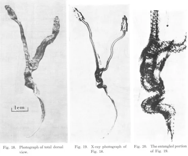
The specimem which was collected in Tsukechi-Cho, Ena-Gun, Gifu-Ken, in October 9, 1968, was 1-2 months old and preserved in ethanol. Because of the evaporation of the ethanol, the specimen was found dry and hard. So it was impossible to anatomize the detail. The sex of the specimen was unknown, too. By the X-ray observation, the vertebra column of the specimen devided at the caudal portion and entangled at the position.
Fig. 18. Photograph of total dorsal view.
Fig. 19. X-ray photograph of Fig. 18.
Fig. 20. The entangled portion of Fig. 19.
Since the famous experiments on the monozygotic
twin, diplositas anterior,
diplositas posterior
and
diplositas ant. et post. of Triton taeniatus
had been worked by Spemann (1928), many
experimental-embryological investigations on the double monster have been done.
Diplositas anterior, diplositas posterior
and diplositas ant. et post. develop by the incomplete separation of a single egg. Generally, the appearance
of the various monsters is influenced by the separated grade of a single egg, and the form and the number
of the internal organs vary with the separated grade, too.
On the deepest grade, the most part of the
body is separated, as mentioned Agkistrodon
halys.
Abbreviation
of figures
a…atrium; d…duodenum; g…gall bladder; h…heart; k…kidney; l…liver; l. a…left atrium;
l. l…left lung; o…oesophagus; p…pancreas; r…rectum; r.a…right atrium; r. l…right lung;
s…stomach; t…testis; tr…trachea; v…ventricle; v. l…vestigial lung; 10V…10th vertebra;
20V…20th vertebra; FV…fused vertebra; (1)=left; (r)=right.
Acknowledgement
The author's
most grateful appriciation
is expressed
to Professor
Hajime
Fukada, Kyoto Kyoiku
University, for his expert guidance with continued encouragement
and a good deal of important suggestion
Vol. 4, No. 1-4, 1971 11
and invaluable advice in the course of the present study. The author also wishes to express his sincerest gratitude to Professor Schuitiro Oidumi, Aoyama Gakuin University, for his constant encouragement given in the course of the present study, and for the improvement of this manuscript. The author would like to express his cordial thanks to Dr. Shigehiro Hirano, Kyoto University, for his kindness to supply the litera-tures and to Dr. Shigeru Kobayashi, Niigata University, for his suggestion and advice.
The author thanks Mr. Teruyasu Kanamori, Misakubo Primary School, and Mr. Teruo Yamaguchi, Tsukechi Junior High School, for their supplying the specimens.
References
Gederstrom, J. A., 1931: A two headed turtle. J. Heredity, Vol. 22, pp. 173-138. Cunningham B., 1927: Two-headed snakes. Sci. Monthly, Vol. 25, pp. 559-561. Gates, W. H., 1929: A two-headed snake. J. Heredity, Vol. 20, pp. 555-556.
Heasman, W. J., 1933: The anatomy of a double-headed snake. J. Anat., Vol. 67, pp. 331-345. Kuroda, N., 1919: A two-headed snake. Zool. Magazine, Vol 31, p. 255.
Kuwano, H., 1902: A two-headed tortoise. Zool. Magazine, Vol. 14, pp. 121-140.
Nakamura, K., 1938: Studies on some Specimens of Double Monster of Snakes and Tortoise. Mem. Coll. Sci. Kyoto Imp. Univ., Ser. B, Vol. 14, pp. 171-191.
Niimi, T., 1965:
On a dichotomous snake, Elaphe climacophora (Boie).
Acta Herpet. Jap., Vol. 1,
pp. 31-32.
Sato, T., 1953: Anatomical Notes on a doubl-headed snake. Collect. and Breed., Vol. 15, pp. 120.
Takara, T., 1963: Habu. Koyusha, Naha, Okinawa.
Yoshinaga, T., 1901: A two-headed snake. Zool. Magazine, Vol. 13, p. 369.