Time-course Observation of Patients with Arthrosis
of the Temporomandibular Joint on MRI
Naoki MAEDA
1), Hidehiko HOSOKI
1), Midori YOSHIDA
1), Hideki SUITO
1),
Oyunbat DASHPUNTSAG
1), Fumihiro MATSUMOTO
2), Yoshizo MATSUKA
3),
Eiji TANAKA
4), Eiichi HONDA
1)Keywords:TMJ, follow-up, morphology, displacement
Abstract:Patients with arthrosis of the temporomandibular joint are examined by MRI, but fewer cases are followed over time by performing MRI several times. The objective of this study was to investigate the presence or absence and types of changes in the structure of the mandibular condyle and articular disc on MR images over time in patients examined by MRI for the diagnosis of arthrosis of the temporomandibular joint in order to clarify the usefulness of performing MRI several times. MR images for patients who underwent MRI several times based on the clinical diagnosis of arthrosis of the temporomandibular joint were analyzed.
Twenty-three patients were examined by MRI twice or more. The shortest and longest intervals between the first and final examinations were 75 and 2,588 days, respectively, and the mean was 735 days. Changes in the bone structure of the mandibular condyle were noted on the first MRI in approximately 63 % and new changes were noted on the final examination in approximately 24 %. In addition, the articular disc position in the closed or open mouth had changed on the later examination in approximately 15 %.
Performing MRI several times along the clinical course was found to be useful to diagnose the disease stage, judge the treatment effects, and help predict the outcome in patients with arthrosis of the temporomandibular joint.
Original Article
1)Department of Oral and Maxillofacial Radiology, Institute of biomedical sciences, Tokushima University Graduate School 2)Department of Oral Medicine, Institute of biomedical sciences, Tokushima University Graduate School
3)Department of Stomatgnathic Function and Occlusal Reconstruction, Institute of biomedical sciences, Tokushima University
Graduate School
INTRODUCTION
Patients with arthrosis of the temporomandibular joint complain of pain of the joint, and noise and disturbance upon joint movement. Imaging is performed to objectively diagnose the causes of these symptoms, and plain radiography1, 2),
contrast radiography3), tomographic radiography, CT4), and
MRI5) are used. At present, MRI is considered useful to
diagnose disorders of the temporomandibular joint including the articular disc5).
The position and morphology of the articular disc and findings related to joint effusion on MRI have been reported, and the association between the MRI and clinical findings has been investigated1, 6-16); however, few patients are examined
by MRI several times during the clinical course. Accordingly, the diagnosis was made based on the findings of the first MRI in most previous reports on patients with temporomandibular joint disorder, and patients examined by MRI several times over a prolonged time have been analyzed in only a few studies. Furthermore, there are no reports analyzing bone structure changes in the temporomandibular joint and shape and position of the articular disc in the closed and opened mouth position.
The objective of this study was to investigate the presence or absence and types of changes in the structure of the mandibular condyle and articular disc in MR images over time in patients diagnosed with arthrosis of the temporomandibular joint to clarify the usefulness of performing MRI several times.
MATERIALS AND METHODS
MR images of patients who visited the outpatient clinic of Tokushima University Hospital between April 2004 and December 2014 (10 years and 9 months) for temporomandibular joint pain, trismus, or joint noise during jaw movement, who were clinically diagnosed with arthrosis of the temporomandibular joint, and who were examined by MRI several times were analyzed.
1) Acquisition method
For MRI, the 1.5-Tesla MR apparatus (Signa EXCITE X1, GE Healthcare Japan, Tokyo) was used with surface coils for the bilateral temporomandibular joints. Using horizontal cross-sectional MR images of the head, cross-sections in the directions vertical to (sagittal section) and parallel (coronal section) with the major axis of the mandibular condyle were set. The acquisition conditions were: T2-weighted imaging 3,500/81 (TR/TE); proton density-weighted imaging, 1,500/22 (TR/TE); FOV, 120 × 120 mm; matrix, 256 × 256; slice thickness, 3 mm, 7 slices for each side. The single acquisition times were 2 minutes and 13 seconds and 1 minute and 39
seconds, respectively. The sagittal view was acquired in the closed and open mouth positions, and the coronal view was acquired only in the closed mouth position. For acquisition in the open mouth position, a 9-mm-thick foam polyethylene bite piece for retention was placed between the upper and lower incisors depending on the size of the mouth.
2) Imaging diagnosis and analysis
MR imaging diagnoses were made by 2 specialized physicians certified by both the Japanese Society for Temporomandibular Joint and Japanese Society for Oral and Maxillofacial Radiology. When the diagnosis for each examination item was inconsistent between the 2 physicians, it was discussed and judged. When the opening degree was different among multiple examinations, slice images with the best matching surrounding anatomical structures were selected and compared.
Changes in the bone structure of the mandibular condyle, positions of the articular disc when the mouth was closed and open, and the presence or absence of morphological changes were classified as follows:
Time-course changes in the bone structure of the mandibular condyle were classified into 4 groups. Bone structure abnormalities were judged to be erosion, flattening, concavity, osteophyte, or eburnation.
Group 1: No abnormality was noted in the bone structure on any MRI examination.
Group 2: No abnormality was noted in the bone structure on the first MRI examination, but an abnormality was detected on the later MRI examination.
Group 3: An abnormality was noted on all MRI examinations, but no marked difference was noted between the examinations. Group 4: An abnormality in the bone structure noted on the first MRI examination had markedly changed on the later MRI examination.
The articular disc position was classified into 3 groups based on the classification previously reported by us: anterior position, slightly anterior position, and normal position15). The
anterior position means that the articular disc was displaced forward, and not located between the mandibular condyle and temporal bone, whereas the slightly anterior position means that the articular disc was displaced forward, but a part of the disc was present between the mandibular condyle and temporal bone. The disc morphology was classified based on the biconcavity, even thickness, and enlargement of the posterior band referring to previous reports6-8, 14), and
morphologies not fitting any classification were regarded as others.
RESULTS
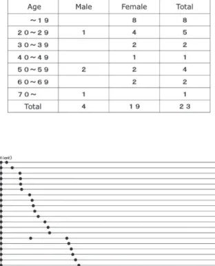
Of 896 patients with arthrosis of the temporomandibular joint examined by MRI in the 10-year-and-9-month period, MRI was performed twice or more for 23 patients (46 joints) comprising 4 males and 19 females aged 13-75 years (mean: 34 years old) at the time of the first examination. This is equivalent to 2.6 % of the total. The shortest and longest intervals between the first and final examinations were 75 days (approximately 2.5 months) and 2,588 days (approximately 7 years and 1 month), respectively, and the mean was 735 days (approximately 2 years).
The distribution of the sex and age of the patients is shown in Table 1. There were many females younger than 20 years old.
The interval between MRI examinations for each patient is shown in Figure 1. The time to the second and later examinations from the first examination is presented. The time to the second and later examinations was not constant and it ranged from 1 to 3 years in many cases.
Regarding time-course changes in the mandibular condylar bone structure in the 23 patients, 17 (37.0 %), 0, 18 (39.1 %), and 11 (23.9 %) joints were classified into Groups, 1, 2, 3, and 4, respectively, i.e., the bone structure changed with time in 11 of all joints. Example images of the presence or absence of bone structure change between MRI examinations are shown in Figure 2.
The distribution of the acquisition interval in each group is shown in Table 2. No change in the bone structure was noted for more than 2 years in some joints, but changes were noted between examinations with an interval of 6 months to one year in others.
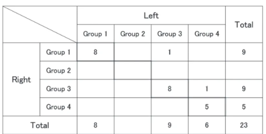
The relationship of changes in the bone structure between the bilateral mandibular condyles is shown in Table 3. Both mandibular condyles were included in the same group in 21 of the 23 patients. The abnormal bone structure noted on the first examination had newly changed on both sides on the later examination in 5 patients.
The articular disc positions in the closed mouth are shown in Table 4. The position differed between the first and final examinations in 2 cases: the position changed from slightly anterior to anterior in one and from normal to anterior in the other. The inspection intervals for these two patients were 1 year and 2 months and 2 years and 10 months. No marked change was noted in other patients.
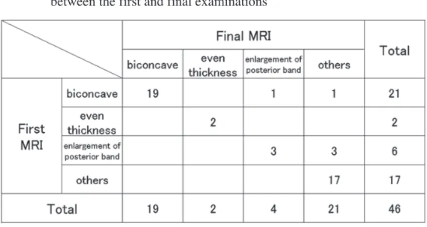
The articular disc morphologies in the closed mouth position are shown in Table 5. Of the biconcave discs on the first examination, one changed to enlargement of the posterior band and another changed to others. Of the 6 joints with enlargement of the posterior band on the first examination, half (3 joints) changed to others. Classification
Table 1 Distribution of the sex and age of the patients
Fig. 1 Interval between MRI examinations. The vertical axis shows each patient and the horizontal axis represents the period. Time to the second and later examinations is presented as circles setting the baseline as the time of the first examination.
Fig. 2 Example of bone structure change. The first MRI and the final MRI of the same case are shown. There is no change in the bone structure between the two examinations on the left side case. In the case on the right side, a significant degenerative change is observed in the condylar head.
of morphologies included in others was difficult. The intervals for the 5 patients who had changes ranged from 9 months to 7 years.
The articular disc positions in the open mouth are shown in Table 6. The position was different from that on the first examination in 7 joints, and it was displaced forward,
excluding one patient. The examination interval of the 7 patients who had changes was 1 year and 2 months to 2 years and 10 months.
The classifications of the articular disc morphology in the open mouth position are shown in Table 7. Approximately 20 % of the biconcave discs on the first examination changed Table 2 The presence or absence of time-course changes in the bone structure
of the mandibular condyle and interval between MRI examinations
Table 3 Relationship between the bilateral mandibular condyles in bone structure changes
Table 4 Articular disc position in the closed mouth: Comparison between the first and final examinations
to others. The examination interval of the six patients with changes ranged from 1 year and 2 months to 2 years and 6 months.
DISCUSSION
Many patients with arthrosis of the temporomandibular joint
are treated with splint therapy, drug therapy, and conservative treatment such as improvement of lifestyle. Some patients respond to treatment and symptoms improve within a short time, but aggravation and remission of symptoms may repeat for a prolonged time in others. When symptoms disappear or alleviate, many patients stop visiting the hospital before being Table 5 Articular disc morphology in the closed mouth position: Comparison
between the first and final examinations
Table 6 Articular disc position in the open mouth: Comparison between the first and final examinations
Table 7 Articular disc morphology in the open mouth position: Comparison between the first and final examinations
diagnosed by attending physicians. Including such patients, MRI examination is rarely repeated several times following the treatment course for patients with arthrosis of the temporomandibular joint, and MRI was repeated for only 23 (2.6 %) of 896 patients in the present study. Furthermore, the Tokushima University Hospital has a limited number of MRI examinations that can be carried out in one day, and the timing to perform the second inspection may be missed because the reservation requires days to confirm. To clarify the usefulness of performing MRI examination several times, we evaluated the images from patients examined by MRI two times or more during the treatment course.
The shortest and longest intervals between the first and final examinations for the 23 patients were 2.5 months and approximately 7 years, respectively. The intervals were not constant and there may have been many causes other than judgment of the treatment effects such as aggravation of symptoms and restart of treatment. It is clinically difficult to keep the examination interval constant. The mean interval was approximately 2 years, which may be slightly long for observation of changes in the bone structure in the temporomandibular joint and confirmation of the articular disc condition.
Of the pathologies of arthrosis of the temporomandibular joint, those that can be diagnosed by MRI are arthrosis deformans of the temporomandibular joint (type IV) and temporomandibular joint disc disorder (type III) in the pathological classification (2013 edition) established by the Japanese Society for Temporomandibular Joint. In the present study, the presence or absence of mandibular condylar deformity at the time of examination and changes between examinations were investigated in patients with arthrosis deformans of the temporomandibular joint, and the articular disc position and morphology in the closed and open mouth positions and changes between examinations were examined in patients with temporomandibular joint disc disorder. In patients in whom no abnormality was noted in the mandibular condylar bone structure on the first examination, no change was noted in the later examination. Although the patients and modality are different, the results are similar with those reported by Hosoki et al.16). It is thought that those
with no change in bone structure on the initial examination are less likely to exhibit changes afterwards. Of the patients in whom an abnormality was noted on the first examination, the abnormality had progressed in approximately 38 % on the later examination, suggesting that performing MRI several times can aid in judging whether the structure and morphology of the mandibular condyle are stable or will change.
Regarding the articular disc position in the closed mouth, the position changed from normal to anterior and from
slightly anterior to anterior in one each of the 46 joints. An abnormal articular disc position on the first examination did not change to normal in any patient. The articular disc position in the open mouth changed from that in the first examination in 7 joints. One of these recovered to the normal position, but aggravation to the anterior position was noted in the other 6 joints, and this may have been because the duration of treatment was originally long in patients examined several times and the clinical symptoms may not have been resolved or may have aggravated in many of these patients. Therefore, the patients in this study may have tended to have an unfavorable course. However, as reported by Badel et al.17) and Kircos et al.18), articular disc displacement without
symptoms in the temporomandibular joint is not rare. Badel et al. reported that an abnormal articular disc position was noted in approximately 20 % of asymptomatic subjects. Therefore, it cannot be concluded whether the abnormal articular disc position is directly related with changes in clinical symptoms. Regarding the articular disc morphology in the closed mouth position, the normal biconcave morphology changed to other morphologies in 2 joints. In the open mouth position, normal morphology became abnormal in 5 joints. These could be predicted to some extent from the presence or absence of abnormal articular disc position, but progression of the disease stage was confirmed in the images acquired during the examinations.
Of the 23 patients, the shortest examination interval demonstrating bone structure change was 9 months, and the shortest examination interval to observe a change in the joint disc shape was also 9 months. Thus, it will likely take time for the change to appear in the bone structure and the joint disc shape; however, according to our results, the change can occur even in 9 months. Therefore, performing MRI over time may become an index.
In this study, we were unable to carry out statistical analyses based on the clinical course including examination interval, the change in symptoms during that interval, and the treatment method because the number of patients considered was very small. The condition is likely to be stable for joints with no change between 2 separate examinations, whereas joints with a change are likely to continuously change. Thus, not only the patient condition but also the future condition can be predicted by performing examinations several times during the treatment course. In the future, diagnostic criteria for staging and prognostic predictions that correlate changes in clinical symptoms are desired.
CONCLUSION
The presence or absence of changes in the bone structure between multiple MRI examinations was judged based on the
size, contour, and internal signal intensity of the mandibular condyle. Changes were noted in 11 (23.9 %) of the 46 joints. The articular disc position changed in the closed or open mouth in 7 joints (15.2 %), and the mandibular condylar bone structure had changed on the later examination in approximately 24 %. New changes were noted on the final examination in approximately 38 % of the joints in which abnormal bone structure was noted on the first examination, and the articular disc position in the closed or open mouth had changed on the later examination in approximately 15 % of the joints.
Performing MRI examination several times was suggested to predict the stage of disease and prognosis at diagnostic imaging of patients with temporomandibular joint disease. This research was conducted with the approval of the Ethics Committee of Tokushima University. (No.2282)
CONFLICT OF INTEREST : The authors have no conflict of interest to declare.
ACKNOWLEDGEMENT
We are grateful to the technologists in charge of MRI in the Radiology Department of Tokushima University Hospital.
REFERENCES
1) Toller P A: The transpharyngeal radiography of arthritis of the mandibular condyle. Brit J Oral Surg 7, 47-54 (1969)
2) Mongini F: The importance of radiography in the diagnosis of TMJ dysfunctions; A comparative evaluation of transcranial radiographs and serial tomography. J prosthet Dent 45, 186-198 (1981)
3) Heffez L and Jordan S: A classification of temporomandibular joint disc morphology. Oral Surg Oral Med Oral Pathol 67, 11-19 (1989)
4) Thompson J R, Christiansen E, Hasso A N and Hinshaw D B Jr: Temporomandibular joints: high-resolution computed tomographic evaluation. Radiology 150, 105-110 (1984)
5) Tasaki M M and Westesson P L: Temporomandibular joint: diagnostic accuracy with sagittal and coronal MR imaging. Radiology 186, 723-729 (1993)
6) Milano V, Desiate A, Bellino R and Garofalo T: Magnetic resonance imaging of temporomandibular disorders: classification, prevalence and interpretation of disc displacement and deformation. Dentomaxillofac Radiol 29, 352-361 (2000)
7) Incesu L, Ta¸skaya-Yılmaz N, Ögütcen-Toller M and Uzun E: Relationship of condylar position to disc
position and morphology. Eur J Radiol 51, 269-273 (2004)
8) Orsini M G, Yatani H, Kuboki T and Yamashita A: Relationship between temporomandibular joint disc position and configuration on magnetic resonance imaging. Oral radiol 12, 39-47 (1996)
9) Müller-Leisse C, Augthun M, Bauer W, Roth A and Günther R: Anterior disc displacement without reduction in the temporomandibular joint: MRI and associated clinical findings. J Magn Reson Imaging 6, 769-774 (1996)
10) Westesson P L and Brooks S L: Temporomandibular joint: relationship between MR evidence of effusion and the presence of pain and disk displacement. Am J Roentgenol 159, 559-563 (1992)
11) Tasaki M M, Westesson P L, Isberg A M, Ren Y F and Tallents R H: Classification and prevalence of temporomandibular joint disk displacement in patients and symptom-free volunteers. Am J Orthod Dentfac Orthop 109, 249-262 (1996)
12) Ogura I: Magnetic resonance imaging characteristics of temporomandibular joint pain during opening and biting in patients with disc displacement. Oral Surg Oral Med Oral Pathol Oral Radiol Endod 102, 669-672 (2006) 13) Foucart J M, Carpentier P, Pajoni D, Marguelles-Bonnet
R and Pharaboz C: MR of 732 TMJs: anterior, rotational, partial and sideways disc displacements. Eur J Radiol 28, 86-94 (1998)
14) Maeda N: Analysis of articular disc morphology with closed and open mouth positions by magnetic resonance imaging in patients with temporomandibular disorders. Shikoku dent.res 27, 11-25 (2014) (in Japanese)
15) Maeda N, Kawaguchi S, Kudo T, Hosoki H, Uemura S: The position of the condyle and the disc in temporomandibular joint dysfunction; comparison between tomography and magnetic resonance imaging. Dental Radiology 39, 1-7 (1999) (in Japanese)
16) Hosoki H. Takagi Y. Iwasaki H. Uemura S. A radiological follow-up study on the patients with temporomandibular joint (TMJ) arthrosis. J. Jpn Soc T.M.J. 2, 48-58 (1990) (in Japanese)
17) Badel T1, Pandurić J, Marotti M, Kern J, Krolo I: Metric analysis of temporomandibular joint in asymptomatic persons by magnetic resonance imaging. Acta Med Croatica. 62, 455-60 (2008)
18) Kircos LT1, Ortendahl DA, Mark AS, Arakawa M: Magnetic resonance imaging of the TMJ disc in asymptomatic volunteers. J Oral Maxillofac Surg. 45, 852-854 (1987)