Abstract [Background] Pulmonary disease caused by nontuberculous mycobacteria has a variable clinical course. Although this is possibly the result of not only host factors, but also bacterial factors, many questions remain to be answered regarding these manifestations. [Methods] To assess the relationship between the progression of pulmonary Mycobacterium avium disease and bacterial factors, we performed variable number tandem repeats (VNTR) typing analysis of M.avium tandem repeats (MATR) in M.avium isolates from 46 patients with different clinical courses, and furthermore, examined the association between disease progres-sion and a pMAH135 plasmid derived from M.avium. [Results] In patients whose treatment was initiated because of worsened chest radiograph findings and/or clinical symptoms within 18 months after being diag-nosed with pulmonary M.avium disease, the detection rate of 6 genes located in pMAH135 was 35.3_47.1% for 17 isolates. However, in untreated patients with a stable condition, these rates were 10.3_13.8% in 29 isolates. MATR-VNTR typing analysis showed that isolates from patients with worsened disease and those with stable disease are clustered differently. In cluster III, the number of isolates from patients with wors-ened disease was higher than that from patients with stable disease (p=0.019), and furthermore, the num-ber of isolates carrying pMAH135 genes was higher than that not carrying pMAH135 genes (p≦0.001). [Conclusion] These results indicate an association between the progression of pulmonary M.avium disease and pMAH135. The presence of pMAH135 genes might be a useful prognostic indicator for pulmonary
M.avium disease and may serve as one criterion for treatment initiation.
Key words : Mycobacterium avium subsp. hominissuis, Disease progression, Variable number tandem repeats, pMAH135 plasmid
1Department of Microbiology, Faculty of Pharmacy, Meijo Univer-sity;2Department of Pharmacy, National Hospital Organization Toyohashi Medical Center; Departments of 3Respiratory Medicine and 4Clinical Research, National Hospital Organization Higashi Nagoya National Hospital
Correspondence to : Kei-ichi Uchiya, Department of Microbiology, Faculty of Pharmacy, Meijo University, 150 Yagotoyama, Tempaku-ku, Nagoya-shi, Aichi 468_8503 Japan.
(E-mail: kuchiya@meijo-u.ac.jp)
(Received 2 Sep. 2015 / Accepted 26 Oct. 2015) −−−−−−−−Original Article−−−−−−−−
ASSOCIATION BETWEEN A pMAH135 PLASMID
AND THE PROGRESSION OF PULMONARY DISEASE
CAUSED BY MYCOBACTERIUM AVIUM
1, 2
Makoto MORIYAMA,
3, 4Kenji OGAWA,
3, 4Taku NAKAGAWA,
1
Toshiaki NIKAI, and
1Kei-ichi UCHIYA
Introduction
Nontuberculous mycobacteria (NTM) infection is thought to be caused by NTM residing in the environment, including residential soil or bathrooms1) 2), and the prevalence of NTM disease is increasing worldwide. In Japan, the prevalence per 100,000 population has increased greatly from 0.82 in 1971 to 5.9 in 20013). Today, the rate is estimated to be around 15.0, which is substantially higher than the rates seen in the United States and Europe4)_6).
Pulmonary disease caused by NTM is divided into two major disease types, the nodular bronchiectatic type and the fibrocavitary type, with the former being more prevalent7)_9). However, the mechanisms involved in the development and exacerbation of pulmonary NTM disease have yet to be elucidated. Although some patients remain stable without treatment, others have deteriorating symptoms despite drug
therapy, demonstrating the disease’s diverse clinical course8)9). Therefore, it would be helpful from the viewpoint of treat-ment if the clinical course of pulmonary NTM disease could be predicted. The establishment of pulmonary NTM disease or the variable clinical course involves bacterial factors10) in addition to host-related risk factors including decreases in the levels of estrogen11), a major female sex hormone, or the presence of polymorphisms in NRAMP1 (encoding natural resistance-associated macrophage protein 1)12).
With regard to mycobacterial plasmids, previous studies isolated pVT213) and pLR714) from M.avium. Given their relatively small size of 4.8_16 kb and their lack of virulence genes, the significance of these plasmids is currently unknown. Our previous study has shown the presence of a circular plasmid derived from M.avium strain TH135, designated pMAH13515). This novel plasmid codes genes that are asso-ciated with pathogenicity of mycobacteria and its resistance
Polymerase chain reaction and sequence analysis
The presence of pMAH135 genes in M.avium isolates was determined by using specific polymerase chain reaction (PCR) primers, as described previously15). The resulting PCR products were purified using a GenElute PCR DNA purifica-tion kit (Sigma-Aldrich, St. Louis, MO), and direct sequencing analysis was performed using the same primers as those used for PCR. The resulting nucleotide sequences were compared with the sequence data of pMAH135. The suitability of the present DNA samples for screening clinical isolates by PCR was determined by amplification of the hsp65 gene―the gene used to identify subspecies of M.avium isolates.
Molecular typing
Variable number tandem repeats (VNTR) typing analysis using M.avium tandem repeats (MATR) was carried out using 15 VNTR loci (MATR-1 to MATR-16, except for MATR-10) and the corresponding primer set as described previously19). After the number of base pairs in the target VNTR loci was estimated by agarose gel electrophoresis in relation to molec-ular weight markers, the number of repetitions of various VNTR loci in each strain was determined and regarded as an allele profile. The Manhattan distance was determined on the basis of each obtained allele profile, and the genotypic diversity of M.avium isolates was analyzed with a Fitch-Margoliash algorithm using the PHYLIP software (version 3.68). The phylogenetic distribution was generated accord-ing to the genotypic diversity of isolates, usaccord-ing the FigTree software (version 1.3.1).
Statistical analysis
Fisher’s exact test was used for categorical variables and Student’s t-test was used for continuous variables. The genetic distances estimated from the Manhattan distance matrix data of M.avium isolates were analyzed using a Mann-Whitney comparison test described previously20). All statistical anal-ysis was performed using the GraphPad Prism version 5.0 (GraphPad Software, San Diego, CA). P values <0.05 were considered significant.
Results Characterization of subjects
Of those who had been diagnosed with pulmonary M.avium disease compatible with ATS/IDSA diagnostic criteria, pa-tients who had few microbiological findings in their sputum specimens or bronchial washes and patients with hardly any subjective symptoms or mild clinical or pulmonary symptoms did not undergo treatment immediately, but were followed up for 18 months with regular checkups. The patients were subsequently divided into two groups: a deteriorated disease group with 17 patients who had worsened chest radiograph findings, clinical symptoms, and/or microbiological findings during the observation period, and a stable disease group with 29 patients who did not have disease exacerbation.
to antimicrobial agents.
In this study, we examined an association between the pMAH135 plasmid and disease progression in a group of patients with different symptoms of pulmonary disease.
Material and Methods Subjects
We have performed this study using M.avium isolates from patients who did not undergo treatment immediately after diagnosis with pulmonary M.avium disease in strains used in a previous study16). These isolates comprised 46 clinical isolates provided by nine National Hospital Organization hospitals throughout Japan: 5 isolates from Asahikawa Med-ical Center, 3 from Nishi-Niigata Chuo National Hospital, 15 from Ibarakihigashi National Hospital, 6 from Tenryu Hospital, 3 from Kinki-chuo Chest Medical Center, 8 from Toneyama National Hospital, 1 from Nishibeppu National Hospital, 3 from Fukuoka Higashi Medical Center, and 2 from Minami Kyushu National Hospital. M.avium subsp.
hominissuis strain TH135 was isolated from the sputum of a patient with pulmonary M.avium disease at Higashi Nagoya National Hospital of the National Hospital Organization. Of those who had been diagnosed with pulmonary M.avium disease compatible with the diagnostic criteria of the Ame-rican Thoracic Society and the Infectious Diseases Society of America (ATS/IDSA)17) between July 2008 and September 2009, patients with few microbiological findings in their sputum specimens or bronchial washes and patients with hardly any subjective symptoms or mild clinical or pulmo-nary symptoms were followed-up regularly for 18 months. Treatment was initiated if conditions worsened in relation to those at diagnosis during the follow-up period, as judged by exacerbation of chest radiograph findings (including chest computed tomographic images), clinical symptoms and/or microbiological findings, and such patients were categorized into the deteriorated disease group (n=17). Patients in a stable condition were categorized into the stable disease group (n=29). M.avium isolates were obtained from the sputa of patients in both groups. Only one strain from each patient was analyzed in this study.
This study was approved by the Ethics Review Committee for Human Research of the Higashi Nagoya National Hospital, and written informed consent was obtained from all patients. Identifi cation of subspecies of M.avium, growth condition, and DNA isolation
The subspecies of M.avium clinical isolates was identified as M.avium subsp. hominissuis by sequence analysis of the 3´ fragment of the hsp65 gene18). The organism was grown in Middlebrook 7H9 liquid medium supplemented with 10 % oleic acid/albumin/dextrose/catalase enrichment (Difco Laboratories, Detroit, MI) at 37゜C. DNA was extracted using InstaGene Matrix (Bio-Rad Laboratories, Hercules, CA) according to the manufacturer’s instructions.
Table 2 Detection rates of pMAH135 genes in M.avium isolates from deteriorated and stable pulmonary disease patients
Locus tag Deteriorated disease patientsa (n=17) Stable disease patientsb (n=29) P value MAH_p01 MAH_p47 MAH_p49 MAH_p59 MAH_p85 MAH_p143 7 (41.2%) 8 (47.1%) 8 (47.1%) 6 (35.3%) 6 (35.3%) 8 (47.1%) 4 (13.8%) 4 (13.8%) 4 (13.8%) 3 (10.3%) 3 (10.3%) 4 (13.8%) 0.07 0.019 0.019 0.058 0.058 0.019 aStrains from the sputa of patients with deteriorated pulmonary disease
bStrains from the sputa of patients with stable pulmonary disease
Table 1 Demographic and clinical characteristics of patient groups Characteristic Deteriorated disease
group (n=17) Stable disease group (n=29) P value Age, years Mean±SD Range Sex, n (%) Male Female Underlying disease, n (%) Type of disease, n (%) Fibrocavitary disease Nodular bronchiectatic disease Solitary lung nodule
Other 65.4±11.8 44_84 7 (41.2) 10 (58.8) 7 (41.2) 7 (41.2) 9 (52.9) 0 ( 0) 1 ( 5.9) 68.1±12.9 34_84 9 (31.0) 20 (69.0) 15 (51.7) 10 (34.5) 17 (58.6) 1 ( 3.4) 1 ( 3.4) 0.48 0.53 0.55 0.76 0.77 1.0 1.0 SD, standard deviation
Age, sex, and clinical characteristics of the subjects are summarized in Table 1. The deteriorated disease group was composed of 7 men and 10 women (mean age 65.4±11.8 years). The stable disease group was composed of 29 persons who were not significantly different from the deteriorated disease group with respect to age (68.1±12.9 years, p=0.48), sex (9 men and 20 women, p=0.53), or proportion with underlying disease (deteriorated group, 41.2%; stable group, 51.7%; p=0.55). In addition, although more subjects had nodular bronchiectatic disease than fibrocavitary disease, there were no significant differences in the radiographic type of disease between the deteriorated disease and stable disease groups (fibrocavitary disease, 7 vs. 10, respectively, p=0.76; nodular bronchiectatic disease, 9 vs. 17, respectively, p=0.77).
Screening of clinical isolates for genes located in pMAH135 In the previous study, the complete genome sequence of
M. avium strain TH135 was determined21). Furthermore, we have demonstrated the presence of a circular plasmid, desig-nated pMAH13515). This plasmid consists of 194,711 nucle-otides and 164 coding sequences (CDSs). Of the pMAH135 CDSs, attention must be paid to those that encode proteins involved in mycobactin biosynthesis22)23) and the type VII secretion system24)_26), both of which are important to mycobacterial virulence as well as to proteins with putative
conserved domains of the multidrug efflux transporter27). To examine the relationship between the progression of pulmonary M.avium disease and pMAH135, we screened 46 clinical isolates provided by nine National Hospital Organ-ization hospitals throughout Japan for 6 CDSs (MAH_p01, MAH_p47, MAH_p49, MAH_p59, MAH_p85, and MAH_ p143) located in pMAH13515). MAH_p01 encodes the repA gene, which is thought to be the replication origin of pMAH 135. The remaining 5 CDSs are involved in the pathogenicity of mycobacteria and its resistance to antimicrobial agents15).
M. avium isolates were isolated from the sputa of 17 patients in the deteriorated disease group and 29 patients in the stable disease group. In this study, PCR was carried out to examine the presence of 6 pMAH135 genes using specific primers, yielding PCR products whose size and sequence match those of CDSs located on the pMAH135. As shown in Table 2, pMAH135 genes were found in 35.3_47.1% of isolates from the deteriorated disease group compared with 10.3_13.8% of isolates from the stable disease group. In particular, the detection rate of MAH_p47, MAH_p49, and MAH_p143 was significantly higher in isolates from the deteriorated disease group than in those from the stable disease group. These findings show that pMAH135 genes were more prevalent in isolates from the deteriorated disease group than in those from the stable disease group.
Fig. 1 Mycobacterium avium tandem repeats_variable number tandem repeat (MATR-VNTR) profiling of clinical isolates. (A) Dendrogram constructed from the results of MATR-VNTR typing analysis, showing the presence of pMAH135 genes for M.avium isolates, including 46 clinical strains (bold: strains from sputa of patients with deteriorated pulmonary disease; roman: strains from sputa of patients with stable pulmonary disease) and strain TH135 (italic). The dendrogram was created from distance matrix files by Fitch-Margoliash analysis according to MATR-VNTR markers. Scale bar indicates Manhattan distance. M.avium isolates were classified into clusters I to III by MATR-VNTR typing analysis. +, positive; −, negative. (B) MATR-VNTR typing analysis of
M. avium isolates. The numbers of tandem repeat units at 15 minisatellite loci are shown for each M.avium isolates.
2.0 I II III Isolate MAH_p01 − − − − − − − − − − − − − − − − − − + − − − − − − − − − − − − − − − − − + + + + + + + + + + + MAH_p47 − − − − − − − − − − − − − − − − − − + − − − + − − − − − − − − − − − − − + + + + + + + + + + + MAH_p49 − − − − − − − − − − − − − − − − − − + − − − + − − − − − − − − − − − − − + + + + + + + + + + + MAH_p59 − − − − − − − − − − − − − − − − − − + − − − − − − − − − − − − − − − − − − − + + + + + + + + + MAH_p85 − − − − − − − − − − − − − − − − − − + − − − − − − − − − − − − − − − − − − − + + + + + + + + + MAH_p143 − − − − − − − − − − − − − − − − − − + − − − + − − − − − − − − − − − − − + + + + + + + + + + + MATR-1 2 2 2 2 2 2 2 2 2 2 2 2 2 1 1 2 2 2 3 3 2 1 1 2 2 1 1 2 2 2 2 2 1 1 1 1 1 1 1 1 1 1 1 1 1 1 1 MATR-2 2 2 3 0 1 0 0 0 0 0 0 0 0 0 0 0 0 0 0 0 0 0 1 0 1 0 0 0 1 1 1 1 0 1 1 1 1 1 1 1 2 2 1 1 1 1 1 MATR-3 2 2 2 1 1 1 1 1 1 1 1 1 1 5 5 5 5 5 5 5 5 5 5 1 1 2 2 5 5 5 5 5 2 5 5 5 1 1 1 1 1 1 1 1 1 2 2 MATR-4 0 0 0 1 1 2 1 1 1 1 1 2 1 1 1 1 1 1 1 1 1 1 2 1 1 1 1 1 2 2 1 1 1 1 1 1 2 2 2 2 2 2 2 2 2 0 3 MATR-5 3 3 3 2 2 2 2 2 2 2 2 2 2 2 2 2 2 2 2 2 2 2 2 2 2 2 2 2 2 2 2 2 2 2 2 2 1 1 2 2 2 2 2 2 2 2 2 MATR-6 2 1 1 2 2 1 2 2 2 2 2 1 2 2 2 2 2 1 1 1 1 1 1 1 1 1 1 2 1 1 2 2 1 1 1 1 1 1 1 1 1 1 1 1 1 1 1 MATR-7 3 3 1 2 2 2 2 2 2 2 2 2 2 6 6 6 6 6 7 4 4 6 6 6 6 5 5 2 2 2 2 1 1 1 1 1 1 1 1 1 1 1 1 1 1 1 1 MATR-8 4 4 4 2 2 2 2 2 2 2 2 2 2 2 2 2 2 1 2 2 1 2 1 2 2 2 2 2 2 2 2 2 2 2 2 2 1 2 1 1 1 1 1 1 2 2 2 MATR-9 2 2 2 2 3 3 2 3 2 2 2 2 2 2 2 2 2 2 2 2 2 3 2 2 3 2 3 3 2 2 2 2 2 3 3 3 3 3 3 3 3 3 3 3 3 3 3 MATR-11 2 5 2 2 2 2 2 2 2 2 2 2 2 2 2 2 2 2 2 2 2 2 2 2 2 2 1 2 2 2 2 2 2 2 1 1 1 1 1 1 1 1 1 1 1 1 1 MATR-12 3 3 3 3 3 3 3 3 3 3 3 3 3 3 3 3 3 3 3 3 3 3 3 3 3 3 3 3 3 3 3 3 3 3 3 3 3 3 3 3 3 3 3 3 3 3 3 MATR-13 2 2 2 0 2 2 2 2 2 2 2 2 2 0 0 2 2 0 0 0 0 0 2 0 0 0 2 2 2 2 2 2 0 0 0 0 0 0 0 0 0 0 0 0 0 0 0 MATR-14 2 4 4 3 3 2 3 2 2 2 2 2 3 2 2 2 2 2 2 2 2 3 3 2 2 2 3 2 2 3 3 3 2 3 2 2 3 3 2 2 2 2 3 3 3 3 3 MATR-15 4 4 4 2 2 2 2 2 2 2 2 2 2 2 2 2 2 2 2 2 2 4 2 2 2 2 2 2 2 2 2 2 2 2 2 2 2 2 2 2 2 2 2 2 2 2 2 MATR-16 2 2 2 3 3 1 1 3 3 3 3 3 3 3 3 3 3 3 3 3 3 3 3 3 3 3 3 3 3 3 3 3 3 3 3 3 2 2 2 2 2 2 2 2 2 2 2 2 A B TH135 TR-M-2 TR-M-4 T one-16 T one-1 T one-13 T one-12 IH-801 IH-483 TR-M-3 IH-217 NN-122 IH-532 T one-5 IH-145 MK20-1 HF-1 DH-7 IH-660 IH-560 Kin-28 TR-M-5 DH-5 IH-945 TR-M-1 NB-36042 Tone-15 Tone-7 Tone-6 DH-6 TR-M-6-4 IH-550 IH-149 IH-065 Kin-27 HF-4 HF-3 IH-788 IH-687 IH-799 MK20-5 DH-8-4 NN-108 Kin-16 NN-127 DH-2 IH-068 Locus tag Minisatellite locus
Relation between pulmonary M.avium disease progression and pMAH135 based on VNTR genotyping
VNTR typing analysis using MATR of the clinical isolates from patients in both groups was performed to examine the relationship between the progression of pulmonary M.avium disease and VNTR genotype. MATR-VNTR analysis is a rapid and highly discriminating subtyping method for M.
avium19). The tandem repeat loci or repetitive units used in VNTR typing analysis are interspersed throughout chromo-somes with multiple sequences of 10- to 100-bp minisatel-lites. The number of repetitions in the sequence is known to vary specifically among bacterial strains and thus allows for their discrimination28).
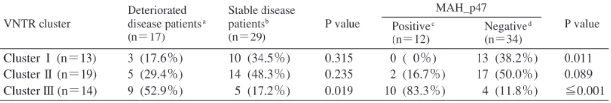
As shown in Fig. 1, the M.avium isolates examined in this study were classified roughly into three clusters: cluster I, cluster II, and cluster III (including strain TH135). Clusters I, II, and III accounted for 17.6% (3/17), 29.4% (5/17), and 52.9% (9/17) of the isolates from the deteriorated disease group (Table 3), respectively, showing that the proportion of cluster III isolates was significantly larger than that from the
stable disease group (p=0.019). Furthermore, to examine relationships between VNTR genotype and disease progres-sion in pulmonary M.avium disease, we compared the genetic distances of clinical isolates from the deteriorated and the stable disease group as previously described20). M.avium strain TH135 was used as a reference to evaluate the Manhattan distance of each M.avium isolate. Among the 46 M.avium isolates, the genetic distance of each clinical isolate from the reference strain was associated with the highest likelihood of disease progression (subjects with deteriorated disease vs. subjects with stable disease, p=0.035; Fig. 2).
Next, we examined the relationship between disease pro-gression and the presence of pMAH135 genes based on VNTR genotyping. As shown in Fig. 1, pMAH135 genes were also conserved in cluster III strains, albeit with a few exceptions. The number of isolates with MAH_p47 in cluster III was significantly higher than the number of isolates without MAH_p47 (p≦0.001; Table 3), and a similar result was obtained for isolates with other pMAH135 genes (Fig. 1). Therefore, MATR-VNTR analysis showed that isolates
Fig. 2 Relationships between VNTR genotype and disease progression in pulmonary M.avium disease. The genetic distance was calculated as the Manhattan distance of M.
avium isolates from the reference strain TH135. The results are shown for M.avium isolates from subjects with dete-riorated or stable pulmonary M.avium disease. Horizontal lines indicate the mean value of genetic distance for the group; P values were calculated using a Mann-Whitney non-parametric comparison test.
Table 3 Relationships between isolates with pMAH135 genes and isolates from deteriorated and stable pulmonary disease patients according to VNTR genotype
VNTR, variable number tandem repeats
aStrains from the sputa of patients with deteriorated pulmonary disease bStrains from the sputa of patients with stable pulmonary disease cStrains with gene-encoding MAH_p47
dStrains without gene-encoding MAH_p47 VNTR cluster Deteriorated disease patientsa (n=17) Stable disease patientsb (n=29) P value MAH_p47 P value Positivec (n=12) Negatived (n=34) Cluster I (n=13) Cluster II (n=19) Cluster III (n=14) 3 (17.6%) 5 (29.4%) 9 (52.9%) 10 (34.5%) 14 (48.3%) 5 (17.2%) 0.315 0.235 0.019 0 ( 0%) 2 (16.7%) 10 (83.3%) 13 (38.2%) 17 (50.0%) 4 (11.8%) 0.011 0.089 ≦0.001 Deteriorated disease group (n=17)
Distance from strain TH135
p=0.035 0 5 10 15 20 Stable disease group (n=29) carrying pMAH135 genes and isolates from patients with
deteriorated disease are significantly grouped in cluster III, indicating that these strains have a similar VNTR genotype. Taken together with Table 2, these findings indicate an association between pMAH135 genes and the progression of pulmonary disease.
Discussion
The development and exacerbation of pulmonary NTM disease depend not only on host factors but also on bacterial factors. Maekura et al. reported that pulmonary disease patients with serotype 4 M.avium strains had significantly poorer prognosis than those with other serotypes10). In recent studies, VNTR typing analysis of M.avium, M.abscessus, and M.massiliense isolates from patients with NTM lung disease revealed that isolates from patients with progressive lung disease and those with stable lung disease are clustered differently20)29), indicating an association between the VNTR genotype and lung disease progression. These results suggest the involvement of bacterial factors in the progression of pulmonary NTM disease, but no specific bacterial factors have been identified. In this study, the screening of clinical isolates from patients with different symptoms for pMAH135 genes, and VNTR typing analysis revealed an association between pMAH135 and the progression of pulmonary M.
avium disease.
The clinical course of patients with pulmonary NTM disease is diverse, with some patients being stable without any treatment and others having deteriorating symptoms despite drug therapy, leading to severe lung damage8)9). The relationship between the VNTR genotype and the progression of pulmonary M.avium disease shown in this and previous studies20) suggests that MATR-VNTR analysis is able to predict the progression of pulmonary M.avium disease. Fur-thermore, the findings in this study showed the possibility that it is much easier to predict the progression of pulmonary
M. avium disease by examining the presence of pMAH135 genes than by performing MATR-VNTR analysis because of the strong correlation between the presence of pMAH135 genes and the VNTR genotype which is associated with the progression of pulmonary M.avium disease (Fig. 1, Table 3).
However, to statistically reveal the association between pMAH135 and the progression of pulmonary M.avium disease and the association between pMAH135 and VNTR genotype in the progression of pulmonary M.avium disease, it is nec-essary to investigate the presence of pMAH135 in addition to pMAH135 genes by increasing the number of patients. Treatment of pulmonary M.avium disease includes drug therapy and surgery. Although clarithromycin-based multidrug therapy is recommended for pulmonary M.avium disease, it is associated with two issues. Firstly, there are possible adverse effects of long-term treatment (12_24 months) and of drug toxicity in patients, and secondly, the efficacy of the drug therapy above certain levels is unknown17)30)31). Consequently, predicting the clinical course of pulmonary M.avium disease is a highly useful clinical indicator for determining the appro-priate treatment approach. Furthermore, the timing of treat-ment initiation influences outcomes and is thus considered
important. However, no clear criteria for determining the timing of treatment are currently available. The results of this study suggest that the presence of pMAH135 genes may serve as one criterion. The present study contains a number of limitations. First, it is possible that criteria for determining the timing of treatment varied among institutions. In addition, the ratios between the deteriorated and stable disease groups and the detection rates of pMAH135 genes in clinical isolates varied between institutions, necessitating further investiga-tion. Comparison and analysis of the whole genome in clinical isolates to identify specific bacterial factors in strains from patients with worsened disease may not only clarify the role of pMAH135 as an exacerbating factor, but also reveal other bacterial factors involved in disease exacerbation.
In conclusion, the progression of pulmonary disease due to M.avium is associated with specific VNTR genotypes in
M. avium. In addition, this study indicates an association between pulmonary disease progression and pMAH135, which codes for genes associated with M.avium pathogenicity and resistance to antimicrobial agents. This indicates the significance of bacterial factors in the disease progression. Taken together, these results indicate that the presence of pMAH135 genes might be a useful prognostic indicator for pulmonary M.avium disease and a criterion for determining the timing of treatment, suggesting the clinical utility of pMAH135 genes.
Acknowledgments
We are grateful to Drs. Satoru Fujiuchi, Katsuhiro Kuwabara, Yuka Sasaki, Emiko Toyota, Ryoji Maekura, Kazunari Tsuyuguchi, Masahiro Shirai, Takefumi Saitoh, Seiji Kawabata, Yoshiaki Tao, Shuichi Takigawa, and Mitsunori Sakatani for kindly providing us with clinical isolates and clinical information. We also thank Ms. Megumi Iwata for technical assistance. This work was supported in part by JSPS KAKENHI Grant Number 15K08049 (to K.U.).
Confl ict of interest
The authors declare that there is no conflict of interest related to this article.
References
1 ) Nishiuchi Y, Maekura R, Kitada S, et al.: The recovery of Mycobacterium avium-intracellulare complex (MAC) from the residential bathrooms of patients with pulmonary MAC. Clin Infect Dis. 2007 ; 45 : 347 351.
2 ) Maekawa K, Ito Y, Hirai T, et al.: Environmental risk factors for pulmonary Mycobacterium avium-intracellulare complex disease. Chest. 2011 ; 140 : 723 729.
3 ) Kajiki A: Non-tuberculous mycobacteriosis. What has been coming out. Kekkaku. 2011 ; 86 : 113 125.
4 ) Dailloux M, Abalain ML, Laurain C, et al.: Respiratory infections associated with nontuberculous mycobacteria in non-HIV patients. Eur Respir J. 2006 ; 28 : 1211 1215.
5 ) Bodle EE, Cunningham JA, Della-Latta P, et al.: Epi-demiology of nontuberculous mycobacteria in patients without HIV infection, New York City. Emerg Infect Dis. 2008 ; 14 : 390 396.
6 ) Moore JE, Kruijshaar ME, Ormerod LP, et al.: Increasing reports of non-tuberculous mycobacteria in England, Wales and Northern Ireland, 1995 2006. BMC Public Health. 2010 ; 10 : 612.
7 ) Tanaka E, Amitani R, Niimi A, et al.: Yield of computed tomography and bronchoscopy for the diagnosis of Myco-bacterium avium complex pulmonary disease. Am J Respir Crit Care Med. 1997 ; 155 : 2041 2046.
8 ) Stout JE, Hamilton CD: Mycobacterium avium complex disease. In: Tuberculosis and nontuberculous mycobacteria infections. David Schlossberg, ed., ASM Press, Washington, D.C., 2011, 531 564.
9 ) Weiss CH, Glassroth J: Pulmonary disease caused by non-tuberculous mycobacteria. Expert Rev Respir Med. 2012 ; 6 : 597 613.
10) Maekura R, Okuda Y, Hirotani A, et al.: Clinical and prognostic importance of serotyping Mycobacterium avium-Mycobacterium intracellulare complex isolates in human immunodeficiency virus-negative patients. J Clin Microbiol. 2005 ; 43 : 3150 3158.
11) Tsuyuguchi K, Suzuki K, Matsumoto H, et al.: Effect of oestrogen on Mycobacterium avium complex pulmonary infection in mice. Clin Exp Immunol. 2001 ; 123 : 428 434. 12) Tanaka G, Shojima J, Matsushita I, et al.: Pulmonary
Mycobacterium avium complex infection: association with NRAMP1 polymorphisms. Eur Respir J. 2007 ; 30 : 90 96. 13) Jucker MT, Falkinham JO 3rd: Epidemiology of infection
by nontuberculous mycobacteria IX. Evidence for two DNA homology groups among small plasmids in Mycobacterium avium, Mycobacterium intracellulare, and Mycobacterium scrofulaceum. Am Rev Respir Dis. 1990 ; 142 : 858 862. 14) Beggs ML, Crawford JT, Eisenach KD: Isolation and
se-quencing of the replication region of Mycobacterium avium plasmid pLR7. J Bacteriol. 1995 ; 177 : 4836 4840. 15) Uchiya K, Takahashi H, Nakagawa T, et al.:
Characteriza-tion of a novel plasmid, pMAH135, from Mycobacterium avium subsp. hominissuis. PLoS One. 2015 ; 10 : e0117797. 16) Nakagawa T, Takahashi H, Ichikawa K, et al.: Multicenter
study on clinical features and genetic characteristics of Mycobacterium avium strains from patients in Japan with lung disease caused by M. avium. Kekkaku. 2012 ; 87 : 687 695.
17) Griffith DE, Aksamit T, Brown-Elliott BA, et al.: An official ATS/IDSA statement: Diagnosis, treatment, and prevention of nontuberculous mycobacterial diseases. Am J Respir Crit Care Med. 2007 ; 175 : 367 416.
18) Turenne CY, Semret M, Cousins DV, et al.: Sequencing of hsp65 distinguishes among subsets of the Mycobacterium avium complex. J Clin Microbiol. 2006 ; 44 : 433 440. 19) Inagaki T, Nishimori K, Yagi T, et al.: Comparison of a
variable-number tandem-repeat (VNTR) method for typing Mycobacterium avium with mycobacterial interspersed repetitive-unit-VNTR and IS1245 restriction fragment length polymorphism typing. J Clin Microbiol. 2009 ; 47 : 2156 2164.
20) Kikuchi T, Watanabe A, Gomi K, et al.: Association between mycobacterial genotypes and disease progression in Myco-bacterium avium pulmonary infection. Thorax. 2009 ; 64 : 901 907.
21) Uchiya K, Takahashi H, Yagi T, et al.: Comparative genome analysis of Mycobacterium avium revealed genetic diversity in strains that cause pulmonary and disseminated disease. PLoS One. 2013 ; 8 : e71831.
22) Rodriguez GM: Control of iron metabolism in Mycobac-terium tuberculosis. Trends Microbiol. 2006 ; 14 : 320 327.
23) De Voss JJ, Rutter K, Schroeder BG, et al.: Iron acquisition and metabolism by mycobacteria. J Bacteriol. 1999 ; 181 : 4443 4451.
24) Abdallah AM, Gey van Pittius NC, Champion PA, et al.: Type VII secretion-mycobacteria show the way. Nat Rev Microbiol. 2007 ; 5 : 883 891.
25) Yu X, Xie J: Roles and underlying mechanisms of ESAT-6
in the context of Mycobacterium tuberculosis-host inter-action from a systems biology perspective. Cell Signal. 2012 ; 24 : 1841 1846.
26) Abdallah AM, Savage ND, van Zon M, et al.: The ESX-5 secretion system of Mycobacterium marinum modulates the macrophage response. J Immunol. 2008 ; 181 : 7166 7175. 27) Putman M, van Veen HW, Konings WN: Molecular
prop-erties of bacterial multidrug transporters. Microbiol Mol Biol Rev. 2000 ; 64 : 672 693.
28) Supply P, Mazars E, Lesjean S, et al.: Variable human minisatellite-like regions in the Mycobacterium tuberculosis genome. Mol Microbiol. 2000 ; 36 : 762 771.
29) Shin SJ, Choi GE, Cho SN, et al.: Mycobacterial genotypes are associated with clinical manifestation and progression of lung disease caused by Mycobacterium abscessus and Mycobacterium massiliense. Clin Infect Dis. 2013 ; 57 : 32 39.
30) Glassroth J: Pulmonary disease due to nontuberculous mycobacteria. Chest. 2008 ; 133 : 243 251.
31) Thomson RM, Yew WW: When and how to treat pulmo-nary non-tuberculous mycobacterial diseases. Respirology. 2009 ; 14 : 12 26.