Efficacy of Laminoplasty in Improving Sensory Disturbances in Patients with Cervical Spondylotic Myelopathy: A Prospective Study
Takeshi Inoue, Shigeru Soshi, Makoto Kubota, Keishi Marumo
-
OBJECTIVE: Upper extremity sensory disturbances are primary symptoms that affect the quality of life (QOL) of patients with cervical spondylotic myelopathy. Although laminoplasty is 1 of the surgical options, its effects on sensory disturbances have remained unclear. We aimed to determine whether surgical intervention would improve the sensory disturbances of patients with cervical spon- dylotic myelopathy.
-
METHODS: We conducted a prospective clinical trial of 101 patients who had undergone open door laminoplasty.
For an objective sensory assessment, we measured the current perception thresholds (CPTs) in the patients’ fore- arms and palms using PainVision PS-2100. For a subjective sensory assessment, numbness in the upper extremities was rated using a visual analog scale (VAS). Using the VAS scores, the patients were divided into those with improvement and without improvement. Their self-reported 36-item short-form health survey and Japanese Orthopae- dic Association cervical myelopathy evaluation question- naire scores were compared.
-
RESULTS: The postoperative CPTs in relationship to the preoperative CPTs at 3, 6, and 12 months was 99.3%, 98.1%, and 93.8% in the forearm and 93.6%, 90.6%, and 87.8% in the palm, respectively. The corresponding postoperative
numbness VAS scores were 63.8%, 50.5%, and 48.0%. At 12 months postoperatively, the 36-item short-form health sur- vey physical and role component summary scores, cervical spine function effectiveness rates, upper and lower ex- tremity function, and QOL items in the Japanese Ortho- paedic Association cervical myelopathy evaluation questionnaire were significantly higher in the improvement group.
-
CONCLUSIONS: Our findings have indicated that improvement in postoperative subjective sensory distur- bances will occur relatively earlier and will be signifi- cantly greater than the improvement in objective sensory disturbances. Furthermore, improvement in the subjective sensory disturbances contributes to functional spinal cord recovery and patients’ health-related QOL.
INTRODUCTION
C ervical spondylotic myelopathy (CSM) is 1 of the most common neurologic disorders among geriatric pop- ulations. It is a progressive degenerative disease charac- terized by cervical spinal cord dysfunction. The common CSM symptoms include sensory disturbances of the extremities,
Key words
-Cervical spondylotic myelopathy
-JOACMEQ
-Laminoplasty
-Numbness
-PainVision
-Sensory disturbance
-SF-36
Abbreviations and Acronyms CI: Confidence interval CPT: Current perception threshold CSM: Cervical spondylotic myelopathy GH: General health
IQR: Interquartile range
JOA: Japanese Orthopaedic Association
JOACMEQ: Japanese Orthopaedic Association cervical myelopathy evaluation questionnaire
OPLL: Ossification of the posterior longitudinal ligament OR: Odds ratio
PCS: Physical component summary PF: Physical functioning
QOL: Quality of life
RCS: Role/social component summary RE: Role limitations due to emotional problems RP: Role limitations due to physical health problems SF-36: 36-Item short-form health survey
VAS: Visual analog scale
Department of Orthopaedic Surgery, The Jikei University School of Medicine, Tokyo, Japan To whom correspondence should be addressed: Takeshi Inoue, M.D.
[E-mail:inoue@jikei.ac.jp]
Citation: World Neurosurg. (2020) 134:e581-e588.
https://doi.org/10.1016/j.wneu.2019.10.141
Journal homepage:www.journals.elsevier.com/world-neurosurgery Available online:www.sciencedirect.com
1878-8750/$ - see front matterª2019 Elsevier Inc. All rights reserved.
clumsiness of the hands, gait abnormalities, and urinary dysfunction.
1The treatment options include decompression surgery, and several studies have demonstrated that cervical laminoplasty will provide satisfactory results.
2-7Sensory distur- bances, such as numbness, will often present as the initial CSM symptoms and will often be localized in the upper limbs.
8However, because it is difficult to quantitatively assess sensory disturbances, few data on their postoperative changes are available. Although the Japanese Orthopaedic Association
9(JOA) and modified JOA
10scores have been commonly used in the clinical assessment of CSM, the objective and quantitative evaluation of sensory disturbances using the JOA score has been challenging, and the pre- and postoperative differences cannot be assessed effectively. However, sensory disturbances will frequently persist after surgery, affecting patients’ activities of daily living and quality of life (QOL). Thus, the assessment of sensory disturbances is vital.
The present study’s aims were to prospectively examine the postoperative improvements in subjective and objective sensory disturbances in patients with CSM after laminoplasty. In addition, we aimed to prospectively evaluate the effect of sensory distur- bance improvement on patient satisfaction, physical disability, and general health.
METHODS Subjects
A total of 101 patients with CSM (78 men and 23 women; mean age, 65.512.8 years), who had undergone open door laminoplasty from May 2009 to November 2016 at our medical center, were enrolled in the present study (Figure 1). Patients with both single-
and multiple-level spinal cord compression lesions were included.
Patients with a history of cervical surgery, cerebral palsy, thoracic myelopathy, ossification of the posterior longitudinal ligament (OPLL), cervical disc herniation, cervical radiculopathy, spinal cord tumor, spinal cord injury, or spinal fusion surgery were excluded from the present study.
Surgical Technique for Modified Open Door Laminoplasty
A single surgeon performed modified open door laminoplasty as initially described by Itoh and Tsuji.
11In brief, the head was elevated at a 20
e30angle with a Mayfield cranial stabilizing device to attain mild
flexion of the cervical spine. A posteriormidline incision was made, followed by an incision of the ligamentum nuchae. The muscles from the C3 to C6 spinous processes were detached, and the muscles attached to C2 and C7 were preserved. Muscle detachment was performed slightly lateral to the laminaefacet junction. Gutters were created on the inner edges of the facet joints on both the open and the hinge sides. A dome-shaped partial laminectomy was performed on the caudal side of C3 and cranial side of C7 on a case-by-case basis.
Once the gutters had been completed, the lamina was opened, and the ligamentum
flavum and the epidural adhesion tissue onthe open side were severed as necessary. A small burr hole was created on the open side, sutures were passed through, and a hydroxyapatite spacer was placed to preserve an open laminal position. A closed drain was installed, and the wound was closed by suturing the ligamentum nuchae.
On postoperative day 2, the patient was permitted to sit, stand, and walk. In principle, a cervical collar was not used. Cervical spine range of motion training and isometric muscle strength- ening were initiated during the early postoperative period.
Assessment of Sensory Disturbances
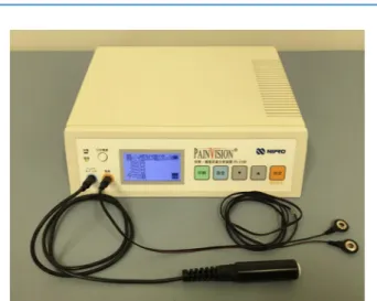
PainVision PS-2100 (Nipro, Osaka, Japan;
Figure 2) was used toperform an objective sensory assessment by selectively stimulating sensory nerves with a pulsed current (A b and A d
Figure 1. Flow chart showing the number of patients enrolled and excluded from the present study. CSM, cervical spondylotic myelopathy.
Figure 2. Photograph of the PainVision PS-2100 unit used to calculate the current perception threshold.
fibers). The PainVision PS-2100 device (Nipro) includes an elec-
trical stimulation system and a control system and can measure both the perception threshold and the pain intensity. The current perception threshold (CPT) mode was used for objective sensory assessment. The CPT measures the minimum perceived current by generating a weak stimulating current, which gradually increases.
When the stimulating current is perceived for the
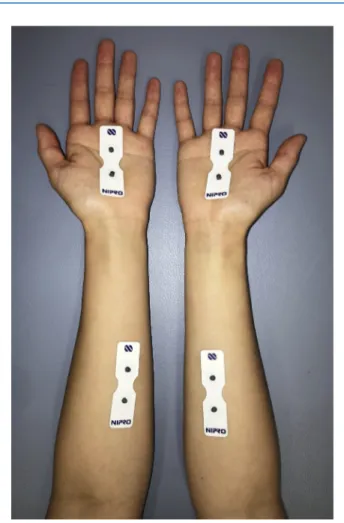
first time, thesubject uses a stop switch button to end the measurement. The point at which a current has initially been perceived represents the CPT. In the present study, PainVision was used to measure the CPT in the proximomedial
flexion side sections of the bilateralforearms and palms. The CPT test sites were identical for all the patients. A weak electrical current was applied to the skin via an electrode. CPT was defined as the minimum threshold that could be perceived as a stimulus. PainVision generated a superficial pulsed current (50 Hz; 0e150 m A; pulse amplitude, 0.3 ms) that gradually increased in intensity and measured the participant’s electrical stimulation threshold (Figure 3). We measured the CPT 3 times in each section and calculated the bilateral average.
For the subjective sensory assessment, numbness in the upper extremities was rated by the patients using a visual analog scale (VAS) for numbness with a score from 0 mm (no pain/numbness) to 100 mm (most intense pain/numbness imaginable). The CPT and VAS numbness were measured preoperatively and at 3, 6, and 12 months postoperatively. Using the change in the VAS numb- ness score at 12 months postoperatively, the patients were divided into an improvement group (n
¼64) and a nonimprovement group (n
¼36). In accordance with the
findings from a previousstudy,
11the improvement group included patients who had had either 1) a
20-mm decrease in the postoperative VAS numb-ness score compared with the preoperative VAS numbness score or 2) a preoperative VAS numbness score of
10 mm with apostoperative VAS numbness score was
<10 mm. One patient wasexcluded from the analysis because both the pre- and the post- operative VAS scores were
10 mm.Assessment According to Patient Perspectives
The assessments were performed using the Japanese Orthopaedic Association cervical myelopathy evaluation questionnaire (JOAC- MEQ)
12and the 36-item short-form health survey (SF-36).
13The JOACMEQ is a patient-based assessment method, in which the patients answer 24 questions related to their QOL and their cer- vical, upper extremity, lower extremity, and bladder function. Each item was scored from 0 to 100, after which the assessments were performed. The effectiveness rate was determined by comparing the pre- and postoperative JOACMEQ scores for each item. The surgical intervention was considered effective if the score had increased by
20 points or if the preoperative score had been<90points and the postoperative score was
90 points.12We obtained scores for the 8 SF-36 subscales (physical func- tioning [PF], role limitations due to physical health problems [RP], bodily pain [BP], general health [GH], vitality [VT], social functioning [SF], role limitations due to emotional problems [RE], and mental health [MH]) and the summary scores of the three components (physical component summary [PCS], mental component summary [MCS], and role/social component summary [RCS]).
13Both the JOACMEQ and SF-36 were used as self- assessment tools preoperatively and at 3, 6, and 12 months postoperatively.
Statistical Analysis
Statistical analyses were performed using SPSS, version 22.0, for Windows (IBM Japan, Tokyo, Japan). All data are presented as mean standard deviation or the median and interquartile range (IQR). The Mann-Whitney
Utest and c
2test were used to analyze the differences between the 2 groups. Repeated measures analyses of variance were performed in the same group using the Wilcoxon signed rank test. We performed logistic regression analysis to identify the preoperative factors associated with nonimprovement in numbness. Univariate logistic regression analysis was per- formed, followed by multivariate logistic regression analysis including the variables with
P <0.2 in the univariate analysis (forward selection method, likelihood ratio). The 95% confidence intervals (CIs) of odds ratios (ORs) were estimated, and risk ratios
<5% were considered significant.
Figure 3. Photography of the measurement sites for the PainVision unit. Proximomedial sections of the flexion side of the bilateral forearms and palms were used to calculate the current perception threshold.
RESULTS
The preoperative median CPT was 14.6 m A (IQR, 7.2 m A) in the forearm and 31.6 m A (IQR, 15.5 m A) in the palm. The preoperative median VAS score for numbness was 77 mm (IQR, 48 mm). The postoperative median CPT at 3, 6, and 12 months in relationship to the preoperative CPT was 99.3% (IQR, 49.8%), 98.1% (IQR, 40.8%), and 93.8% (IQR, 43.2%) in the forearm and 93.6% (IQR, 35.9%), 90.6% (IQR, 33.8%), and 87.8% (IQR, 33.8%) in the palm, respectively. In contrast, the postoperative median VAS score for numbness at 3, 6, and 12 months in relationship to the preoper- ative VAS for numbness was 63.8% (IQR, 55.2%), 50.5% (IQR, 71.3%), and 48.0% (IQR, 72.6%), respectively. By comparing the objective and subjective assessments, we noted that the subjective symptoms scores had improved to a greater extent compared with the objective CPT values at all measurement points (Table 1).
According to the VAS scores for numbness at 12 months, the patients were divided into an improvement group (n
¼64) and nonimprovement group (n
¼36). The SF-36 and JOACMEQ scores were compared between the 2 groups. Although the preoperative VAS scores for numbness in the upper extremities were greater in the improvement group than in the nonimprovement group, no
statistically significant differences were found between the 2 groups in terms of sex, age, preoperative forearm and palm CPT, and VAS scores for numbness in the lower extremities (Table 2).
When the preoperative SF-36 subscale scores were compared be- tween the 2 groups, the GH score was significantly greater in the improvement group than in the nonimprovement group (Table 3).
The postoperative PCS and RCS scores were also significantly greater in the improvement group than in the nonimprovement group at all assessment points. Additionally, although all subscale scores in the improvement group at 12 months postoperatively were greater than the preoperative scores, the increase in the nonimprovement group was limited to PF, RP, and RE (Table 3). The preoperative JOACMEQ item scores did not differ between the 2 groups. The effectiveness rate at 6 and 12 months postoperatively were significantly greater in the improvement group than in the nonimprovement group for upper extremity function, QOL items (at all assessment points), cervical spine function, and lower extremity function (
Table 4).
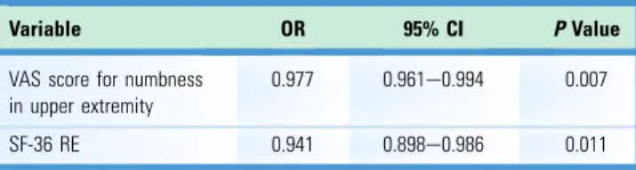
Univariate and multivariate analyses were used to analyze the preoperative factors contributing to the differences between the 2 groups. Age, sex, forearm and palm CPT, VAS scores for numbness in the upper and lower extremities, SF-36 score, 3-component SF-36 summary scores, JOACMEQ score, and the duration of the symp- toms before surgery were set as independent variables. Multivariate analysis indicated that greater preoperative VAS scores in the upper extremities were associated with a lower proportion of the non- improvement group (OR, 0.977; 95% CI, 0.961e0.994;
P¼0.007), and greater preoperative SF-36 RE scores were associated with a lower proportion of the nonimprovement group (OR, 0.941; 95% CI, 0.898e0.986;
P¼0.011;
Table 5).Table 1.
Current Perception Threshold and Visual Analog Scale Score for Numbness at Each Assessment Point*
Variable Preoperatively
Postoperatively
3 Months 6 Months 12 Months
Objective sensory assessment Forearm CPT
(mA)
14.6 (7.2) 13.8 (5.7) 14.5 (5.7) 13.4 (8.3)
Postoperative versus preoperative CPT (%)
NA 99.3 (49.8) 98.1 (40.8) 93.8 (43.2)
Palm CPT (mA) 31.6 (15.5) 29.4 (16.6) 29.9 (14.3) 28.7 (12.2) Postoperative
versus preoperative CPT (%)
NA 93.6 (35.9) 90.6 (33.8) 87.8 (33.8)
Subjective sensory assessment VAS score for
numbness (mm)
77 (48) 43 (50) 36 (56) 33 (54)
Postoperative versus preoperative VAS (%)
NA 63.8 (55.2) 50.5 (71.3) 48.0 (72.6)
Data presented as median (interquartile range).
CPT, current perception threshold; NA, not applicable; VAS, visual analog scale.
*The comparison of the postoperative CPT in relation to the preoperative CPT at 3, 6, and 12 months in the forearm and in the palm with the postoperative VAS score for numbness in relation to the preoperative VAS score at 3, 6, and 12 months using the Mann-WhitneyUtest revealedPvalues of<0.001 for all measurement points.
Table 2.
Baseline Data for Improvement and Nonimprovement Groups
Variable Improvement Nonimprovement PValue*
Patients 64 36
Sex 0.8
Male 50 27
Female 14 9
Age (years) 65.314.1 65.510.2 0.73
Preoperative forearm CPT (mA)
16.810.9 17.310 0.48
Preoperative palm CPT (mA)
36.312.1 32.212.6 0.085
Preoperative VAS score for numbness in upper extremity (mm)
75.324 60.830.3 0.019
Preoperative VAS score for numbness in lower extremity (mm)
40.636.4 41.932.1 0.82
Data presented as n or meanstandard deviation.
CPT, current perception threshold; VAS, visual analog scale.
*Mann-WhitneyUtest.
DISCUSSION
In the present study, we performed an objective assessment of the pre- and postoperative differences in sensory disturbances using PainVision combined with a subjective assessment of numbness using a VAS. We included only patients with CSM (excluding cervical disc herniation and OPLL) who had undergone the same surgical procedure performed by a single surgeon at the same facility. The assessment focused on the patients’ sensory distur- bances. We found that the subjective sensory assessment had improved more than had the objective sensory assessment.
To the best of our knowledge, no previous prospective studies have assessed sensory disturbances using an objective approach, including detailed analyses. The quantitative assessment of sensory disturbances can be difficult, and previous assessments have used the patients’ subjective experiences (i.e., VAS, numerical rating scale). We observed improvement in the 8 SF-36 subscale scores at 12 months postoperatively compared with the preoperative scores in the improvement group. In addition, the effectiveness rates for all items of the JOACMEQ, except for bladder function, were
Table 3.Scores for the 36-Item Short-Form Health Survey
Subscales in the Improvement and Nonimprovement Groups
SF-36 Score
Group PValue
Improvement Nonimprovement A* By Cz
PF NA <0.001 0.033
Preoperatively 19.224.0 12.521.6 0.23 12 Months
postoperatively
37.220.9 19.719.1 <0.001
RP NA <0.001 0.027
Preoperatively 23.818.3 19.514.0 0.35 12 Months
postoperatively
42.015.9 25.516.0 <0.001
BP NA <0.001 0.79
Preoperatively 38.510.8 36.812.8 0.28 12 Months
postoperatively
46.911.3 36.910.2 <0.001
GH NA <0.001 0.42
Preoperatively 44.69.3 39.610.5 0.031 12 Months
postoperatively
49.110.9 38.910.8 <0.001
VT NA <0.001 0.15
Preoperatively 41.211.7 38.310.6 0.21 12 Months
postoperatively
49.612.8 40.612.3 0.0015
SF NA <0.001 0.64
Preoperatively 34.717.4 33.614.2 0.77 12 Months
postoperatively
47.014.0 35.015.0 <0.001
RE NA <0.001 0.012
Preoperatively 30.718.6 23.715.3 0.076 12 Months
postoperatively
45.315.9 30.515.3 <0.001
MH NA <0.001 0.083
Preoperatively 40.912.1 38.210.7 0.24 12 Months
postoperatively
49.514.1 42.113.0 0.0097
PCS NA <0.001 0.19
Preoperatively 27.816.3 23.017.5 0.16 3 Months
postoperatively
38.413.5 30.415.2 0.0095
6 Months postoperatively
40.113.1 27.412.9 <0.001
12 Months postoperatively
39.515.2 26.115.0 <0.001
Continues
Table 3.
Continued
SF-36 Score
Group PValue
Improvement Nonimprovement A* By Cz
MCS NA 0.54 0.29
Preoperatively 53.911.0 52.811.7 0.53 3 Months
postoperatively
54.311.3 53.312.0 0.82
6 Months postoperatively
54.110.6 52.113.1 0.45
12 Months postoperatively
53.911.1 50.913.0 0.25
RCS NA <0.001 0.012
Preoperatively 28.620.1 25.913.8 0.54 3 Months
postoperatively
35.716.3 25.616.7 0.0059
6 Months postoperatively
40.214.8 30.618.2 0.0097
12 Months postoperatively
44.414.5 32.416.2 <0.001
SF-36, 36-Item short-form health survey; PF, physical function; NA, not applicable; RP, role limitations due to physical health problems; BP, bodily pain; GH, general health; VT, vitality; SF, social functioning; RE, role limitations due to emotional problems; MH, mental health; PCS, physical component summary; MCS, mental component summary;
RCS, role component summary.
*Pvalues from comparison between the improvement and nonimprovement group scores at each assessment point using the Mann-WhitneyUtest.
yPvalues from comparison between the preoperative and 12-month postoperative scores in the improvement group using the Wilcoxon test.
zPvalues from comparison of the preoperative and 12-month postoperative scores in the nonimprovement group using the Wilcoxon test.
significantly greater in the improvement group than in the non- improvement group. Furthermore, the PCS and RCS scores in the SF-36 were significantly greater at all assessment points in the improvement group compared with those in the nonimprovement group, as were the effectiveness rates for upper extremity function in the JOACMEQ scores and QOL items. Compared with the non- improvement group, those in the improvement group had greater cervical spine and lower extremity function at 6 and 12 months postoperatively. To the best of our knowledge, no previous study has performed assessments as frequently as those in the present study (i.e., preoperatively and 3, 6, and 12 months postoperatively).
3,4,14,15Thus, we consider our data valuable. These results suggest that the superiority or inferiority of the improvement in sensory disturbances could be an index of neural viability. Furthermore, the sensory disturbance itself could influence the patients’ health- related QOL.
The ascending sensory tracts in the spinal cord include the lateral spinothalamic tract (which transmits pain and tempera- ture), ventral spinothalamic tract (tactile sensations), and poste- rior funiculus (epicritic and deep sensations—the sense of vibration and position).
16Detailed examination of these tracts will effectively identify the cross-sectional spread of the lesion into the spinal cord. Seichi et al.
17reported that the distribution of sensory disturbances in the upper extremities is more reliable for a neurological level diagnosis than muscle weakness and the deep tendon reflexes. PainVision can selectively stimulate the A b and A d
fibers.18,19The minimum perceivable stimulus (i.e., pain, temperature, tactile sensation) current was considered the CPT.
20The benefit of this test compared with conventional methods is that sensory disturbances can be noninvasively and painlessly quantified, enhancing patients’ understanding of the
Table 4.Preoperative Japanese Orthopaedic Association
Cervical Myelopathy Evaluation Questionnaire Scores and Efficacy Rates Stratified by Group
Variable
Group PValue
Improvement Nonimprovement A* By
Cervical spine function
Preoperatively 72.023.7 63.626.7 0.13 3 Months
postoperatively (%)
43.4 27.3 0.13
6 Months postoperatively (%)
56.9 21.9 0.0017
12 Months postoperatively (%)
61.7 37.1 0.028
Upper extremity function
Preoperatively 65.228.0 59.024.2 0.19 3 Months
postoperatively (%)
68.6 41.2 0.012
6 Months postoperatively (%)
71.7 39.4 0.0030
12 Months postoperatively (%)
69.2 33.3 0.0012
Lower extremity function
Preoperatively 47.434.0 43.627.9 0.56 3 Months
postoperatively (%)
56.6 50.0 0.55
6 Months postoperatively (%)
65.5 36.4 0.0080
12 Months postoperatively (%)
64.2 32.4 0.0037
Bladder function
Preoperatively 67.026.3 70.321.5 0.77 3 Months
postoperatively (%)
45.3 24.2 0.050
6 Months postoperatively (%)
45.8 27.6 0.10
12 Months postoperatively (%)
40.0 29.4 0.31
Continues
Table 4.
Continued
Variable
Group PValue
Improvement Nonimprovement A* By
QOL
Preoperatively 41.519.5 38.716.4 0.52 3 Months
postoperatively (%)
40.6 19.4 0.031
6 Months postoperatively (%)
43.8 20.0 0.018
12 Months postoperatively (%)
44.3 11.1 <0.001
QOL, quality of life.
*Pvalues from comparison of the preoperative Japanese Orthopaedic Association Cer- vical Myelopathy Evaluation Questionnaire item scores between the improvement and nonimprovement groups using the Mann-WhitneyUtest.
yPvalues from comparison of the efficacy rates of each Japanese Orthopaedic Associ- ation Cervical Myelopathy Evaluation Questionnaire item between the improvement and nonimprovement groups using thec2 test.
test, and does not require unique skills.
18In the present study, we observed some differences in the postoperative improvement of the objective and subjective assessments of sensory disturbances, which we attributed to differences between the 2 sensory pathways and differences in spinal plasticity.
16Furthermore, our
findings were in line with a previous study,which detected faster improvement of the subjective than the objective outcome measures after lumbar spine surgery.
21Laminoplasty has been reported as an important option for the treatment of CSM in numerous studies
3-7and has been used more frequently in Japan than elsewhere owing to the greater incidence of OPLL. Furthermore, the suitability of laminoplasty for single-level stenosis has continued to be debated. Chiba et al.
3conducted a minimum 10-year follow-up survey of 80 patients (CSM, 27 patients; OPLL, 53 patients) after open door laminoplasty and reported a CSM improvement rate using the JOA scores of 57.
9% at 3 years postoperatively and 55.7% in the
final survey,demonstrating favorable maintenance of the results. For the pa- tients with OPLL, the improvement rates were 63.3% and 47.9% at 3 years postoperatively and at the
final survey, respectively,demonstrating a reduction in improvement over time. A study by Seichi et al.
4reported the long-term results (10 years) after double-door laminoplasty. They had excluded cases complicated by athetoid cerebral palsy.
4They reported that long-term stability was maintained in 78% of the 35 patients with OPLL and nearly 100% of the 25 patients with CSM. However, these assessments had only included the physician’s subjective evaluation using JOA scores.
The assessment systems for cervical disorders can be grouped into single-item and comprehensive assessment systems. The scoring systems with a single item include the VAS and the Nurick scale.
22,23Comprehensive scoring systems include the JOA scale, modified JOA scale, and SF-36. The JOA scale was created by a deep understanding of cervical myelopathy, and the assessment was implemented by a medical practitioner.
9However, recently, more emphasis has been given to patient-based evaluation or value- based medicine. Thus, the JOACMEQ,
12a patient-based evalua- tion method, has been proposed, with mean scores determined from healthy individuals stratified by age and sex.
24The SF-36 has been the most widely used self-reported health status survey in the
world.
14,25It was carefully constructed from a psychometric aspect and is an internationally accepted measure of health status owing to its proven scientific reliability and validity. A recent prospective study using the JOACMEQ and SF-36, by Fujiwara et al.,
14reported no significant differences in the surgical treatment outcomes or functional recovery prognosis between the CSM and the OPLL groups. The study also reported a negative correlation between axial neck pain and JOACMEQ-assessed cervical spine function.
14Zhou et al.
15compared the patient-based SF-36 assess- ment with the physician-implemented modified JOA assessment score and found that improved modified JOA scores correlated with improvements in PF, RP, and SF at 1 year postoperatively. The present study has demonstrated that for patients who have exhibited postoperative improvement in the upper extremity VAS score for numbness, neurological recovery will also have been achieved at a relatively early stage after surgery. Moreover, the multivariate anal- ysis results indicated that the preoperative SF-36 RE scores (i.e., psychological factors in daily life before surgery) contributed to the postoperative improvement in numbness. Previous investigations have shown an association between psychological factors and sur- gery outcomes in other settings. Thus, Visser et al.
26have demonstrated a correlation between depression and poorer preoperative and postoperative total knee arthroplasty scores.
Furthermore, Flanigan et al.
27have observed an association between psychological distress, fear-avoidance behavior, poor perceived self-efficacy, or pessimistic personality traits and elective orthopedic surgery outcomes. The reasons for these correlations have not yet been definitively elucidated. However, they might be related to differences in the motivation for treatment, active participation in therapeutic activities, pain perception, and dis- crepancies between expectations for the surgical outcome and actual recovery. However, the
findings of a correlation betweenpsychological factors and postoperative numbness improvement should be discussed critically and examined in further studies for confirmation.
The present study had several limitations. Although we used Pain- Vision as an objective sensory test for pain, temperature, and tactile sensation, we did not examine the correlation between the PainVision results and the results of conventional objective sensory tests.
CONCLUSIONS
Our
findings have indicated that patients exhibiting postoperativeimprovement in upper extremity numbness can also achieve spinal cord function recovery relatively early in the postoperative period.
These data could greatly contribute to medical professionals’ un- derstanding of CSM and help them provide better explanations to their patients.
ACKNOWLEDGMENTS
Editorial support in the form of medical writing was provided by Editage (available at:
www.editage.com), a Division of CactusCommunications.
Table 5.
Multivariate Analysis Results*
Variable OR 95% CI PValue
VAS score for numbness in upper extremity
0.977 0.961e0.994 0.007
SF-36 RE 0.941 0.898e0.986 0.011
OR, odds ratio; CI, confidence interval; VAS, visual analog scale; SF-36, 36-item short-form health survey; RE, role limitations due to emotional problems.
*Only observation items withP0.2 on univariate analysis were included as inde- pendent variables in the multivariate analysis.
REFERENCES
1. Bakhsheshian J, Mehta VA, Liu JC. Current diag- nosis and management of cervical spondylotic myelopathy.Global Spine J. 2017;7:572-576.
2. Hirabayashi K, Watanabe K, Wakano K, Suzuki N, Satomi K, Ishii Y. Expansive open-door lam- inoplasty for cervical spinal stenotic myelopathy.
Spine. 1983;8:693-699.
3. Chiba K, Ogawa Y, Ishii K, et al. Long-term re- sults of expansive open-door laminoplasty for cervical myelopathy -average 14-year follow-up study.Spine. 2006;31:2998-3005.
4. Seichi A, Takeshita K, Onishi I, et al. Long-term results of double-door laminoplasty for cervical stenotic myelopathy.Spine. 2001;26:479-487.
5.Cho SK, Kim JS, Overley SC, Merrill RK. Cervical laminoplasty: indications, surgical considerations, and clinical outcomes.J Am Acad Orthop Surg. 2018;
26:e142-e152.
6. Kaye ID, Hilibrand AS, Morrissey PB, Vaccaro AR.
Laminoplasty is the preferred procedure for a posteriorly based multilevel surgery in a patient with a neutral spine and cervical spondylotic myelopathy: true or false?Clin Spine Surg. 2018;31:
1-5.
7. Heller JG, Edwards CC II, Murakami H, Rodts GE.
Laminoplasty versus laminectomy and fusion for multilevel cervical myelopathy: an independent matched cohort analysis.Spine. 2001;26:1330-1336.
8. Hilton B, Tempest-Mitchell J, Davies B, Kotter M.
Assessment of degenerative cervical myelopathy differs between specialists and may influence time to diagnosis and clinical outcomes. PLoS One.
2018;13:e0207709.
9. Hirabayashi K, Miyakawa J, Satomi K, Maruyama T, Wakano K. Operative results and postoperative progression of ossification among patients with ossification of cervical posterior longitudinal ligament.Spine (Phila Pa 1976). 1981;6:
354-364.
10. Benzel EC, Lancon J, Kesterson L, Hadden T.
Cervical laminectomy and dentate ligament sec- tion for cervical spondylotic myelopathy.J Spinal Disord. 1991;4:286-295.
11. Itoh T, Tsuji H. Technical improvements and re- sults of laminoplasty for compressive myelopathy
in the cervical spine.Spine (Phila Pa 1976). 1985;10:
729-736.
12. Fukui M, Chiba K, Kawakami M, et al, Subcom- mittee of the Clinical Outcome Committee of the Japanese Orthopaedic Association on low back pain and cervical myelopathy evaluation. JOA back pain evaluation questionnaire (JOABPEQ)/JOA cervical myelopathy evaluation questionnaire (JOACMEQ): the report on the development of revised versions April 16, 2007.J Orthop Sci. 2009;
14:348-365.
13. Ware JE, Sherboune CD. The MOS 36-item short- form health survey (SF-36): I. Conceptual frame- work and item selection. Med Care. 1992;30:
473-489.
14. Fujiwara H, Oda T, Makino T, Moriguchi Y, Yonenobu K, Kaito T. Impact of cervical sagittal alignment on axial neck pain and health-related quality of life after cervical laminoplasty in pa- tients with cervical spondylotic myelopathy or ossification of the posterior longitudinal ligament:
a prospective comparative study.Clin Spine Surg.
2018;31:E245-E251.
15. Zhou F, Zhang Y, Sun Y, et al. Profiles of and correlation between objective and subjective outcome assessments following open-door lam- inoplasty for cervical spondylotic myelopathy.Chin Med J (Engl). 2014;127:2659-2663.
16. Nathan PW, Smith MC, Cook AW. Sensory effects in man of lesions of the posterior columns and of some other afferent pathways. Brain. 1986;109:
1003-1041.
17. Seichi A, Takeshita K, Kawaguchi H, et al.
Neurologic level diagnosis of cervical stenotic myelopathy. Spine (Phila Pa 1976). 2006;31:
1338-1343.
18. Wang D, Zhang K, Han S, Yu L. PainVision apparatus for assessment of efficacy of pulsed radiofrequency combined with pharmacological therapy in the treatment of postherpetic neuralgia and correlations with measurements.Biomed Res Int. 2017;2017:5670219.
19. Ohtori S, Kawaguchi H, Takebayashi T, et al.
PainVision apparatus is effective for assessing low back pain.Asian Spine J. 2014;8:793-798.
20. Katims JJ. Electrodiagnostic functional sensory evaluation of the patient with pain: a review of the neuroselective current perception threshold and pain tolerance threshold.Pain Dig. 1998;8:219-230.
21. Gautschi OP, Joswig H, Corniola MV, et al. Pre- and postoperative correlation of patient-reported outcome measures with standardized Timed Up and Go (TUG) test results in lumbar degenerative disc disease. Acta Neurochir (Wien). 2016;158:
1875-1881.
22. Nurick S. The pathogenesis of the spinal cord disorder associated with cervical spondylosis.
Brain. 1972;95:87-100.
23. Nurick S. The natural history and the results of surgical treatment of the spinal cord disorder associated with cervical spondylosis.Brain. 1972;
95:101-108.
24. Tanaka N, Konno S, Takeshita K, et al. An outcome measure for patients with cervical myelopathy the Japanese Orthopaedic Association cervical myelopathy evaluation questionnaire [JOACMEQ]: an average score of healthy volun- teers.J Orthop Sci. 2014;19:33-48.
25. Singh A, Gnanalingham K, Casey A, Crockard A.
Quality of life assessment using the short form-12 (SF-12) questionnaire in patients with cervical spondylotic myelopathy comparison with SF-36.
Spine (Phila Pa 1976). 2006;31:639-643.
26. Visser MA, Howard KJ, Ellis HB. The influence of major depressive disorder at both the preoperative and postoperative evaluations for total knee arthroplasty outcomes.Pain Med. 2019;20:826-833.
27. Flanigan DC, Everhart JS, Glassman AH. Psycho- logical factors affecting rehabilitation and out- comes following elective orthopaedic surgery.J Am Acad Orthop Surg. 2015;23:563-570.
Conflict of interest statement: The authors declare that the article content was composed in the absence of any commercial or financial relationships that could be construed as a potential conflict of interest.
Received 24 July 2019; accepted 22 October 2019 Citation: World Neurosurg. (2020) 134:e581-e588.
https://doi.org/10.1016/j.wneu.2019.10.141
Journal homepage:www.journals.elsevier.com/world- neurosurgery
Available online:www.sciencedirect.com 1878-8750/$ - see front matterª2019 Elsevier Inc. All rights reserved.