1
Clarification of the sterilization mechanism of antimicrobial photodynamic therapy for Candida albicans
(Candida albicansに対する抗菌的光線力学的療法の殺菌メカニズムの解明)
伊澤 万貴子1), 大塚 一聖2), 小峯 千明3)
1)日本大学松戸歯学部口腔健康科学講座 顎顔面矯正学分野
2)日本大学松戸歯学部保存修復学講座
3)日本大学松戸歯学部口腔健康科学講座 歯科臨床検査医学分野
(指導:福本 雅彦 教授)
2
Title: Clarification of the sterilization mechanism of antimicrobial photodynamic therapy for Candida albicans
Makiko Izawa
1), Issei Otuka
2), Chiaki Komine
3)
1)Department of Oral Health Science, Division of Maxillofacial Orthodontic, Nihon University School of Dentistry at Matsudo
2)Department of Operative Dentistry, Nihon University School of Dentistry at Matsudo
3)Department of Oral Health Science, Division of Laboratory Medicine for Dentistry, Nihon University School of Dentistry at Matsudo
Corresponding author: Makiko Izawa
Department of Oral Health Science, Division of Maxillofacial Orthodontic, Nihon University School of Dentistry at Matsudo
2-870-1 Sakaecho-Nishi, Matsudo, Chiba 271-8587, Japan Phone number: +81-47-360-9624; Fax number: +81-47-360-9624 E-mail address: [email protected]
(Director: Professor Masahiko Fukumoto)
3
<Abstract>
Background:There has been a continuing increase in dental fungal infections in recent
years, and according to studies of the frequency of occurrence of fungal infections that
look at the frequency of different causative fungi, there has been a particular increase in
infections caused by Candida and Aspergillus species. The conventionally used
antifungal drugs can not reach the depth of the biofilm, and as a result, they have the
disadvantage of causing resistant bacteria.
Therefore, we focused on antimicrobial photodynamic therapy (a-PDT), which is
receiving attention because of the absence of such side effects, and examined its
fungicidal mechanism against C. albicans.
Objective:The aims of the present study were two-fold: (1) to investigate the
relationship between the amount of 1O2 generated using the electron spin resonance
(ESR) spin-trapping technique and the fungicidal effects on C. albicans; and (2) to
observe the destruction of the cell wall after a-PDT by scanning electron microscopy
(SEM). Thus, two experiments were performed.
4
Materials and Methods:The first experiment used a 0.01% aqueous solution of
methylene blue (MB) and ultrapure water as a control. Both were irradiated with a diode
laser, and the amount of ¹O2 generated was measured using ESR spectroscopy. Then, the
number of colony-forming units per milliliter (CFU/mL) of C. albicans present after
incubation under different sets of conditions was determined, and the experimental groups
were defined as follows: with laser-irradiation, L(+); without laser-irradiation, L(-);
containing MB, M(+); and not containing MB, M(-). These were then combined to form
four groups: L(+)M(+); L(+)M(-); L(-)M(+); and L(-)M(-). The second experiment
followed the first with observation of the cell wall of C. albicans by SEM.
Results:The irradiation of MB with a 660 nm diode laser was caused an irradiation
time-dependent increase in the generation of 1O2, and that the C. albicans sterilization
rate increased proportionally. Observation of SEM images of C. albicans exposed to 1O2
showed that the surface of the fungal cells fused, and the normal morphology of single,
independent cells was lost in an irradiation time-dependent fashion, meaning that fusion
5
was dependent on the amount of 1O2 generated, and images of cells beginning to fuse
together and of irregular, bumpy shapes were observed.
Conclusion: The present findings clarified the relationship between 1O2 generation via
excited MB and the fungicidal effect on C. albicans. Moreover, it was considered that
C. albicans might be sterilized by 1O2 attacked to surface layer.
<key words>
Antimicrobial photodynamic therapy, methylene blue, diode laser (λ=660 nm), singlet oxygen, Candida albicans
6
<和文対訳>
近年、真菌症は増加傾向にあり、歯科領域においてカンジダおよびアスペルギル
ス属が起因となる感染症の増加が特に見られる。従来使用されている抗真菌薬
はバイオフィルムの深さまで到達することができず、結果として耐性菌を生じ
させてしまうなどの欠点があった。そこで我々はそのような副作用のない事で
脚光を浴びている抗菌的光線力学的療法(a-PDT)に着目し、C. albicans に対
する殺菌メカニズムについて検討を行った。本研究は(1)電子スピン共鳴(ESR)
spin-trapping法を利用し、a-PDTから発生した一重項酸素(1O2)量とC. albicans
の殺菌効果の関係性について検討、(2)走査型電子顕微鏡(SEM)を用いてa-
PDT後のC. albicansの菌体表面を観察することを目的として行った。
本研究では0.02%のメチレンブルー(MB)水溶液を用い、純水(PW)を対照
として両者に半導体レーザーを照射し、生成された1O2の量をESR 分光法にて
測定した。実験群はレーザー照射の有無をL (+)、L (-)、0.02% MBの有無を
M(+)、M(-)と定義し、これらを組み合わせてL(+)M(+); L(+)M (-); L
7
(-) M(+); L(-) M(+)の4つのグループに分け行った。次に4つのグルー
プそれぞれにC. albicansを作用させ、インキュベートした後のC. albicansの1
ミリリットル当たりのコロニー形成単位の数(CFU/mL)を測定した。その後
SEMによるC. albicansの細胞壁の観察を行った。結果は、MBに660 nm半導
体レーザーを照射することにより1O2は照射時間依存的に発生量が増加し、それ
に比例してC. albicansの殺菌率も増加することを認めた。1O2に暴露された C.
albicansのSEM画像を観察すると照射時間依存的に真菌細胞の表面が融合し、
単一の独立した正常な形態が失われ、融合し始めた像や不規則な凹凸を呈する
像が観察された。これらのことから、本研究により、a-PDTにより励起された
MBから発生した1O2がC. albicansに対し濃度依存的に殺菌効果を示し、1O2に
よりC. albicansの表層が侵害されることにより殺菌に至ることが推測された。
8
<Introduction>
Dental diseases such as dental caries, periodontitis, endodontic disease and denture
candidiasis are mostly infectious diseases caused by oral microorganisms 1-3). As
microorganisms in oral cavity have been shown to be involved in systemic diseases such
as endocarditis, aspiration pneumonia and diabetes 4-6), how to control microorganisms is
the most important topics in clinical practice.
There has been a continuing increase in fungal infections in recent years, and according
to studies of the frequency of occurrence of fungal infection that look at the frequency of
different causative fungi, there has been a particular increase in infections caused by
Candida and Aspergillus species.
The background of increasing fungi is improvements in detection techniques and
detection sensitivity, at the same time as (1) an increase in the use of broad-spectrum
antibiotics and steroid medications that have side effects of oral candidiasis (OC) and oral
dryness, (2) an increase in the number of high-risk patients, and (3) an increase in the
number of elderly people 7).
9
OC is a disease encountered with a relatively high frequency in dental treatment. It is
caused by Candida species, particularly Candida albicans (C. albicans), which are part
of the resident oral microbial flora. C. albicans is an opportunistic pathogen that normally
has weak pathogenicity and proliferative ability, but which multiplies and demonstrates
pathogenicity, causing an outbreak of opportunistic infection, if the body’s immunity is
compromised 7). C. albicans exhibits dimorphism, having a filamentous form comprising
hyphae or pseudohyphae and a yeast form 8,9). It is usual for such fungi to invade mucous
membranes as hyphae that adhere strongly to the mucous membrane, and with repeated
recurrence, the infection progresses to intractable OC 10). At present, Japan is rapidly
becoming a super-aging society, and changing disease patterns in dentistry are being seen
as a result. This is leading to a need for dental clinics to take on the responsibility of
treating elderly people who are receiving primary nursing care at home or in facilities.
There is a high rate of OC in such patients, and expertise in OC will therefore be essential
for dentists. In addition, there is an increased risk of fungal pneumonia among elderly
people due to pulmonary aspiration of Candida species, and the risk is further increased
10
if swallowing function is reduced as a result of cerebral infarction or other disease. There
is, therefore, considerable attention being paid to the importance of oral care for elderly
persons 7,10).
Although the conventional treatment of OC applied to topical or systemic antifungal
agents (azole, polyenes), it resulted in the development of resistant Candida species 11).
Additionally, the organization of microorganisms in biofilms is a protective shell,
enabling the survival of these pathogens even in unfavorable conditions and providing
high resistance to antifungal agents 12). Considering the increased incidence of resistant
pathogens to conventional antifungal treatments and drug toxicity, studies have searched
for strategies to control fungal species 13). In recent years, novel methods of disinfection
for use in treating dental caries, periodontal disease, endodontic disease 14-18) and OC 19-
23) have become available. It is well known that the application of photodynamic therapy
(PDT), including antimicrobial PDT (a-PDT), can be used as a disinfection method 24-26).
Many studies have demonstrated that the use of a-PDT for bactericidal and fungicidal
effect requires that many variables be taken into account when developing a-PDT protocol,
11
including light parameters, photosensitizers, and light delivery techniques 27-29). Many
investigators have demonstrated that C. albicans is effectively sterilized by a-PDT in the
following manner: (1) photosensitizing agents (PS) attach to the cell membrane of C.
albicans, (2) irradiation with light at a specific wavelength matched to the peak absorption
of PS leads to the generation of singlet oxygen (1O2), and (3) fungidal death via
destruction of the cell walls is induced by 1O2 30-33). However, although the oxidizing
power of 1O2 has been shown to induce fungicidal effects, the details of the relationship
between the amount of generated 1O2 and the degree of fungicidal effects have not yet
been clarified. Moreover, there is no report that visually observed cell wall destroyed by
a-PDT.
1O2 is a reactive oxygen species (ROS). Nakano et al 34) and Tatsuzawa et al 35) reported
that 1O2 was toxic to prokaryotic cells and is almost completely nontoxic to eukaryotic
cells. In addition, Silva et al 36) reported that moderate angiogenesis, fibrogenesis, or
noninflammatory cells were observed in the animal models treated with PDT. On the
other hand, the methylene blue (MB) used in this study may cause biotoxicity due to cell
12
staining. George and Kishen 37) reported that 10 μmol/L MB, when irradiated for 20
minutes with a 30-mW diode laser, killed 30% of fibroblasts. Therefore, to safely apply
a-PDT, the amount of generated 1O2 should be carefully considered.
The aims of the present study were two-fold: (1) to investigate the relationship between
the amount of generated 1O2 using the electron spin resonance (ESR) spin-
trapping technique and the fungicidal effects on C. albicans (2) to observe the destruction
of cell wall after a-PDT by using scaning electon microscopy (SEM).
13
<Materials and Methods>
Reagents and Laser Source
Pure water (PW: Ultrapure Water for Molecular Biology) was purchased Merck Millipore
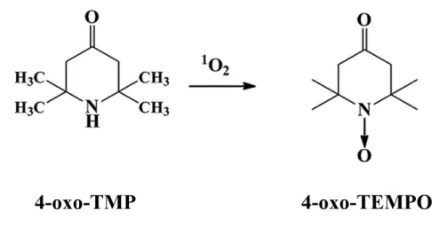
(Tokyo, Japan), MB and 2,2,6,6-tetramethyl-4-piperidone (4-oxo-TMP) were purchased
from FUJIFILM Wako Pure Chemical Industries, Ltd. (Osaka, Japan). MB was used as a
PS in this study. The 2,2,6,6-tetramethyl-4-piperidone-N-oxyl (4-oxo-TEMPO) was
purchased from Sigma Aldrich (St. Louis, MO, USA). All other reagents were analytical
grade. A diode laser (=660 nm, 200 mW in CW) supplied by Osada Electric Co. Ltd.
(Tokyo, Japan) was used as the irradiation source. A diode laser was used in a non-contact
mode, and delivered with distance from the tip (φ300 m) of the quartz fiber to the
surface of bacterial suspension being 3 cm. The laser irradiation time periods were set to
600, 1,200 and 1,800 seconds and the power densities were 106, 212 and 318 W/cm2,
respectively.
Experiment 1: The relationship between the amount of generated 1O2 and the fungicidal effects on C. albicans
14
The 0.02% MB aqueous solution was used in this study. PW was used as a control. The
0.02% MB and control were irradiated with a diode laser. The amount of generated ¹O2
was measured using ESR spectroscopy (JES FA-200, JEOL, Tokyo, Japan). The 0.02%
MB and 40 mM 4-oxo-TMP were mixed in test tubes (φ12 mm) to make final
concentrations of 0.01% MB and 20 mM 4-oxo-TMP. Immediately, the mixtures were
irradiated with the diode laser for 600, 1,200 and 1,800 seconds. Subsequently, the
mixture was transferred into an ESR flat cell and then was measured using an ESR
spectrometer. The ESR measurements were conducted under the following conditions:
magnetic field, 335 ± 5 mT; modulation width, 0.025 mT; time constant, 0.1 seconds;
microwave power, 4.00 ± 0.05 mW; sweep width, 5 mT; sweep time, 2 minutes; and
amplitude, 100. The signal intensities were normalized to a MnO marker and the
concentrations of the stable radical products (4-oxo-TEMPO) were determined using an
external standard based on the signal height 38).
Finally, the amount of generated 1O2 was evaluated according to the 1O2 specific
oxidation from 4-oxo-TMP to 4-oxo-TEMPO (Figure 1), which is detectable with ESR.
15
C. albicans was obtained from the American Type Culture Collection (ACTT18804). A
suspension of C. albicans from culture grown on brain heart infusion (Becton, Dickinson,
and Co., NJ, USA) at 37°C for 24 hours was prepared in sterile physiological saline. The
final concentration was adjusted to 1 × 10⁷ cells/ml of the suspension and 0.01% MB in
2 mL of saline. Immediately after mixing the test tubes, the mixtures were irradiated with
stirring for 600, 1,200 and 1,800 seconds using a diode laser. After the laser irradiation
was complete, 10-fold serial dilutions were prepared and 100 L aliquots of each dilution
were seeded in duplicate onto Sabouraud dextrose agar (Difco, MI, USA) plates and
incubated for 48 hours at 37°C. Finally, the number of colony-forming units per milliliter
(CFU/mL) present after incubation was determined. The experimental groups were
defined as follows: with laser-irradiation, L(+); without laser-irradiation, L(-); containing
MB, M(+); and not containing MB, M(-). These were combined to form four groups:
L(+)M(+); L(+)M(-); L(-)M(+); and L(-)M(-).
Experiment 2: The observation of cell wall fracture after Experiment 1
16
The each experimental group mixtures, which is involved C. albicans, were centrifuged
for 10 minutes at 1,300 x g. The cell pellet was fixed in 2.5% glutaraldehyde for 1hour
and dehydrated in several ethanol washes (10, 25, 50, 75, and 90% for 20 minutes and
100% for 1hour). Then, the cell pellet was incubated at 37℃ for 24 hours to dry, and
transferred to aluminium stubs and covered with Au-Pd for 120 seconds at 40 mA. After
metalization, the cell wall of C. albicans was examined and photographed by SEM (S-
3400N, Hitachi, Japan), operating at 15 kV, at ×10.0 k magnification 39).
Statistical Analysis
The results of experiment 1 were analyzed with one way analysis of variance (ANOVA).
When appropriate, ANOVA was followed by post-hoc Tukey’s test to compensate for
multiple comparisons (α=0.05).
17
<Results>
Experiment 1: The relationship between TEMPOL and signal intensity ratio based on
MnO marker increased in a concentration-dependent manner (Figure 2). Figure 3 showed
the typical ESR-spectra of 0.01% MB irradiated by the diode laser for 600, 1,200 and
1,800 seconds. The ESR spectra displayed a 1:1:1 triplet signal characteristic of 4-oxo-
TEMPO having a hyperfine splitting constant (aN=1.608 mT) 38). Figure 4 showed the
amount of 1O2 generated from 0.01% excited MB. The amount of generated 1O2 was
increased by the laser irradiation in a time-dependent manner. A positive correlation was
observed between the amount of generated 1O2 and the laser irradiation time (R2=0.999).
According to the equation of linear relationship between the amountof 4-oxo-TEMPO
and the irradiation time, the amount of 1O2 generated from 0.01% excited MB during 600,
1,200 and 1,800 seconds of irradiation was about 82.7, 159.4 and 245.3M, respectively.
Figure 5 showed the numbers of CFU/mL of C. albicans in groups L(-)M(-), L(+)M(-),
L(-)M(+) and L(+)M(-). In group L(+)M(+), the number of CFU/mL was significantly
reduced in association with the laser irradiation time compared to the other groups
18
(p<0.05). On the other hand, no fungicidal effects were observed in groups, L(-)M(-),
L(+)M(-) or L(-)M(+). In brief, the amount of generated 1O2 necessary to kill C. albicans
(> 99.99%) was at least about 245.3 M.
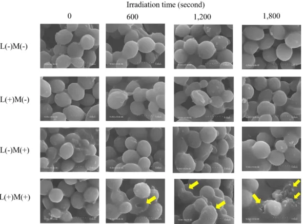
Experiment 2: In the observation of the surface of the fungal cells after irradiation, in
groups L(-)M(-), L(+)M(-), and L(-)M(+), no structural damage was seen, and there were
no surface changes, so that the characteristic shape was preserved (Figure 6). In group
L(+)M(+), the surface of the fungal cells was fused in an irradiation time-dependent
fashion, meaning that fusion was dependent on the amount of 1O2 generated. Cells
beginning to fuse together and the formation of irregular bumpy shapes were observed.
In the SEM images following 1,800 seconds of irradiation, there were images of cells that
had fused and undergone further breakdown in morphology, becoming amorphous lumps
of material.
19
<Discussion>
C. albicans is a dimorphic fungus present in the regular flora of the mouth, skin, and
pharynx of healthy people. When it shows pathogenicity, it undergoes a transition from
the yeast form to the filamentous form, establishing itself in host tissues and multiplying,
causing damage to target tissues. Progression of the infection can lead to fungemia or
systemic infection 40,41).In recent years, it has been thought that this ability to transition
to the filamentous form is a major factor in pathogenicity in deep-seated mycosis 42-44).
There is an increasing range of options available for the treatment of fungal infections,
but there is nothing currently that can offer dramatic effects. Furthermore, there are
numerous problems, such as the side effects of drugs 45).
On the other hands, a-PDT mechanism damages fungal cells when ROS penetrate the
cell walls and membranes, the allowing displacement of the PS into the cell. Then,
oxidizing species generated by the excitiation of light induce the photodestruction of
internal cellular organelles, leading to cell death. Thus, the 1O2 generated by the
excitiation of PS is non-specific oxidizing agent against which there is no defence 46,47).
20
The present study therefore aimed to use 1O2, which has few side effects, as a safe method
to kill C. albicans and also to clarify the mechanism by which death occurs.
The results show that irradiation of MB as PS with a 660 nm diode laser caused an
irradiation time-dependent increase in the generation of 1O2, and that the C. albicans
sterilization rate increased proportionally. Observation of SEM images of C. albicans
exposed to 1O2 showed that the surface of the fungal cells fused, and the normal
morphology of single, independent cells was lost in an irradiation time-dependent fashion,
meaning that fusion was dependent on the amount of 1O2 generated. Cells beginning to
fuse together and the formation of irregular bumpy shapes were observed. This suggests
a mechanism whereby the surface layer of C. albicans is disrupted by a-PDT, leading to
the death of the fungus. At the same time, it has been reported that the half-line of 1O2 in
the cell system is just 2 s, so that even if 1O2 were generated in the extracellular fluid of
eukaryotic cells, the 1O2 would be deactivated and transform to its ground state of
molecular oxygen before it could enter the cells; it would therefore be unable to reach
mitochondrial respiratory chain enzymes in the cells and attack them. In other words, in
21
eukaryotic cells, which do not have respiratory enzyme systems in the cell membrane, but
instead have them in the mitochondria within the cell, as long as the 1O2 cannot reach the
respiratory system within the cell, this mechanism of cell destruction cannot operate 34,35).
C. albicans is eukaryotic, and therefore the above finding should be valid for this species.
Consequently, there should be no sterilization effect, but a sterilization effect was found
in the present study. We considered that there are two reasons. As one reason, the PS used
in this study was MB, which is known for its use as a cell staining agent, and MB stains
the cell wall of C. albicans 48). The cell wall of fungi plays a number of roles in the
biological activity of the cell. Then, as well as protecting the cell against stress from the
physical environment, it is involved in functions such as retaining the morphology of the
cell, the intake of nutrients from the outside world, and the exchange of materials with
the outside world. Therefore, appears that 1O2 generated from within the cell wall itself
disrupts the cell wall so that these functions are lost, leading to the death of the cell. As
another reason, although C. albicans has some kinds of antioxidants enzymes, such as
catalase, superoxide dismutase and glutathione peroxidase 49-51), because they are
22
ineffective against 1O2, internal cellular organelles were also injured after surface layer
destruction by 1O2, leading to the death of the cell. However, C. albicans was not
completely sterilized. As the reason, C. albicans may have some defense mechanisms, so
further studies are needed. Hsieh YH reported that although PDT alone effectively
eradicated C. albicans biofilms, when combined with fluconazole, PDT significantly
inhibited C. albicans to greater extent 52). We also consider that the development of
resistant bacteria is the most feared in OC, therefore it is necessary that developing
methods of therapeutic system to break down the biofilm with PDT and penetrate the
antifungal drug deeply.
In conclusion, the present findings clarified the relationship between 1O2 generation via
excited MB and the fungicidal effect on C. albicans. Moreover, it was considered that C.
albicans might be sterilized by 1O2 attacked to surface layer.
23
<Acknowledgments>
We thank Profs. Tsujimoto Y, Hiratsuka K and Fukumoto M for assistance in preparation
this manuscript.
This work was supported by JSPS KAKENHI Grant Number 19K19076 to Komine C.
<Conflict of Interest>
No potential conflicts of interest were disclosed.
24
<Reference>
1. Wilson M: Susceptibility of oral bacteria biofilm to antimicrobial agents. J Med
Microbiol, 44 : 79-87, 1996.
2. Soukos NS, Goodson JM: Photodynamic therapy in the control of oral
biofilms. Periodontol 2000, 55 : 143-166, 2011.
3. Rodrigues CF, Silva S, Henriques M: Candida glabrata: a review of its features and
resistance. Eur J Clin Microbiol Infect Dis, 33 : 673-688, 2014.
4. Nery EB, Meister F Jr., Ellinger A, et al: Prevalence of medical problems in
periodontal patients obtained from three different populations. J Periodontology,
58 : 564-568, 1987.
5. Terpenning MS, Taylor GW, Lopatin DE, et al: Aspiration pneumonia: dental and
oral risk factors in an older veteran population.J Am Geriatr Soc, 49 : 557-563,
2001.
6. Taylor GW: Bidirectional interrelationships between diabetes and periodontal
diseases: an epidemiologic perspective. Ann Periodontal, 6 : 99-112, 2001.
25
7. Sakaguchi H: Treatment and Prevention of Oral Candidiasis in Elderly Patients.
Med Mycol J, 58 : 43-49, 2017.
8. Yamamoto T: Oral Candidiasis: Clinical Features and Control. The Official Journal
of Japanese Society of Laboratory Medicine, 58 : 1027-1034, 2010.
9. Sakaguchi H: Recent Findings of Oral Candidiasis.J Jpn Soc Dent Prod, 27 : 3-8,
2013.
10. Nagaosa S, Shinagawa T, Sakaguchi H, et al: A Study of Oral Microbial Flora of
Elderly Stroke Patients-Around the Candida-. J J Gerodont, 16 : 14-21, 2001.
11. Sanglard D, Coste A, Ferrari S: Antifungal drug resistance mechanisms in fungal
pathogens from the perspective of transcriptional gene regulation. FEMS Yeast
Res, 9 : 1029-1050, 2009.
12. Seneviratne CJ, Silva WJ, Samaranayake YH, et al: Architectural analysis, viability
assessment and growth kinetics of Candida albicans and Candida glabrata. Arch
Oral Biol, 54 : 1052–1060, 2009.
13. Carmello JC, Alves F, G Basso F, et al: Treatment of Oral
26
Candidiasis Using Photodithazine®- Mediated Photodynamic Therapy In Vivo,
PLoS One. 2016 Jun 2 ; 11 : e0156947. doi: 10.1371/journal.pone.0156947, 2016.
14. Bonsor SJ, Nichol R, Reid TM, et al: Microbiological evaluation of photo-activated
disinfection in endodontics (an in vivo study). Br Dent J, 200 : 337-341, 2006.
15. Konopka K, Goslinski T: Photodynamic therapy in dentistry. J Dent Res, 86 : 694-
707, 2007.
16. George S, Kishen A: Augmenting the antibiofilm efficacy of advanced noninvasive
light activated disinfection with emulsified oxidizer and oxygen carrier. J Endod,
34 : 1119-1123, 2008.
17. Takasaki A, Aoki A, Mizutani K, et al: Application of antimicrobial photodynamic
therapy in periodontal and peri-implant diseases. Periodontol 2000, 51 : 109-140,
2009.
18. Rolim JP, de-Melo MA, Guedes SF, et al: The antimicrobial activity of
photodynamic therapy against Streptococcus mutans using different
photosensitizers. J Photochem Photobiol B, 106 : 40-46, 2012.
27
19. Mima EG, Pavarina AC, Dovigo LN, et al: Susceptibility of Candida albicans to
photodynamic therapy in a murine model of oral candidiosis. Oral Surg Oral Med
Oral Pathol Oral Radiol Endod, 109 : 392-401, 2010.
20. Dovigo LN, Carmello JC, de Souza Costa CA, et al: Curcumin-mediated
photodynamic inactivation of Candida albicans in a murine model of oral
candidiasis. Med Mycol, 51 : 243-251, 2013.
21. Khademi H, Torabinia N, Allameh M, et al: Comparative evaluation of
photodynamic therapy induced by two different photosensitizers in rat experimental
candidiasis. Dent Res J, 11 : 452-459, 2014.
22. Freire F, Costa AC, Pereira CA, et al: Comparison of the effect of rose bengal- and
eosin Y-mediated photodynamic inactivation on planktonic cells and biofilms of
Candida albicans. Lasers Med Sci, 29 : 949-955, 2014.
23. Freire F, de Barros PP, da Silva Ávila D, et al: Evaluation of gene expression SAP5,
LIP9, and PLB2 of Candida albicans biofilms after photodynamic inactivation.
Lasers Med Sci, 30 : 1511-1518, 2015.
28
24. Chan Y, Lai CH: Bactericidal effects of different laser wavelengths on
periodontopathic germs in photodynamic therapy. Laser Med Sci, 18 : 51-55, 2003.
25. Maisch T: Anti-microbial Photodynamic therapy: useful in the future. Laser Med,
22 : 83-91, 2007.
26. Ng R, Singh F, Papamanou DA, et al: Endodontic photodynamic therapy ex vivo. J
Endod, 37 : 217-222, 2011.
27. Soukos NS, Chen PS, Morris JT, et al: Photodynamic treatment of endodontic
disinfection. J Endod, 32 : 979-984, 2006.
28. Foschi F, Fontana CR, Ruggiero K, et al: Photodynamic inactivation of
Enterococcus faecalis in dental root canals in vitro. Lasers Surg Med, 39 : 782-787,
2007.
29. Fimple JL, Fontana CR, Foschi F, et al: Photodynamic treatment of endodontic
polymicrobial infection in vitro. J Endod, 34 : 728-734, 2008.
30. Teichert MC, Jones JW, Usacheva MN, et al: Treatment of oral candidiasis with
methylene blue-mediated photodynamic therapy in an immunodeficient murine
29
model. Oral Surg Oral Med Oral Pathol Oral Radiol Endod, 93 : 155-160, 2002.
31. Fabio CA, Yolanda MB, Carmen GM, et al: Use of photodynamic therapy and
chitosan for inactivation of Candida albicans in a murine model. J Oral Pathol
Med, 45 : 627-633, 2016.
32. Carmello JC, Alves F, Ribeiro A, et al: In vivo photodynamic inactivation of
Candida albicans using chloro-aluminum phthalocyanine. Oral Diseas, 22 : 415-
422, 2016.
33. Silva MP, dos Santos TA, de Barros PP, et al: Action of antimicrobial photodynamic
therapy on heterotypic biofilm: Candida albicans and Bacillus atrophaeus. Lasers
Med Sci, 31 : 605-610, 2016.
34. Nakano M, Kambayashi Y, Tatsuzawa H, et al: Useful 1O2 (1Δg) generator, 3-(4’-
methyl-1’-naphthyl)-propionic acid, 1’,4’-endoperoxide (NEPO), for dioxygenation
of squalene (a skin surface lipid) in an organic solvent and bacterial killing in
aqueous medium. FEBS Lett, 432 : 9-12, 1998.
35. Tatsuzawa H, Maruyama T, Misawa N, et al: Inactivation of bacterial respiratory
30
chain enzymes by singlet oxygen. FEBS Lett, 439 : 329-333, 1998.
36. Silva LA, Novaes AB Jr., de Oliveira RR, et al: Antimicrobial photodynamic
therapy for the treatment of teeth with apical periodontitis: a histopathological
evaluation. J Endod, 38 : 360-366, 2012.
37. George S, Kishen A: Advanced noninvasive light-activated disinfection: assessment
of cytotoxicity on fibroblast versus antimicrobial activity against Enterococcus
faecalis. J Endod, 33 : 599-602, 2007.
38. Nakamura K, Ishiyama K, Ikai H, et al: Reevaluation of analytical methods for
photogenerated singlet oxygen. J Clin Biochem Nutr, 49 : 87-95, 2011.
39. Costa AC, de Campos Rasteiro VM, Pereira CA, et al: Susceptibility of Candida
albicans and Candida dubliniensis to erythrosine- and LED-mediated
photodynamic therapy. Arch Oral Biol, 56 : 1299-1305, 2011.
40. Jacobsen ID, Wilson D, Wächtler B, et al: Candida albicans dimorphism as a
therapeutic target. Expert review of anti-infective therapy, 10 : 85-93, 2012.
41. Mayer FL, Wilson D, Hube B et al: Candida albicans pathogenicity mechanisms.
31
Intravital, 4 : 119-128, 2013.
42. Lo HJ, Köhler JR, DiDomenico B, et al: Nonfilamentous C. albicans Mutants Are
Avirulent. Cell, 90 : 939-949, 1997.
43. Saville SP, Lazzell AL, Monteagudo C, et al: Engineered control of cell
morphology in vivo reveals distinct roles for yeast and filamentous forms of
Candida albicans during infection. Eukaryotic Cell, 2 : 1053-1060, 2003.
44. Shareck J, Belhumeur P: Modulation of morphogenesis in Candida albicans by
various small molecules. Eukaryotic Cell, 10 : 1004-1012, 2011.
45. Izumikawa K, Kohno S: Current Status of Drug Delivery Systems for Antifungal
Drugs. Drug Delivery System, 27 : 93-105, 2012.
46. Donnelly RF, MC Carron PA, Tunney MM: Antifungal photodynamic therapy.
Microbiol Res, 163 : 1-12, 2008.
47. Gonzales F, Maisch T: Photodynamic inactivation for controlling Candida albicans
infections. Fungal Biol, 116 : 1-10, 2012.
48. Robert IL, Martin JC: Interaction of Candida albicans with Human Leukocytes
32
and Serum. J Bacteriol, 98 : 996-1004, 1969.
49. Nakagawa Y, Koide K, Watanabe K, et al: The Expression of the Pathogenic Yeast
Candida albicans Catalase Gene in Response to Hydrogen Peroxide. Maicrobiol.
Immunol, 43 : 645-651, 1999.
50. Martchenko M, Alarco A-M, harcus D, et al: Superoxide Dismutases in Candida
albicans:Trenscriptional Regulation and Functional Characterization of the Hyphal-
induced SOD5 Gene. Mol cell Biol, 15 : 456-467, 2004.
51. Komalapriya C, kaloriti D, Tillmann AT, et al: Integrative Model of Oxidative
Stress Adaptation in the Fungal pathogen Candida albicans. PLoS One, 10 :
e0137750. doi: 10.1371/journal.pone.0137750, 2015.
52. Hsieh YH, Zang JH, Chuang WC, et al: An in Vitro Study on the Effect of
Combined Treatment with Photodynamic and Chemical Therapies on Candida
albicans. Int J Mol Sci, 19 : 337-346, 2018.
33
Figure 1 From 4-oxo-TMP to 4-oxo-TEMPO by generated 1O2
A 4-oxo-TMP is a scavenger with high reactivity with 1O2. As a result, stable 4-oxo-
TEMPO free radical is produced as a reaction product.
34
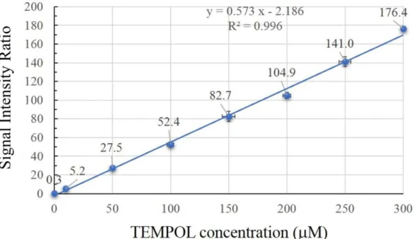
Figure 2 External standard curve of the TEMPOL
A positive correlation was observed between each concentrations of TEMPOL and
signal intensity (R2=0.996). The equation of the line is y = 0.573x-2.186. The data
points indicate the mean values (n=6) with standard deviation bars.
35
Figure 3 ESR-spectra obtained after laser irradiation
The typical ESR-spectra of the control and 0.01% MB after laser irradiation for 600,
1,200 and 1,800 seconds. The white and black circles indicate the Mn2+ marker and the
nitroxide radical, respectively.
36
Figure 4 The relationship between laser irradiation time and 4-oxo-TEMPO
The amount of generated 1O2 increased with laser irradiation. A positive correlation was
observed between the amount of generated 1O2 and 0.01% MB (R2=0.999).
The equation of the line is y = 39.784x-37.63. The data points indicate the mean values
(n=6) with standard deviation bars.
37
Figure 5 Numbers of CFU/mL in the suspension after a-PDT
In the L(+)M(+) group, the number of C. albicans cells decreased with a >4-log
reduction within 1,800 seconds. The data points indicate the mean values (n=6) with
standard deviation bars. The numbers of CFU/mL decreased significantly in the
L(+)M(+) group at 600, 1,200 and 1,800 seconds, compared to all other groups
(p<0.05).
38
Figure6 Typical SEM images of C. albicans surface on each conditions
The yellow arrows indicated fused cells and bumpy shapes.
The experimental groups were defined as follows: with laser-irradiation, L(+); without
Laser-irradiation, L(-); containing MB, M(+); and not containing MB, M(-).