Accumulated exposure dose of multiple radiological examinations at an emergency center
救命救急センターにおける複数放射線検査の 累積被ばく線量について
弘前大学大学院保健学研究科保健学専攻
提出者氏名: 成田 将崇
所 属: 医療生命科学領域 放射線生命科学分野
指導教員: 細川 洋一郎
Table of contents
Abbreviations ... 2
Introduction ... 3
Materials and Methods ... 5
Results ... 14
Discussion ... 17
Conclusion ... 20
Acknowledgements ... 21
References ... 22
Abstract ... 24
Abbreviations
ALARA : as low as reasonably achievable AP : anterior posterior
CBCT : cone beam CT CRA : cranial
CT : computed tomography FOV : field of view
FPD : flat panel detector HU : hounsfield units
ICRP : international commission on radiological protection IVR : intervention radiology
kV : kilo-voltage Lat : lateral
mA : milli-ampere
RAOCRA : right anterior oblique cranial position SID : source image receptor distance
TLD : thermoluminescent dosimeter
WA : weight fraction of active bone marrow WM : weight fractions of mineralized tissue
3D-DSA : three-dimensional digital subtraction angiography
Introduction
The International Commission on Radiological Protection (ICRP) recommends that exposure doses to medical radiation should be as small as possible to reduce both exposure and the number of people exposed (as low as reasonably achievable, ALARA).
In the 1997 Recommendation, the Commission recommended a system of dose limitation, the main features are as follows: “ (a) no practice shall be adopted unless its introduction produces a positive net benefit; (b) all exposures shall be kept as low as reasonably achievable (ALARA), economic and social factors being taken into account; and (c) the dose equivalent to individuals shall not exceed the limits recommended for the appropriate circumstances by the Commission” (ICRP Publication 26,1977,p3)1). ALARA is considered to be an important part of radiation protection. In the 2007 Recommendation, “the principle of optimization of Protection: the likelihood of incurring exposure, the number of people exposed, and the magnitude of their individual doses should all be kept as low as reasonably achievable, taking into account economic and societal factors” (ICRP Publication 103,2007,p14) 2).
The principle of the application of dose limits states that we should decrease total dose reasonably as low as possible to any individual to refer to the dose limits recommended by the Commission. Measures should be taken to comply with the basic concepts of optimization2,3).
At the Hirosaki University Hospital, patients with various symptoms have been transported to our emergency center since the establishment of the Advanced Critical Care Center in April 2010. We experienced the number of radiological examinations conducted at the emergency center over approximately 3 months, from July to September 2013. There were 50 CT cases in 302 total cases, and most examinations were plane head CTs. There were 120 cases that received two imaging types using mobile X-ray imaging and CT imaging; 13 cases received mobile X-ray imaging, CT imaging, and DSA examinations, which have a large radiation exposure dose. Emergency center physicians extensively use radiographic equipment in the acute evaluation of seriously injured blunt
trauma patients. At our emergency center, radiographs of the chest and pelvis of patients with major trauma are commonly taken shortly after transportation to the emergency department, followed by computed tomography (CT) of the head, neck, chest, abdomen, and pelvis. This series of examinations has been informally referred to as the "pan scan."
CT has become an essential diagnostic tool for treatment, particularly in trauma patients4). In addition, numerous DSA of the head may be performed depending on the injuries sustained. When bleeding is confirmed by CT examination, DSA of the brain/body trunk is performed to identify the rupture site and confirm the size and shape of the aneurysm.
In some cases, coil embolization for the aneurysm or arterial embolization is performed.
New technologies have the potential to manage radiation risks properly, but also enables the performance of more complex procedures that may expose patients and staffs to higher radiation doses. For example, although the inclusion of cone beam CT (CBCT) in an intervention radiology (IR) examination offers advantages to patients at the emergency, it contributes to an increased radiation dose5,6).
IR provides important benefits to public health, but the use of ionizing radiation has inherent risks that must be evaluated and minimized. Nishizawa7) and Maruyama8) reported the radiation doses received from various radiological examinations. The relationship between exposure dose and radiation damage has been described previously9-13). However, there are no reports on the accumulated exposure dose due to radiological examinations in patients at emergency center.
When several examinations are performed, as in the case for an emergency center patient, it is also important to understand the doses received by individual organs and the effective doses in order to optimize patient protection. There is no upper limit for medical exposure, because the benefit of the exposure may be potentially larger than the risk of exposure. However, to adhere to ALARA and to prevent radiation damage, it is important to understand the radiation dose received from imaging. The purpose of this study was to evaluate each organ dose using radiological imaging of a phantom and to examine the risk of radiation damage from radiography in patients at emergency center.
Materials and Methods Equipment
Thermolucent dosimeters (TLDs) can vary in sensitivity and X-ray dependence.
To calibrate the TLDs by exposing them to X-rays of the same effective energy, each TLD was irradiated together with a Radcal model 9015 radiation dosimeter (Radcal, California, USA).
Each TLD element was calibrated based on the dose absorbed by soft tissue. For calibration, the TLD and the Radcal dosimeter were placed in the radiation field of the mobile X-ray imaging apparatus and the DSA apparatus, and calibration constants for each TLD were obtained. However, as it was difficult to irradiate the tube fixed within the CT apparatus, a calibration constant was obtained using a C-arm type fluoroscope (KXO-80Z Ultimax-i DREX-UI80, Toshiba Medical Systems, Tochigi, Japan). In addition to the tantalum filter attached to the equipment, a half-value layer was measured by applying a 10-mm A85 aluminum plate to the tube irradiation port. It was possible to produce a line quality similar to the half-value layer of 8.7 mm of Al of the CT apparatus.
By irradiating both the TLD and the Radcal dosimeter, a calibration constant with the same linear quality as the CT apparatus was obtained.
Each examination was conducted under the following parameter. The CT unit used in this experiment included two 32-slice multidetector-row CT (SOMATOM Definition, Siemens Medical Solutions, Erlangen, Germany). The general scan parameters for the head of the phantom were: helical acquisition; tube voltage, 120 kilo-voltage (kV); tube current, CARE DOSE 4D (ref. 350mA); pitch, 0.8; rotation time, 1.0 s; field of view (FOV), 250 mm; slice thickness, 5.0 mm (64 × 0.6 mm).
CT imaging of the trunk from the neck assumed identification of the bleeding site, and plane imaging from the neck to the pelvic area and Dynamic 2-phase imaging were performed. The scan parameters were: tube voltage, 120 kV; tube current, CARE DOSE 4 D (ref. 180-200 mA); pitch, 1.2; rotation time, 1.0 s; FOV, 350 mm; slice thickness, 5.0 mm (64 × 0.6 mm). The DSA unit used in this experiment was an AXIOM
Artis dBA Twin (Siemens Medical Solutions). It has two X-ray tubes on the C-arm, allowing the patient to be viewed from various angles. DSA images were acquired using an X-ray tube and the following parameters: tube voltage, 73 kV; tube current, 160 mA;
exposure time, 100 ms; and X-ray filter, 0.1 mmCu.
In our hospital, the arterial phase is exposed at 4 frame/s and the venous phase is exposed at 2 frame/s, but the imaging parameters are the same in both phases. Parameters such as tube voltage vary depending on the normal imaging angle, but the most used imaging parameter was used as the parameter in this experiment. Exposure angles included: 1) cranial-caudal view, 15 degrees (CRA); 2) right anterior oblique, 20 degrees;
cranial view, 15 degrees (RAOCRA); and 3) lateral (Lat): FOV, 32 cm; CRA and RAOCRA, 42 cm. The number of DSA imaging times was determined by referring to the number of 6-vessel exposure frames performed in clinical practice. The number of frames taken by dosimetry was calculated as 257 frames on the front of the head, 424 frames on the side, and 167 frames in the oblique position. The number of frames on the lateral side of the DSA oblique imaging was included.
The head 3D-DSA images were obtained by rotating the C-arm with the flat panel fixed at 180 degrees. In clinical examination, 126 frame mask images were acquired, and 126 frames were taken again after injecting the contrast medium. The contrast agent cannot be used in the phantom. To obtain the exposure dose, 126 frames were acquired twice, as in clinical practice. The tube voltage of the 3D-DSA was 70 kV, the tube current was 170 mA, and the exposure time was 9.1 ms/frame. The total number of exposures was 252 frames.
The mobile X-ray unit used in this experiment was a Mobilett XP (Siemens Medical Solutions). The tube voltage was 70 kV, the tube current was 12 mA, and the SID was 110 cm. We performed three-directional X-ray imaging of the front of the head, cranial-caudal view, 30 degrees of the head, and the left side of the head. A human body equivalent phantom (Kyoto Kagaku Company, Kyoto, Japan) was used for measurement.
The phantom was assumed to be male because the breast was not attached. A breast
phantom was made using Mix-DP (Soft tissue equivalent material) phantom. The breast thickness was 3 cm, which is the average breast thickness of a Japanese woman. The CT value of the breast gland was measured, and the CT value was 35-45 Hounsfield units (HU). The breast phantom had a CT value of 38 HU, which was comparable.
Half Value Layer Measurement
An ionization chamber dosimeter, model 9015 (6 cc chamber, Radcal Corp.), and a semiconductor type dosimeter (IBA Corporation) were used to measure the half-value layer. An A85 aluminum plate (purity 99.8%) was used as an absorbing plate.
We used the tube installed in the device and the tube voltage used for imaging as the half-value layer of the DSA device. The Al plate was not installed on a carbon patient bed, but the value was measured using styrol foam fixed on the X-ray tube with no absorbent material (Figure 1). In consideration of the influence of backscattered rays from flat panel
Figure 1 An ionization chamber dosimeter model 9015 (6-cc chamber, Radcal Corporation, Monrovia, CA, USA) was used for the half-value layer measurement of the
DSA apparatus. The measurement was conducted using an A85 aluminum plate (99.8%
purity, UACJ Corporation, Tokyo, Japan) attached to a tube with styrol foam. In consideration of the influence of backscattered rays from flat panel detector (FPD) the chamber was installed 10 cm away from the FPD surface. The irradiation field was set to be as small as possible (50 × 30 mm), and included the ionization chamber dosimeter.
detector (FPD), the Radcal dosimeter was installed 10 cm from the FPD surface and measurements were conducted. The irradiation field was as small as possible (50 × 30 mm) 14) and covered the ionization chamber dosimeter. Measuring the half-value layer of a CT apparatus usually requires a fixed X-ray tube. In this case, the service mode of the CT apparatus did not allow the X-ray tube to be fixed. As an alternative way of measuring the half-value layer, semiconductor detector were installed at the center of the irradiation field.
Measurements of tissue or organ doses due to medical exposure were performed exclusively using TLD inserted into anthropomorphic phantoms consisting of tissue equivalent materials. The TLD system (TORECK Co, Ltd., Yokohama, Japan) consists of an MSO-S chip (Mg2Sio4:Tb, 2Φ × 12 mm, TORECK Co., Ltd.), TLD holders (5Φ × 16 mm, TORECK Co, Ltd.), and a readout system (Kyokko TLD Reader 2500, Kasei Optonix, LTD). The TLDs were annealed at 500°C for 10 min and the initial values were read using the TLD Reader 2500. After X-ray irradiation, the TLDs were heated at 500°C for 20-30 min and allowed to stand for 20-30 min on the Al plate, then placed in the dark box and cooled.
Dose Evaluation Method
The effective dose, absorbed dose, and air kerma are used in radiological dose evaluation. Here, an absorbed dose conversion coefficient is required to convert the radiation dose measured by the dosimeter to the effective dose or absorbed dose; the
conversion uses the absorbed dose conversion coefficient, which varies according to the quality of the beam and the phantom. The effective energy of each examination was required. The photon mass attenuation coefficient (μ/ρ) and the mass energy absorption coefficient (μen/ρ) in each examination were calculated from the photon attenuation data book and from the effective energy obtained from the half-value layer measurement.
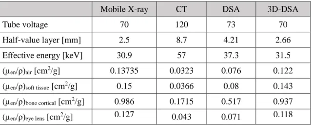
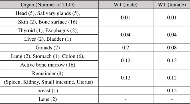
The measured value with respect to tube voltage, half-value layer, and effective energy, and the mass energy absorption coefficients of soft tissue, air, bone cortical, and lens for all examinations were shown in Table 1. To ascertain each organ dose due to exposure to diagnostic X-rays, the TLD was inserted into 44 organs of the human tissue equivalent phantom and the dose measurement of each scan was performed 3 times. A total of 68 dosimeters were inserted into tissues and organs deemed necessary to determine the effective dose by ICRP 103. The number of TLDs implanted in each tissue or organ and the organization weighting factor were indicated in Figure 2 and Table 2-1.
The human tissue equivalent phantom had features from the head to the proximal thigh.
Each anatomical region was scanned using CT, and the organ positions were determined.
For men, the total weighting factor was set to 1 by including a breast tissue weighting coefficient of 0.12 in the gonads.
Table 1. Half-value layer and effective energy in each examination, moreover mass energy absorption coefficients of bio-soft tissue and air, bone cortical, lens.
Mobile X-ray CT DSA 3D-DSA
Tube voltage 70 120 73 70
Half-value layer [mm] 2.5 8.7 4.21 2.66
Effective energy [keV] 30.9 57 37.3 31.5
(µen/ρ)air [cm2/g] 0.13735 0.0323 0.076 0.122
(µen/ρ)soft tissue [cm2/g] 0.15 0.0366 0.08 0.143
(µen/ρ)bone cortical [cm2/g] 0.986 0.1715 0.517 0.937
(µen/ρ)eye lens [cm2/g] 0.127 0.043 0.071 0.118
Figure 2. A thermoluminescent dosimeter projected on a computed tomography (CT) scout image. The dose received by each organ whose position was determined by CT imaging was necessary for effective dose evaluation per International Radiation Protection Committee Publication 103.
Table 2-1 Number of TLDs used and inserted organ and Tissue weighting factor (WT: ICRP Publication 103).
The radiation dose X [R] of the Radcal dosimeter was calculated using the air (μen/ρ) and soft tissue (μen/ρ) values of the mass energy absorption coefficient reported by Hubbell,15) and was converted to the soft tissue absorbed dose Dsoft tissue (mGy) by the
Organ (Number of TLD) WT (male) WT (female)
Head (5), Salivary glands (5),
0.01 0.01
Skin (2), Bone surface (16) Thyroid (1), Esophagus (2),
0.04 0.04
Liver (2), Bladder (1)
Gonads (2) 0.2 0.08
Lung (2), Stomach (1), Colon (6),
0.12 0.12
Active bone marrow (16) Remainder (4)
0.12 0.12
(Spleen, Kidney, Small intestine, Uterus)
breast (1) 0.12
Lens (2) - -
formula
Dsoft tissue [mGy] = 8.76 × ((μen/ρ) soft tissue / (μen/ρ) air) × X [R] (1)
The tissue dose at the organ position where the TLD was inserted was converted to the soft tissue absorbed dose using the effective energy shown in Table 1 using the conversion coefficient of the body soft tissue absorbed dose at the effective energy of the measurement of the Radcal dosimeter and the dose measurement.
The absorbed dose Dorgan of each tissue/organ was calculated from the soft tissue absorbed dose Dsoft tissue corresponding to the measured value of each TLD; it was calculated from the mass energy absorption coefficient (μen/ρ) organ value of each tissue/organ and the mass energy absorption coefficient (μen/ρ) soft tissue value using the following equation:
Dorgan = Dsoft tissue × ((μen/ρ) organ / (μen/ρ) soft tissue) (2)
Multiple TLDs were inserted into the head and salivary glands of the phantom.
As the X-ray tube was installed on the right side surface or the left side surface for head lateral radiography, the dose on the left and right sides may have been increased. A plurality of TLDs were inserted to determine the left and right dose differences in measured values, and the dose values in the head and salivary glands were ascertained
from the average values of the soft tissue absorbed dose.
For Dcolorectal, TLDs were placed in 5 locations of the ascending colon, transverse colon, descending colon, sigmoid colon, and rectum of the phantom. The average dose of the ascending colon and transverse colon was taken as the dose of the proximal portion of the large intestine Dproximal. The colorectal dose Dcolorectal was evaluated based on ICRP Publication 67 using the average dose of the descending colon, sigmoid colon, and rectum as the distal dose of distal colon Ddistal and the following
equation:
Dcolorectal = 0.57 ×Dproximal + 0.43 ×Ddistal (3)
In the lung, liver, and esophagus, the organs were divided in two sections, the dosimeter was placed at their respective centers of gravity, and the average value of the soft tissue absorption dose obtained from each TLD was taken as the organ dose.
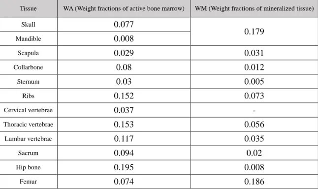
For the marrow dose Dbone marrow, the bone soft tissue absorbed dose Dabs (absorbed dose for active bone marrow) obtained from the TLD placed in the bone marrow part of each bone was divided into a plurality of bones. The weight ratio of individual active bone marrow to total bone marrow weight shown in Table 2-2 was calculated from the following formula:
Dbone marrow = Σ Dabs × WA (4)
where WA is the weight fraction of active bone marrow. The bone surface dose Dbone surface can be expressed with the following equation using the absorbed dose Dabs and the weight fractions of mineralized tissue (WM) at the same measurement point as in the case of red marrow:
Dbone surface = (ΣDabs × WM) × [(μen/ρ) bone cortical / (μen/ρ) soft tissue + 1] / 2 (5)
The effective dose E was calculated by dividing the dose of each tissue/organ calculated by equations (1) to (5) by the following equation:
E = ΣwT × HT (6)
where HT is the equivalent dose, T is the tissue or organ, and wT is the tissue weighting coefficient of the tissue.
Table 2-2. Weight Fractions of Active Bone Marrow and Mineralized Tissue (WA. WM : ICRP Publication 70).
Tissue WA (Weight fractions of active bone marrow) WM (Weight fractions of mineralized tissue)
Skull 0.077
0.179
Mandible 0.008
Scapula 0.029 0.031
Collarbone 0.08 0.012
Sternum 0.03 0.005
Ribs 0.152 0.073
Cervical vertebrae 0.037 -
Thoracic vertebrae 0.153 0.056
Lumbar vertebrae 0.117 0.035
Sacrum 0.094 0.02
Hip bone 0.195 0.008
Femur 0.074 0.186
Results
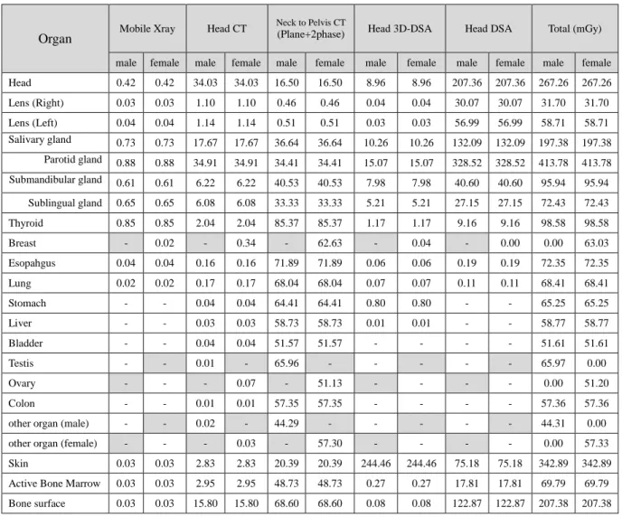
The equivalent dose and effective dose for each examination were calculated using the values in Table 2-2. The measured equivalent doses for all examinations are shown in Table 3. The results are presented per organization according to examination type. The results of equivalent dose were variable. The equivalent dose of the head is the average value of 5 TLDs, and the dose value of the salivary glands is the mean value of the parotid, submandibular, and sublingual glands. The liver, lung, and esophagus are the average values of the two sections of each divided organ.
In this experiment, we primarily measured the head, the dose received by the head and cervical regions showed the highest value. The head dose was 0.42 mGy for mobile imaging, 34.03 mGy for head CT imaging, 16.50 mGy for neck-to-pelvis CT, 8.96 mGy for 3D-DSA, and 207.36 mGy for DSA, totaling 267.26 mGy. The salivary gland dose was determined by averaging the doses of the submandibular and parotid glands, and was 0.73 mGy for mobile imaging, 17.67 mGy for head CT imaging, 36.64 mGy for neck-to-pelvis CT, 10.26 mGy for 3D-DSA, and 132.09 mGy for DSA, totaling 197.38 mGy. The parotid dose of 413.78 mGy before averaging was the highest among all organs measured. The lens dose was 0.03-0.04 mGy in head 3-direction, 1.10-1.14 mGy in head CT, 0.46-0.51 mGy in neck-to-pelvis region CT, and 0.03-0.04 mGy in head 3D-DSA. It was highest in DSA: the right crystalline lens was 30.07 mGy, the left crystalline lens was 56.99 mGy, and the left side lens dose was high. The total lens dose was 31.7-58.71 mGy.
In neck-to-pelvis CT, the dose was measured in multiple body organs, it was particularly high in the thyroid gland. The testes dose was 65.96 mGy and the ovaries dose was 51.13 mGy. The other organs received 44.29 mGy in a male and 57.3 mGy in a female. Also, a dose of 16.50 mGy was measured for the head, which was not included in the imaging range. The breast dose was 0.02 mGy in head 3-direction imaging, 0.34 mGy in head CT imaging, 62.63 mGy in the neck-to-pelvis CT, and 0.04 mGy in 3D-DSA, which was measured in most scans. In DSA, the measurement had a lower limit, and it was not measured. The breast dose was 63.03 mGy. The radiography dose received by the
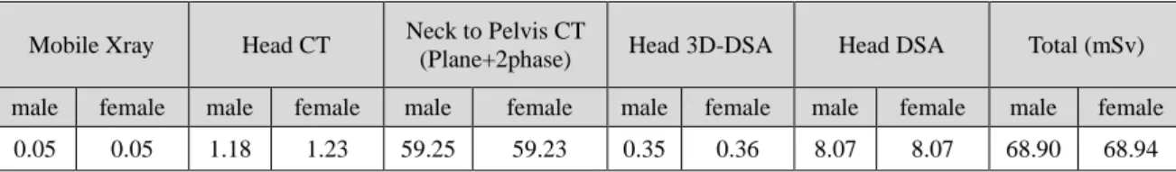
head declined sharply in the area below the manubrium because they were farther from the range of radiation exposure. The calculated effective doses for all examinations are shown in Table 4. The head dose was 0.05 mSv for mobile X-ray, 1.18-1.23 mSv for head CT, 59.23-59.25 mSv for neck-to-pelvis CT, 8.07 mSv for DSA, and 0.35-0.36 mSv for 3D-DSA. The total effective dose was 68.90-68.94 mSv when multiple radiation scans were performed on patients transported to the emergency center.
Table 3 Equivalent dose (mGy) in each organ per procedure
Organ Mobile Xray Head CT Neck to Pelvis CT
(Plane+2phase) Head 3D-DSA Head DSA Total (mGy)
male female male female male female male female male female male female Head 0.42 0.42 34.03 34.03 16.50 16.50 8.96 8.96 207.36 207.36 267.26 267.26 Lens (Right) 0.03 0.03 1.10 1.10 0.46 0.46 0.04 0.04 30.07 30.07 31.70 31.70
Lens (Left) 0.04 0.04 1.14 1.14 0.51 0.51 0.03 0.03 56.99 56.99 58.71 58.71
Salivary gland 0.73 0.73 17.67 17.67 36.64 36.64 10.26 10.26 132.09 132.09 197.38 197.38 Parotid gland 0.88 0.88 34.91 34.91 34.41 34.41 15.07 15.07 328.52 328.52 413.78 413.78 Submandibular gland 0.61 0.61 6.22 6.22 40.53 40.53 7.98 7.98 40.60 40.60 95.94 95.94
Sublingual gland 0.65 0.65 6.08 6.08 33.33 33.33 5.21 5.21 27.15 27.15 72.43 72.43
Thyroid 0.85 0.85 2.04 2.04 85.37 85.37 1.17 1.17 9.16 9.16 98.58 98.58
Breast - 0.02 - 0.34 - 62.63 - 0.04 - 0.00 0.00 63.03
Esopahgus 0.04 0.04 0.16 0.16 71.89 71.89 0.06 0.06 0.19 0.19 72.35 72.35
Lung 0.02 0.02 0.17 0.17 68.04 68.04 0.07 0.07 0.11 0.11 68.41 68.41
Stomach - - 0.04 0.04 64.41 64.41 0.80 0.80 - - 65.25 65.25
Liver - - 0.03 0.03 58.73 58.73 0.01 0.01 - - 58.77 58.77
Bladder - - 0.04 0.04 51.57 51.57 - - - - 51.61 51.61
Testis - - 0.01 - 65.96 - - - - - 65.97 0.00
Ovary - - - 0.07 - 51.13 - - - - 0.00 51.20
Colon - - 0.01 0.01 57.35 57.35 - - - - 57.36 57.36
other organ (male) - - 0.02 - 44.29 - - - - - 44.31 0.00
other organ (female) - - - 0.03 - 57.30 - - - - 0.00 57.33
Skin 0.03 0.03 2.83 2.83 20.39 20.39 244.46 244.46 75.18 75.18 342.89 342.89 Active Bone Marrow 0.03 0.03 2.95 2.95 48.73 48.73 0.27 0.27 17.81 17.81 69.79 69.79 Bone surface 0.03 0.03 15.80 15.80 68.60 68.60 0.08 0.08 122.87 122.87 207.38 207.38
Table 4 Effective dose (mSv) at each examination.
Mobile Xray Head CT Neck to Pelvis CT
(Plane+2phase) Head 3D-DSA Head DSA Total (mSv) male female male female male female male female male female male female 0.05 0.05 1.18 1.23 59.25 59.23 0.35 0.36 8.07 8.07 68.90 68.94
Discussion
In mobile head 3-direction imaging, head 3D-DSA, and head DSA, the dose received by the salivary glands was higher than that of the head, which was the imaging target. Although scout imaging of the CT examination begins at the cervical vertebra in examinations of the head, the actual imaging range is from the cerebellum to the parietal region, and the submandibular and sublingual glands are not included in the imaging range. However, the imaging range in the body axis direction is wide and the cervical spine is included in the imaging range in mobile head 3-direction imaging, head 3D-DSA, and head DSA thought they are not used for image reconstruction of the blood vessels. In addition, because the parotid gland is closer to the X-ray tube than the head, the parotid gland receives a high dose even at the salivary gland; the dose was approximately 2.5 times higher than the TLD value in the center of the head. Mobile imaging of the head side and DSA lateral imaging were conducted in the L-R direction. As a result, the lens dose was closer to the X-ray tube and the dose on the left side was higher than the dose on the right side.
The organs of the trunk received a lower dose than the head because they were farther away from the source of the radiation. During head radiography, the body area below the sternum does not appear to receive enough radiation to contribute to the effective dose.
The TLD is enclosed in a tin filter, and measurement is conducted by detecting radiation through a 1-mm slit in the filter. However, the filter has directional dependency, and even in the preliminary survey, it was too small by approximately 5-6% if the incident angle exceeded 30 degrees. As mentioned above, in mobile head Towne imaging, the angle was set at 30 degrees from the side of the head. Therefore, we suggested that the exposed dose of head Towne imaging method was measured as less than the actual dose, and underestimated the measured value.
The equivalent dose in the measured DSA examination is the value of only the imaging dose, not the fluoroscopic dose. From the results of the area dosimeter in the
investigated case, the ratio of the fluoroscopic dose to the total dose was approximately 3 to 5%. Originally, it was thought that the total of the imaging and the fluoroscopic doses was closer to the clinical exposure dose, fluoroscopic doses was due to the low ratio contributing to exposure dose of DSA, we omitted it in this measurement. The effective dose of head CT imaging was approximately 24 times that of mobile head imaging. It has been previously reported that the effective dose of head CT is 1.1 mSv to 2.0 mSv and the head dose is 41.7 mGy to 71.0 mGy7). The equivalent and effective dose values calculated in this study are considered to be included in the measurement range indicated in the previous study. The head is not included in a CT examination from the neck to pelvic region. The dose ratio of the cranial base and the upper part of the cerebrum was approximately 6 times, and as the distance from the imaging range increased, the dose decreased. It is thought that the dose was measured while not scanning because it was affected by the influence of the scattered rays from the imaging range.
It is impossible for high-energy trauma patients to raise both hands during transportation, CT imaging is performed with the patient's arm lowered. CT imaging parameters are automatically determined by extracting the body outline from the CT scout image. However, when imaging with both arms, the imaging parameter tends to be high because the body contour is widely recognized. Since both arms were not attached to the human body phantom, there is a possibility that the dose will increase further if we assume both arms are lowered.
The breast dose in the DSA examination was below the lower limit of measurement. In head 3-direction imaging, the tube enters from the anterior posterior (AP) direction and has an angle of 30 degrees in the cranio-caudal direction. The breast is not directly irradiated, but receives scattered X-rays, and the breast dose is measured. In DSA, the breast dose was not measured because the tube was behind the head and X-rays were incident from the back of the head in the caudal direction.
After summing all the examinations, the equivalent dose of the head was determined to be 267.26 mGy. This value was lower than the threshold value of 3 Gy of
temporary epilation advocated in ICRP Publication 59. The testicular dose of a CT examination was measured as 65.97 mGy; the right lens was 31.7 mGy and the left was 58.71 mGy. The thyroid gland was measured as 85.37 mGy. ICRP Publication 103 states that the threshold dose of a testis that causes temporary infertility is 150 mGy, and ICRP Publication 118 states that the cataract threshold dose is 500 mGy16). In addition, hypothyroidism develops within 1 year after exposure to high doses of radiation. For adults, the threshold dose is approximately 25 to 30 Gy17). Based on the results of this experiment, the dose does not exceed any threshold dose described in ICRP Publication 103.
In the 1990 Recommendation, the ICRP stated the principle of justification for radiation protection, and any decision to change radiation exposure may mitigate those benefits. As several radiation examinations may be performed depending on the disease, there is a concern that the radiation dose will increase. However, when considering the life expectancy of the patient, it is not realistic to offer treatment without first performing radiological scans. If the advantages and risks of radiation exposure are compared, exposure cannot be stopped completely because the potential medical benefits are high.
The results of this study, it is considered that there is a low possibility of radiation injury, there is a possibility that whole-body CT imaging may be additionally performed, in which case the effective dose may exceed 100 mSv. In order to get maximal benefit and minimal harmful effects, it is necessary to examine appropriate imaging sections and imaging parameters when scanning.
Conclusions
We evaluated organ doses and the effective dose received from mobile X-ray, head CT, neck-to-pelvic, and head DSA examinations using an anthropomorphic phantom. Estimation of patient radiation risks due to multiple radiological examinations is essential to assess imaging justification (dose reduction). Patients are exposed to considerable radiation doses. Estimation of the patient radiation risk helps staff to improve awareness of the exposure consequences of radiological examinations and to keep the patient radiation dose as low as achievable. The risk of multiple radiological examinations is directly proportional to the organ doses, and understanding the radiation dose received from each examination helps to prevent radiation damage. There is no upper limit for medical exposure due to the potential health benefits of the procedures.
The present dose data will lead to the optimization of scan parameters and will also be useful for the estimation of radiation risks for patients of emergency centers who require multiple radiological examinations.
Acknowledgments
I would like to show my greatest appreciation to Professor Hosokawa whose comments and suggestions were of inestimable value for my study. I am also indebted to Associate Professor Kudou and Assistant Professor Terashima who provided technical help and sincere encouragement.
Referrences
1) International Commission on Radiological Protection. Recommendations of the International Commission on Radiological Protection: ICRP Publication 26. Ann ICRP 26. 1:3, Pergamon Press, Oxford UK, 1977.
2) International Commission on Radiological Protection. Recommendations of the International Commission on Radiological Protection: ICRP Publication 103. Ann.
ICRP 103. 1:14, Pergamon Press, Oxford UK, 2007.
3) International Commission on Radiological Protection. Recommendations of the International Commission on Radiological Protection: ICRP Publication 60. Ann ICRP 21. 8:1–3, Pergamon Press, Oxford UK, 1991.
4) Foster BR, Anderson SW, Soto JA: CT angiography of Extremity Trauma. Tech Vasc Inter Radiol, 9(4): 156-166, 2006.
5) Berris T, Gupta R, Rehani MM: Radiation dose from cone-beam CT in neuroradiology applications. AJR Am J Roentgenol, 200: 755–761, 2013.
6) Kyriakou Y, Richter G, Dolfler A, Kalender WA: Neuroradiologic applications with routine C-arm flat panel detector CT: evaluation of patient dose measurements. AJNR Am J Neuroradiol, 29:1930–1936, 2008.
7) Nishizawa K, Maruyama T, Takayama M, Iwai K, Furuya Y: Estimation of effective dose from CT examination. Nippon Acta Radiologica, 55:763–768, 1995.
8) Maruyama T, Iwai K, Nishizawa K, Noda Y, Kumamoto Y: Organ or tissue doses, effective dose and collective effective dose from X-ray diagnosis, in Japan.
Radioisotopes, 45:761–773, 1996.
9) Thorat JD, Hwang PY: Peculiar geometric alopecia and trigeminal nerve dysfunction in a patient after Guglielmi detachable coil embolization of a ruptured aneurysm. J Stroke Cerebrovasc Dis, 16(1):40-42, 2007.
10) Wen CS, Lin SM, Chen Y, Chen JC, Wang YH, Tseng SH: Radiation-induced temporary alopecia after embolization of cerebral arteriovenous malformations. Clin
Neurol Neurosurg, 105:215-217, 2003.
11) Vano E, Arranz L, Sastre JM, Moro C, Ledo A, Gárate MT, Minguez I: Dosimetric and radiation protection considerations based on some cases of patient skin injuries in interventional cardiology. Br J Radiol, 71:510–516, 1998.
12) Cascade PN, Peterson LE, Wajszczuk WJ, Mantel J: Radiation exposure to patients undergoing percutaneous transiuminal coronary angioplasty. Am J Cardio, 59:996
−997, 1987.
13) Shope TB: Radiation induced Skin injuries from Fluoroscopy. Radiographics, 16(5):1195-1199, 1996.
14) Tsusaka M: Half-value layer measurement accuracy and effective energy evaluation.
Japanese Society of Radiological Technology, 51:540, 1995.
15) Hubbell JH, Seltzer SM: Tables of X-ray mass attenuation coefficients and mass energy-absorption coefficients 1 keV to 20 MeV for elements Z=1 to 92 and 48 additional substances of dosimetric interest, NISTIR 5632 National Institute of Standards and Technology. NISTIR , Gaithersdurg Maryland, 1995.
16) International Commission on Radiological Protection: ICRP Statement on Tissue Reactions and Early and Late Effects of Radiation in Normal Tissues and Organs – Threshold Doses for Tissue Reactions in a Radiation Protection Context. ICRP Publication 118. Ann ICRP 41. 1-2:309, Pergamon Press, Oxford UK, 2012.
17) International Commission on Radiological Protection: Nonstochastic Effects of Ionizing Radiation the skeleton. ICRP Publication 41. Ann ICRP 14. 3:45, Pergamon Press, Oxford UK, 1984.
Abstract
Accumulated exposure dose of multiple radiological examinations at an emergency center
救命救急センターにおける複数放射線検査の累積放射線量について
成田 将崇
弘前大学大学院保健学研究科保健学専攻 医療生命科学領域 放射線生命科学分野
国際放射線防護委員会(ICRP)では、放射線検査における線量を達成可能な限り低 くするよう推奨している(as low as reasonably achievable, ALARA)。しかし、救命 救急センターに搬送される患者の中には、ポータブルX線撮影やCT(computed tomography)検査、血管造影検査など複数の放射線検査を施行される患者がおり、
患者への被ばく線量の増加が懸念される。人体等価ファントムに
TLD(thermoluminescent dosimeter)線量計を挿入し、放射線検査での各臓器におけ
る等価線量と実効線量を計測した。ポータブルX線撮影、頭部から骨盤部まで の全身CT、頭部血管造影、これら施行された検査の水晶体合計等価線量は30.07
~56.99mGyであった。この線量はICRP Pub 118に記載されている白内障線量の
閾値500mGyよりも少ない。頭部ポータブルX線撮影の実効線量は0.05mSv、
全身CTは60.43mSv、脳血管造影は8.42mSv、合計実効線量は68.9mSvであっ
た。ALARAの考えをもとにX線量の最適化を行うためには、検査における被ば
く線量を把握する必要がある。測定された線量結果は、救命救急センターで複 数の検査を行った患者の放射線リスクを評価するうえで有用である。