microcircuit dynamics studied with anatomical and behavioral methods
Author Abudukeyoumu Nilupaer Degree Conferral
Date
2019‑03‑31
Degree Doctor of Philosophy Degree Referral
Number
38005甲第27号 Copyright
Information
(C) 2019 The Author.
URL http://doi.org/10.15102/1394.00000810
Okinawa Institute of Science and Technology Graduate University
Thesis submitted for the degree Doctorate of Philosophy
Cholinergic interneurons in striatal microcircuit dynamics studied with anatomical and behavioral
methods
by
Nilupaer Abudukeyoumu
Supervisor: Gordon W Arbuthnott
October, 2018
Declaration of Original and Sole Authorship
I, Nilupaer Abudukeyoumu, declare that this thesis entitled “Cholinergic interneurons in striatal microcircuit dynamics studied with anatomical, and behavioral methods” and the data presented in it are original and my own work.
I confirm that:
• No part of this work has previously been submitted for a degree at this or any other university.
• References to the work of others have been clearly acknowledged. Quotations from the work of others have been clearly indicated, and attributed to them.
• In cases where others have contributed to part of this work, such contribution has been clearly acknowledged and distinguished from my own work.
• None of this work has been previously published elsewhere, with the exception of the following:
1. N. Abudukeyoumu, M. Garcia-Munoz, Y. Nakano, G. W. Arbuthnott (2018).
" Impaired reach-to-grasp responses in mice depleted of striatal cholinergic interneurons." Society for Neuroscience, San Diego, USA (Poster of the Year Award).
2. N. Abudukeyoumu, Hernández-Flores T, M. Garcia-Munoz and G. Arbuthnott (2018). "Cholinergic modulation of striatal microcircuits." European Journal of Neuroscience.
3. Mekhail, S. P, N. Abudukeyoumu, J. Ward, G. Arbuthnott and S. N. Chormaic (2018). "Fiber-bundle-basis sparse reconstruction for high resolution wide-field microendoscopy." Biomed Opt Express 9(4): 1843-1851.
4. N. Abudukeyoumu, Y. Nakano, M. Garcia-Munoz, G. W. Arbuthnott (2017).
" Partial Deletion of Cholinergic Interneurons from Dorsolateral Striatum and
its Role in Behavioral Responses." 12th International Basal Ganglia Society
Meeting, Mérida, Yucatán. México.
5. N. Abudukeyoumu, M. Garcia-Munoz, O. P. Jaidar, G. W. Arbuthnott (2016).
"Striatal cholinergic interneurons: their depletion and its progression." Society for Neuroscience, San Diego, USA.
6. N. Abudukeyoumu, M. Garcia-Munoz, Y. Nakano, O. P. Jaidar and G.
Arbuthnott (2015). "Cholinergic interneurons sprouting in striatum?." British Neuroscience Association, Festival of Beuroscience, 23
rdMeeting, Edinburgh, UK.
7. M. Garcia-Munoz, N. Abudukeyoumu, O. P. Jaidar, Lopez-Huerta, V. G, Y.
Nakano and G. Arbuthnott (2014). "Cortical bursting activity is enhanced by blockade of astrocytic glutamate transporter." Astrocytes in Health and Neurodegenerative Disease. A joint Biochemical Society/British Neuroscience Association Focused Meeting. University College London, Institute of Child Health, UK.
Signature:
Date: October, 2018
Acknowledgements
I would like to extend thanks to the many people in our research Unit, who so generously offered help to the work presented in this thesis.
Special mention goes to my enthusiastic supervisor, Gordon Arbuthnott. My PhD has been an amazing experience with peaks and valleys and I thank Gordon wholeheartedly, not only for his tremendous academic support, patient guidance, encouragement and valuable discussion which contributed greatly to the development of my research but also for giving me wonderful opportunities to participate many international conferences around the world to extend my knowledge in the field. His supervision and great scientific guidance were always helpful.
I am also hugely appreciative to Marianela Garcia-Munoz, especially for sharing her writing and proof-reading expertise willingly, and for being so dedicated to her role as academic editor for all of my manuscripts during publishing.
Similar profound gratitude goes to former postdoc Omar Jaidar, who has been a truly dedicated mentor and technical support to learn many experimental procedures, data analysis and how to efficiently design experiments. I am particularly indebted to Omar for his constant faith in my lab work, I have very fond memories of my time there.
I would like to express my special thanks of gratitude to my colleague Teresa Hernandez Flores for the golden opportunity to work together on the topic of “Cholinergic modulation of striatal microcircuits” which was published recently.
I am also very thankful to the technicians of Brain Mechanism for Behaviour Unit, Yoko Nakano and Maria Carter who contributed greatly to the experiments presented in this thesis. Other unit members special mention goes to Teresa Hernandez Flores for teaching me new calcium imaging method, Violeta Gisselle Lopez Huerta and Esther Lai for giving me feedback for presentations and valuable questions regarding my research.
To professor Jeff Wickens, for allowing me to attend Neurobiology Research Unit
Neuroscience journal club, and for providing me with a fantastic discussion on many topics.
Also, Andrew liu, a technician in the same lab who guided loyally at the beginning of my research by teaching immunohistochemistry and answering my all of the questions. Sho Aoki and Masakazu Igarashi, for going far beyond the call of duty.
I also would like to thank professor Paul Bolam and Kenji Doya, the external and internal examiners at my PhD proposal defence. Paul Bolam made the long trip possible from UK to Okinawa and gave me insightful comments and feedbacks in regard to my proposal and research progress after the examination. Many thanks to professor Kenji Doya whom always ready to help and his constructive suggestions which were available during and after the proposal defence.
Many thanks also go to my thesis committee members Gail trip, Bern Kuhn and Mitsuhiro Yanagida whom were supported me throughout my PhD. Spending extra time to schedule the meetings and give me fully support and valuable discussions during each research progress report.
I appreciate the great supports from our lab administrator Hiroko Chinone, as well as the great supports from OIST student support section staffs and the Dean (Jeff Wickens and Ulf Skoglund). The financial support from government of Japan and Graduate school made it possible my stay in Japan and fully focus on research.
Finally, but by no means least, I would like to thank the great support and love of my
parents, siblings, extended family members and friends made it possible for me to undertake
my PhD. Their great supports once more proved the eternal truth of a proverb: It takes a village
to raise a child. Especially thanks to my mom, Talaykiz, for her continued unbelievable
support and encouragement who experienced all of the ups and downs of my research and
without her I would not have come this far.
List of abbreviations and acronyms
A list of all abbreviations that were used throughout this thesis and their full names provided in alphabetical order presented below:
5-HT Serotonin
A
1G protein coupled adenosine receptor type 1 A
2AG protein coupled adenosine receptor type 2A AAVrh10 adeno associated virus serotype 10
AC Adenylate cyclase/Adenylyl cyclase
ACh Acetylcholine
AChE Acetylcholinesterase
AMPA α-amino-3-hydroxy-5-methyl-4-isoxazolepropionic acid
BG Basal ganglia
BK Potassium current
cAMP Cyclic adenosine monophosphate
Ca
VVoltage gated calcium channel
CCK Cholecystokinin
CE Coefficient of error
ChAT Choline acetyltransferase ChIs Cholinergic interneurons
CRIs Calretinin expressing interneurons
DA Dopamine
DAPI 4′,6-diamidino-2-phenylindole
DAT Dopamine transporter
DLS Dorsolateral striatum
DMS Dorsomedial striatum
eCB endocannabinoid
EnK Enkephalin
EP Entopeduncular nucleus
FAIs Fast adapting interneurons FSIs Fast-spiking interneurons GABA Gamma-aminobutyric acid
GAD Glutamic acid decarboxylase
GFAP Glial fibrillary acidic protein
Glu Glutamate
GP Globus pallidus
GPe External globus pallidus
GPi Internal globus pallidus
HD Huntington’s disease
LDT Laterodorsal tegmental nucleus LTSIs Low threshold-spiking interneurons mAChRs Muscarinic acetylcholine receptors
MG Myasthenia gravis
MSNs Medium spiny neurons
NAc/NAcc Nucleus accumbens
nAChRs Nicotinic acetylcholine receptors NGFIs Neurogliaform interneurons
NMDA N-Methyl-D-aspartic acid or N-Methyl-D-aspartate
NOS Nitric oxide
NPY Neuropeptide Y
OFT` Open field test
P Postnatal age
PD Parkinson’s disease
pDMS Posterior dorsomedial striatum
Pf Parafascicular nucleus
PKC Protein Kinase C
PLC Phospholipase
PPN Pedunculopontine nucleus
PV Parvalbumin
PVIs Parvalbumin-containing interneurons SABIs Spontaneously active bursty interneurons
SK Small conductance calcium activated potassium channels SNc Substantia pars compacta
SNr Substantia nigra pars reticulata
SOMs Glutamate decarboxylase expressing somatostatin interneurons
SP Substance P
STN Subthalamic nucleus
TAN Tonically active neurons
THIs Tyrosine hydroxylase-expressing interneurons
VA Ventral anterior nucleus
vAChT Vesicular acetylcholine transporter vGLUT1 Vesicular glutamate transporter type 1 vGLUT2 Vesicular glutamate transporter type 2
VL Ventral lateral nucleus
VMAT1 Vesicular monoamine transporter 1
VTA Ventral tegmental area
List of Figures
Figure 1.1 The schematic diagram of direct and indirect pathways in basal ganglia circuits. 21 Figure 1.2 Schematic showing different cell types of striatal microcircuits and major inputs to
the striatum that contribute to local microcircuit dynamics. ... 23
Figure 1.3 Classification of neuronal subtypes in the striatum... 25
Figure 1.4 The synaptic connectivity probability between different neuronal cell types within striatal circuits estimated from paired recordings in slices from transgenic mice. ... 33
Figure 1.5 ACh receptor subtypes distinct expression on striatal neurons and signal transduction pathways. ... 37
Figure 2. 1 A schematic diagram for tissue preparation process for stereological systematic random sampling. ... 50
Figure 2. 2 Histological verification of specific lesion of cholinergic interneurons in the striatum. ... 55
Figure 2. 3 The ablation of ChIs in the striatum. ... 58
Figure 2. 4 vAChT positive puncta in the striatum show transient increase after ChAT-sap treatment. ... 61
Figure 2. 5 Histological verification of specific lesion of ChIs in transgenic mice striatum. . 63
Figure 2. 6 Histological verification of cholinergic neurons in PPN. ... 65
Figure 2. 7 Striatal volume comparison in control and ChAT-sap treated groups. ... 67
Figure 2. 8 Control groups volume. ... 68
Figure 2. 9 CE for ChIs counting. ... 70
Figure 2. 10 CE for terminal counting. ... 72
Figure 3. 1 Gradients of function and the subdivision of the striatum. ... 78
Figure 3. 2 Experimental time line. ... 81
Figure 3. 3 Schematic diagram for tissue preparation process for stereological systematic random sampling. ... 84
Figure 3. 4 Two behavior tests used for animals ... 88
Figure 3. 5 The confirmation of ChIs depletion in ChAT- sap injected animals after behavioral training. ... 91
Figure 3. 6 The reach to grasp task performance. ... 93
Figure 3. 7 Shaping paw preference. ... 95
Figure 3. 8 Percentage of paw preferences of mice assessed by reach-to-grasp task. ... 98
Figure 3. 9 Paw preferences changes during the training sessions. ... 99
Figure 3. 10 The distinct reach to grasp task approach in lesioned and control group. ... 100
Figure 3. 11 The various reaching approaches of ChIs lesioned mice. ... 101
Figure 3. 12 Different levels of learners within intact control group. ... 103
Figure 3. 13 Latency to retrieve pellets ... 106
Figure 3. 14 The reach-to-grasp attempts ... 109
Figure 3. 15 The number of pellets achieved with a single reach-to-grasp action. ... 111
Figure 3. 16 The number of animals used in task. ... 112
Figure 3. 17 The confirmation of ChIs depletion in mice trained for behavior experiment. 113 Figure 3. 18 The correlation analysis of ChIs and the success rate. ... 114
Figure 3. 19 The path taken by individual mouse over a 15 minute testing session in the open field based on images taken from above. ... 115
Figure 3. 20 The exploratory behavior of mice in different conditions (1) ... 116
List of Tables
Table 1. 1 Full list of systematic articles included for synaptic connectivity... 33
Table 1. 2 Full list of systematic articles included for ACh receptor subtypes ... 37
Table 2. 1 The ChAT-sap toxin treated group ... 47
Table 2. 2 The Control group... 47
Table 2. 3 The Sham group... 47
Contents
Acknowledgements ... 7
List of abbreviations and acronyms ... 9
List of Figures ... 12
List of Tables ... 14
Chapter 1 General Introduction ... 20
1.1 Basal Ganglia ... 20
1.2 Striatum ... 21
1.3 Striatal cell types ... 24
1.3.1 Medium - sized spiny projection neurons ... 25
1.3.2 Striatal interneurons ... 27
1.3.2.1 Cholinergic interneurons ... 27
1.3.2.2 Other types of interneurons ... 30
1.4 Source of acetylcholine and cholinergic receptors in the striatum ... 34
1.5 Striatal microcircuits ... 38
1.6 Striosomes and matrix ... 39
1.7 Movement disorders related to cholinergic interneurons... 40
Chapter 2 Partial lesion of cholinergic interneurons from dorsolateral striatum and its progression ... 45
2.1 Introduction ... 45
2.2 Materials and methods ... 46
2.2.1 Animals and groups ... 47
2.2.2 Stereotaxic Surgery ... 48
2.2.3 Perfusion ... 50
2.2.4 Sectioning ... 51
2.2.5 Histology ... 51
2.2.6 Estimation of the total number of ChIs and vAChT boutons using the optical fractionator ... 52
2.2.7 Statistical analysis ... 53
2.3 Results ... 54
2.3.1 The confirmation of lesion with dual immunolabelling: ChIs and vAChT ... 54
2.3.2 ChIs decrease in ChAT- sap immunotoxin injected striatum ... 56
2.3.3 vAChT positive sprouts in ChAT-sap immunotoxin injected striatum ... 59
2.3.4 The observation of ChIs lesion in eGFP transgenic mice ... 62
2.3.5 The examination of PPN cholinergic neurons ... 64
2.3.6 Striatal volume loss in ChAT-sap lesioned group as measured by stereology during cell counting. ... 66
2.3.7 Evaluation of ChIs counting accuracy with two different CEs ... 68
2.3.8 Evaluation of vAChT counting accuracy with two different CEs ... 71
2.4 Discussion ... 73
Chapter 3 Impaired reach-to-grasp responses in mice depleted of striatal cholinergic interneurons ... 77
3.1 Introduction ... 77
3.2 Methods ... 79
3.2.1 Animals and groups ... 79
3.2.2 Behavioral habituation and training ... 80
3.2.3 Stereotaxic Surgery ... 82
3.2.4 Perfusion ... 85
3.2.5 Sectioning ... 85
3.2.6 Histology ... 85
3.2.7 Estimation of the total number of ChIs in ChAT-SAP injected and behaviorally trained animals using the optical fractionator ... 86
3.2.8 Quantitative analysis of performance... 88
3.2.8.1 Placement of pellet ... 88
3.2.8.2 1
stattempt ... 88
3.2.8.3 Successful retrieval on the first reach-to-grasp ... 88
3.2.8.4 Success or fail ... 89
3.2.8.5 Grasp count ... 89
3.2.8.6 Failed reaches ... 89
3.2.8.7 Criteria for learning curve within intact control animals ... 89
3.2.9 Statistical analysis ... 90
3.2.10 Ethical approval ... 90
3.3 Results ... 90
3.3.1 Immunostaining for ChAT shows loss of cholinergic interneurons after ChAT- sap toxin administration. ... 90
3.3.2 The quantitative analysis of reach to grasp task performance in different groups ... 91
3.3.3 Paw preferences of mice in different groups ... 94
3.3.4 The variability of reaching-approaches in lesioned and non-lesioned group ... 99
3.3.5 The different levels of performance within intact control group ... 102
3.3.6 The latency to retrieve 5 pellets ... 104
3.3.7 The latency to retrieve first pellet ... 107
3.3.8 The number of reach-to-grasp attempts ... 108
3.3.10 The number of pellets achieved with a single reach-to-grasp action ... 110
3.3.11 The number of mice that did not attend to the reach-to-grasp task ... 112
3.3.12 Significant decrease of ChIs on lesioned compared to control side ... 112
3.3.12 The correlation of ChIs lesion and success rate ... 114
3.3.14 Open field test ... 115
3.4 Discussion ... 118
Chapter 4 Conclusion ... 124
4.1 Thesis summary ... 124
4.2 Impact and future work ... 124
References... 126
Cholinergic interneurons in striatal microcircuit dynamics studied with anatomical and behavioral methods
by
Nilupaer Abudukeyoumu
Submitted to the Graduate School in partial fulfillment of the requirements for the degree of Doctorate of Philosophy at the Okinawa Institute of Science and Technology Graduate
University
Abstract
Basal ganglia (BG) refer to a group of nuclei in the brain’s subcortical regions. They are
associated with cerebral cortex, thalamus and brainstem structures that perform many functions
including motor control, procedural learning and memory. The largest nucleus of the BG is
striatum that has two major neuronal cell types: medium spiny neurons and interneurons. This
thesis focuses on one class of interneurons containing choline acetyltransferase (ChAT), the
cholinergic interneurons (ChIs). This thesis investigated the role of ChIs in striatal microcircuit
dynamics by anatomical and behavior approaches. ChIs participate in voluntary motor control,
associated in learning, procedural memory, action selection, planning and execution of
movement, through strong cholinergic inputs to other striatal neurons. However, the basic
anatomical properties of ChIs after targeted lesion is poorly understood, though others have
studied the behavioral consequences of toxin injection. The immunotoxin ribosome
inactivating protein (saporin, sap) physically linked to choline acetyltransferase (ChAT-sap)
selectively damaged striatal ChIs in toxin-treated mice monitored at 2, 4 and 6 weeks after
application of the toxin. Systematic random cell counting, reach-to-grasp behavior task and
open field test (OFT) was used to explore anatomical and behavioral differences in animals
where the ChIs were destroyed in dorsolateral striatum (DLS). The thesis analyses yielded an
unexpected outcome of specific ChIs lesion in DLS where vesicular acetylcholine transporter
(vAChT) positive terminal numbers increased while the numbers of neurons themselves were
reduced. The increase in vAChT positive terminals might derive from compensatory axonal
sprouting from surviving ChIs, or from afferent axonal terminal fields of cholinergic
mesopontine neurons. But the source was not further investigated in this study. In addition, the
study showed the decreased number of ChIs in injection site with no recovery after 2, 4, and 6
weeks’ time in this study. The thesis also reports the effect of a selective depletion of ChIs
from the DLS in a reach-to-grasp task. The mean percentage of successful grasps for the last 6
training sessions was almost half of the value for intact control, and sham operated mice. These
results indicate that striatal depletion of ChIs impairs success rate, learning, motor skills in the
reach-to-grasp task was observed. These results suggest that the participation of ChIs in striatal
mediated motor learning impacts the function of the whole striatal microcircuitry. The lack of
ChIs also altered rearing behavior (total number and duration), travelled distance and speed of
movement in an open field. In addition, the results are consistent with an important
participation of acetylcholine in striatal mediated behaviors possibly by their significant
innervation from motor cortex.
Chapter 1 General Introduction
1.1 Basal Ganglia
The basal ganglia (BG) are a group of highly interconnected subcortical nucleus of varied embryonic origins, located in the forebrain region of vertebrates. The main components of BG are striatum, substantia nigra pars reticulata (SNr), substantia nigra pars compacta (SNc), subthalamic nucleus (STN), internal globus pallidus (GPi) and external globus pallidus (GPe) (Albin et al., 1989; Herrero et al., 2002). These nuclei are involved in voluntary motor control, associated learning, cognitive functions, procedural memory, action selection, planning and execution of movement (Redgrave et al., 1999; Herrero et al., 2002; Kreitzer, 2009; Doig et al., 2010; Stocco et al., 2010; Gerfen & Surmeier, 2011).
The main components of BG form a circuit with two main input nuclei: the striatum and STN; an intermediate nucleus GP (GP, external segment or GPe in primates); and two output nuclei, the entopeduncular nucleus (EP, internal segment of GP or GPi) and the SNr.
Additionally, the BG receive modulatory dopaminergic inputs from the SNc and ventral tegmental area (VTA). The former innervates the dorsal segment of the striatum while the latter nucleus sends projections to ventral striatum (Albin et al., 1989; Bolam et al., 2000; Herrero et al., 2002; Wichmann & DeLong, 2003; Gerfen & Surmeier, 2011; Havekes et al., 2011).
Basically, the striatum receives glutamatergic projections from all motor, sensory and
associational cortical regions (Bolam et al., 2000; Gerfen & Surmeier, 2011). Moreover, the
thalamus provides a second major excitatory input to the striatum (Doig et al., 2010). GP and
SNr, the output gates of the BG system provide GABAergic input axons to thalamic nuclei that
project back to frontal cortical areas. The input and output nuclei balance behavior initiation
and planning of movement due to cortex and BG system working together as a loop. BG
synapses can be modified by activity through a mechanism that depends on dopamine (DA)
(Wickens et al., 1996; Arbuthnott & Wickens, 2007; Gerfen & Surmeier, 2011) as well as the
participation of other neurotransmitters (Lovinger, 2008). The connectivity within BG and
main input into striatum can be seen in a simplified connectivity schematic diagram focused
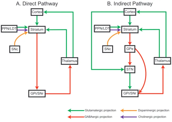
on striatum (Figure 1.1).
Figure 1.1 The schematic diagram of direct and indirect pathways in basal ganglia circuits.
A.In the direct pathway circuit, the striatum receives excitatory glutamatergic inputs from cortex, thalamus (ventral anterior nucleus (VA)/Ventral lateral nucleus (VL) complex) and PPN/LDT mid brain regions. Additionally, striatum also receives modulatory dopaminergic inputs from SNc and cholinergic inputs from PPN/LDT midbrain region. Transiently GABAergic inhibitory projections from the striatum project to tonically active GABAergic inhibitory neurons in the GPi and SNr. which in turn send GABAergic inhibitory projections to thalamus (VA/VL). Finally, thalamus sends glutamatergic excitatory projections to cortex. B. The indirect pathway includes the GPe and STN. The GPe GABAergic inhibitory neurons projects to the STN, that also directly receives glutamatergic excitatory input from cortex. The STN neurons sends excitatory projections to the GPi/STN.
1.2 Striatum
The striatum is the biggest portion of subcortical BG nuclei, known as the neostriatum or striate nucleus is the main entrance to the BG output system that play important role in coordination of motor movement, mood, decision making, motivation cognition, memory and
Glutamatergic projection GABAergic projection
Dopaminergic projection Cholinergic projection
A. Direct Pathway B. Indirect Pathway
Striatum
Thalamus Cortex
PPN/LDT
SNc
GPi/SNr
Striatum
Thalamus Cortex
PPN/LDT
SNc GPe
STN
GPi/SNr
procedural learning (Albin et al., 1989; Graybiel et al., 1994; Hikosaka et al., 2000; Packard
& Knowlton, 2002; Kreitzer & Malenka, 2008).
Structurally, the striatum in primates is divided by white matter (the internal capsule) into two large parts: caudate nucleus and putamen. A significant anatomical difference between primates and rodents is that this kind of separation is not present in BG circuits of rodents (Alexander et al., 1986; Percheron et al., 1987; Wickens, 1993). The inputs to the two compartments are different, caudate nucleus receives input from prefrontal cortex, and putamen receives input from somatosensory and motor cortex. However, even in rodent different areas have different inputs that form different functional and anatomical connections.
For instance, DLS is the target of sensorimotor cortical areas, whereas the dorsomedial striatum integrates information coming from association cortices. The striatum also includes ventral striatum and the nucleus accumbens. Striatum receives two major excitatory glutamatergic inputs from layer V and VI cortical pyramidal neurons (in rat and monkey or also layer II, III in cat) (Jones et al., 1977; Royce, 1982; Rakic et al., 1986; McGeorge & Faull, 1989).
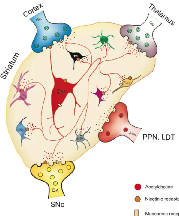
Additional prominent inputs (Figure 1.2) from other parts of BG system into striatum (e.g.
dopaminergic inputs from SNc, glutamatergic inputs from ventral VTA) increase complexity
and diversity of striatal functions in different motor control, reinforcement learning and drug
addictions (Usunoff et al., 1976).
Figure 1.2 Schematic showing different cell types of striatal microcircuits
and major inputs to the striatum that contribute to local microcircuit
dynamics.
1.3 Striatal cell types
The striatal microcircuit complexity (Figure 1.2) is increased by diversity of different neurons, interneurons, and a rich variety of neurotransmitters such as acetylcholine (ACh), DA, gamma-aminobutyric acid (GABA) serotonin (5-HT), substance P (SP) and glutamate (Glu) etc (Lynch et al., 1972; Fonnum et al., 1978).
On a macro level, there are two major cell types involved in striatal circuits (Figure 1.3): spiny cells/medium sized spiny projection neurons (MSNs) constitute 90-95% of striatal neurons and the remaining neurons are a neurochemically and morphologically distinct group of rarer aspiny local circuit neurons called interneurons, which are constitute 5-10% of the neuron population (Graveland & DiFiglia, 1985; Kawaguchi, 1993; Kawaguchi et al., 1995;
Kreitzer, 2009; Gerfen & Surmeier, 2011).
There are about 17 million corticostriatal neurons participate in the innervation of striatal microcircuits in the rat, formed from some 2.8 million neurons, that are unilaterally distributed and make GABAergic inhibitory synapses. Around half of these neurons make the direct pathway (Figure 1.1. A) and the other half form the indirect pathway (Figure 1.1. B) MSNs in rats (Oorschot, 1996; Zheng & Wilson, 2002; Wilson, 2013). Much of the literature about the basal ganglia is centered upon the differences in these two pathways, however, they are not the main focus of this thesis, nor even of the research of the unit in which these experiments were carried out. We will concentrate instead on the interneuron types including the Chi’s that are our major interest in this thesis.
The second major cell type, the interneurons, are sub-divided into two groups: ChIs and GABAergic interneurons (Kawaguchi, 1993; Kawaguchi et al., 1995; Silberberg & Bolam, 2015).
Interneurons (Figure 1.3) have been studied by [3H]-thymidine autoradiography,
morphology, chemical markers expression properties, genetic deletion, optogenetic control,
functional characteristics and electrophysiological properties (Steiner & Tseng, 2010). The
most recent count of the individual types of interneurons includes: ChIs, tyrosine hydroxylase-
expressing interneurons (THIs), low threshold interneurons (LTSIs), parvalbumin-containing
interneurons (PVIs) or fast spiking interneurons (FSIs), neuropeptide Y (NPY), NADPH
diaphorase, nitric oxide (NOS) and glutamate decarboxylase expressing somatostatin interneurons (SOM), calretinin expressing interneurons (CRIs) and spontaneously active bursty interneurons (SABIs) (Kimura et al., 1981; Bredt et al., 1991; Kawaguchi et al., 1995;
Figueredo-Cardenas et al., 1996; Wu & Parent, 2000; Tepper & Bolam, 2004; Kreitzer, 2009;
Ibanez-Sandoval et al., 2010; Tepper et al., 2010; Assous et al., 2018). However, this list is not static; in fact, subclasses of new interneurons types are being discovered and characterized and BAC transgenic mice developed to aid their study (Silberberg & Bolam, 2015). The massive external inputs, local GABAergic MSNs and interneuron circuits work together to achieve organizational and functional physiology of striatum in different behavior tasks.
Figure 1.3 Classification of neuronal subtypes in the striatum.
1.3.1 Medium - sized spiny projection neurons
MSNs are inhibitory neurons that use GABA as their main neurotransmitter. Every MSN has glutamic acid decarboxylase (GAD) (Calabresi et al., 2014) a synthetic enzyme for GABA (Kita & Kitai, 1988), GAD65 is extensively expressed by MSNs (Steiner & Tseng, 2010). MSNs make up approximately 90% of all striatal neurons (the portion of MSNs in different species varies from 90%-95%) (Kemp & Powell, 1971). The MSNs have spherical cell bodies roughly 10-20µm in diameter and up to 7 primary spiny dendrites that are 1.5-2.5µm in diameter in mammals (DiFiglia et al., 1976; Bishop et al., 1982; Graveland & DiFiglia, 1985). Their axons are arborized in a radius of 500µm and mutually inhibit other MSNs by the
Striatal neuron types
Medium spiny neurons Interneurons
iMSNs dMSNs
GABAergic Interneurons
THIs
PV/FSIs LTSIs/PLTSIs
CRIs FAIs SABIs
?
Cholinergic interneurons Type I, II, III, IV
Coexpressing NPY, NGF, SOM, NOS, NADPH.
release of GABA (Wilson & Groves, 1980). MSNs are the main target of cortical inputs and form the only output of striatum. Additionally, MSNs also receive inputs from thalamus and amygdala (DiFiglia et al., 1976; Wilson & Groves, 1980). MSNs are subdivided into two categories: direct pathway medium spiny neurons (dMSNs) and indirect pathway medium spiny neurons (iMSNs) according to the types of receptors, G-protein expression and distinct projections targets (Abbott & Nelson, 2000).
Unlike other GABAergic interneurons, glutamatergic and dopaminergic terminals, MSNs do not exhibit large nAChRs currents (Zhou et al., 2002) leaving mAChR as main target of cholinergic actions in striatal projection neurons. ACh evokes complex excitatory actions by direct modulation of several ionic currents, mainly K
+, Na
+and Ca
2+(Pineda et al., 1995;
Perez-Rosello et al., 2005; Shen et al., 2007; Carrillo-Reid et al., 2009b) and participates in plasticity of the striatum (Wang et al., 2006).
Both dMSNs and iMSNs highly express M1 receptors (Bernard et al., 1992; Yan et al., 2001; Goldberg et al., 2012) that are mainly found in spines on MSNs (Hersch et al., 1994;
Hersch & Levey, 1995; Alcantara et al., 2001). M1 receptors increase neuronal excitability of both MSNs by enhancement of persistent Na
+conductance and by directly or indirectly depressing K
+currents (Akins et al., 1990; Galarraga et al., 1999; Figueroa et al., 2002; Perez- Rosello et al., 2005; Shen et al., 2005; Shen et al., 2007; Carrillo-Reid et al., 2009b; Goldberg et al., 2012; Perez-Ramirez et al., 2015). Moreover, M1 receptors modulate Ca
2+-channels involved in neurotransmitter release and activating Ca
2+-dependent K
+-channels that shape MSNs firing pattern (Dolezal & Tucek, 1999; Galarraga et al., 1999; Perez-Rosello et al., 2005;
Perez-Burgos et al., 2008; Perez-Burgos et al., 2010). Besides its direct modulatory actions on ionic currents, ACh also plays an important role in the regulation of synaptic transmission and striatal plasticity through the endocannabinoid (eCB) system (Oldenburg & Ding, 2011). At inhibitory synapses in MSNs, M1 receptors promote eCB production and retrograde activation of CB1 receptors resulting in suppression of this inhibitory synaptic transmission (Narushima et al., 2007). While at glutamatergic synapses, M1 receptors reduce postsynaptic Ca
V1.3 currents, which promote a decrease in eCB production and a decrease in activation of presynaptic CB1 receptors (Wang et al., 2006).
On the other hand, M4 receptors are selectively expressed by dMSNs (Santiago &
Potter, 2001; Yan et al., 2001; Goldberg et al., 2012) and their activation selectively increase
excitability of dMSNs by enhancing Ca
V1 Ca
2+-channels and increase network activity (Hernandez-Flores et al., 2015). In addition, M4 receptors play an important role in the plasticity of glutamatergic cortistriatal synapses (Shen et al., 2015).
1.3.2 Striatal interneurons
1.3.2.1 Cholinergic interneurons
In general, anatomical studies have revealed ChIs immunoreactive for choline ChAT, with a large multipolar cell body of 23 - 50µm in diameter with 3 to 6 primary dendrites that extend in a radial pattern (Doig et al., 2014) and that arborize up to 1 mm (Kimura et al., 1981;
Bolam et al., 1984b; Wilson et al., 1990). Electron microscopy of rat striatal tissue performed by Doig et al. 2010; 2014, indicates that ChIs receive a prominent inhibitory input and that most of excitatory input is from thalamic afferents; a single ChI receives 8450 ± 694 connections of which the majority are symmetric. Moreover, there are approximately three times more vesicular glutamate transporter type 2 (vGLUT2)-positive thalamic terminals than vesicular glutamate transporter type 1 (vGLUT1)-positive cortical terminals in an individual ChI (Doig et al., 2014). It is important to mention that boutons expressing vGLUT1 (cortical) and vGLUT2 (thalamic) are the highest in the dorsal one-third in the rat striatum (Wouterlood et al., 2012). In addition, the density of vGLUT1 and vGLUT2 in the striatum is highly age dependent. The expression level of vGLUT2 terminals in posterior dorsomedial striatum (pDMS) dramatically decreased in aged animals. By contrast, vGLUT1 density remained unchanged by age (Matamales et al., 2016). However, since vGLUT2 is also expressed in some DA terminals in ventral striatum (Stuber et al., 2010) it is harder to isolate thalamic inputs (Abudukeyoumu et al., 2018).
In spite of the comparative small number of ChIs (Lehmann et al., 1979; Bolam et al., 1984a; Bennett & Wilson, 1999; Bennett et al., 2000; Kreitzer, 2009; Girasole & Nelson, 2015), their long and many branched axons, allow a widespread release of ACh (Bolam et al., 1984a;
Contant et al., 1996; Calabresi et al., 2000; Abudukeyoumu et al., 2018).
Similar to dopaminergic axon varicosities, cholinergic ones form few structurally
defined synaptic connections, therefore favoring a slow cholinergic volume transmission
(Descarries et al., 1997; Zhou et al., 2001; Aznavour et al., 2003; Coppola et al., 2016;
Ovsepian et al., 2016; Dunant & Gisiger, 2017). The integration of a striatal cholinergic tone established by volume and synaptic transmission is considered to act within neuronal networks to change their balance of activity to possibly initiate neuronal ensembles with specific functions (Carrillo-Reid et al., 2009a; Fuxe et al., 2012; Abudukeyoumu et al., 2018).
The spontaneously active firing characteristic of ChIs ensures the basal cholinergic tone (Kawaguchi et al., 1995; Lee et al., 1998; Wilson, 2005). These neurons have high input resistance, a broad action potential duration (Wilson et al., 1990; Tubert et al., 2016), a depolarized, and often changing, resting membrane potential that is often fixed at -60mV with a low holding current (Threlfell et al., 2012). These interneurons also called “tonically active neurons or TANs” and “autonomous pacemakers”, are able to produce action potentials at 2- 10 Hz in the absence of synaptic input (Bolam et al., 1984a; Wilson et al., 1990). Behind this tonic or pacemaking mechanism, is an interplay of several ionic conductances (Wilson et al., 1990; Pisani et al., 2007). Their pacemaker cycle begins with an initial tetrodotoxin-sensitive sodium current-induced depolarization, that leads to calcium influx from Ca
V2channels. This first calcium influx in turn, activates the calcium and voltage-activated big potassium currents (BK). This potassium influx contributes to membrane repolarization and activation of the Cav2.2 current, that in turn, activates the small-conductance calcium-activated potassium current (SK). This second potassium current induces a medium duration after- hyperpolarization (mAHP) of 100 - 200 ms, that defines the spike pattern and spike width (Kawaguchi, 1992; Bennett et al., 2000; Goldberg & Wilson, 2005). A decrease in intracellular calcium levels reduces the SK current and consequently the mAHP. The Ih inward cyclic nucleotide-gated cation current (HCN) repolarizes the membrane to about -60 mV, with a resulting inactivation of the outward potassium A-type KV4 current. At the end of the cycle, depolarization is slowed down, the persistent sodium current is activated, and the threshold for an action potential is reached, beginning a new sequence (Bennett et al., 2000; Goldberg &
Wilson, 2005; Deng et al., 2007; Pisani et al., 2007; Abudukeyoumu et al., 2018).
Another feature of ChIs is a long pause in the tonic firing, that follows bursts of action
potentials. Their intrinsic properties allow ChIs to fire in regular, irregular and in burst fashion
interspersed with long pauses (Bennett et al., 2000; Goldberg & Wilson, 2005; Wilson, 2005)
(Sanchez et al., 2011; Aceves Buendia et al., 2017). During a burst, a subthreshold
accumulation of calcium through Cav1 channels recruits an additional potassium current that
in turn, produces a long-lasting (several seconds) hyperpolarization (sAHP) (Wilson &
Goldberg, 2006; Tubert et al., 2016; Abudukeyoumu et al., 2018).
It is considered that the delta frequency activity of these interneurons results from the combination of synaptic inputs and intrinsic mechanisms (Beatty et al., 2015). A muscarinic dependent coherence between motor cortex and ChIs can be established following optogenetic stimulation at both beta and low gamma frequencies (Kondabolu et al., 2016). The reports on striatal oscillatory activity at different frequencies and the synchronization with other brain regions have been the topics of several recent publications (Brittain & Brown, 2014; Feingold et al., 2015; Sharott et al., 2017; Abudukeyoumu et al., 2018).
Recordings of striatal neurons in behaving primates revealed two cellular striatal populations (Kimura et al., 1984): phasic active neurons that show brief action potentials and low spontaneous activity or MSNs (Wilson & Groves, 1981; Apicella, 2017) and TANs that display a broader action potential and tonic spontaneous firing rate (<12 Hz) (Kimura et al., 1984; Wilson et al., 1990; Aosaki et al., 1995; Apicella, 2002; Doig et al., 2014; Apicella, 2017). Following electrophysiological criteria, TANs were considered as putative ChIs when antidromic stimulation from GP was unable to activate them (Kimura et al., 1990; Kimura et al., 1996). Moreover, in view of their morphological, electrophysiological, regional, functional and immunoreactivity similarities, TANs were identified as ChIs (Wilson et al., 1990; Aosaki et al., 1995; Bennett & Wilson, 1999; Reynolds et al., 2004; Inokawa et al., 2010; Goldberg &
Reynolds, 2011; Bradfield et al., 2013; Schulz & Reynolds, 2013; Atallah et al., 2014;
Abudukeyoumu et al., 2018).
The fact that the firing properties of TANs are similar to some GABAergic interneurons
has created confusion in their proper neuronal differentiation in extracellular recordings (Berke,
2008; Beatty et al., 2012; Gonzales et al., 2013; Gonzales & Smith, 2015; Apicella, 2017). It
would be best to identify all interneurons, including cholinergic, not only associated to their
extracellular electrophysiological characteristics but with other criteria. The systematic
approach to interneuron research being developed (Kepecs & Fishell, 2014; Wamsley & Fishell,
2017) will provide a database of properly genetically classified interneurons (e.g., mRNA-
expression profile, chemical markers expression properties). However, mRNA expression
profiles can also be variable, so a single method might not be sufficient for classification. The
future will likely bring further determination of their individual electrophysiological characteristics and integrative properties (Abudukeyoumu et al., 2018).
1.3.2.2 Other types of interneurons
The recent development in molecular technology and different types of transgenic mice has contributed immensely to the discovery of different cell types, synaptic connections and diversity of interneurons circuitry in the striatum. GABAergic interneurons are noticeably different from ChIs and GABAergic MSNs in morphology, expression of molecular markers, functional characteristics, and action potential shape. The GABAergic interneurons identified until now includes: FSI/PV
+, THI, LTS/PLTS (NPY, NGF, SOM, NOS, NADPH), CRI, FAI and SABI (Figure 1.3).
Fast-spiking interneurons (FSIs) make up around 2.6% of striatal GABAergic cell population. Among the FSI, the best-characterized type is the one that express calcium-binding protein, parvalbumin (PV), which are characterized with a dense axonal arborization of over 600µm of diameter, display a high frequency action potential firing (200-300 Hz) with no spike adaptation and electronically coupling in groups by gap junctions (Kita et al., 1990; Kawaguchi, 1993; Fukuda, 2009; Tepper et al., 2010). The duration of their action potential is short and they are distributed more often in the dorsal striatal area. They receive a powerful cortical input and use GABA as neurotransmitter to form local cell connections to modulate the functional correlation between MSNs (Kubota et al., 1993; Fukuda, 2009).
Tyrosine hydroxylase-expressing interneurons (THIs) are GABAergic neurons in striatum of different species (human, monkey, rat, and mouse) these TH
+positive interneurons also express vesicular monoamine transporter 1 (VMAT1) and DA transporter (DAT) (Betarbet et al., 1997). THIs use GABA as the main neurotransmitters but they do not release DA (Dubach et al., 1987; Betarbet et al., 1997; Meredith et al., 1999; Palfi et al., 2002; Lopez- Real et al., 2003; Tande et al., 2006; Huot & Parent, 2007; Ibanez-Sandoval et al., 2010;
Ibanez-Sandoval et al., 2015). THIs comprise four subtypes (Figure 1.3): Type I, Type II, Type
III, Type IV that constitute 60%, 13%, 6% and 21% of all striatal THIs in the striatum,
respectively. THIs are medium sized with soma ranging from 11.4 to 24µm and the shape of
soma differ such as round, ovoid and pyramidal. Aspiny and varicose dendrites are common
for all four types of THIs. Each different type of THIs has distinct electrophysiological properties such as input resistance, resting potential, action potential and firing rate for review see (Ibanez-Sandoval et al., 2010; Ibanez-Sandoval et al., 2015). TH
+projection neurons can be found in SNc and VTA brain regions, which do use DA as neurotransmitter while the TH
+interneurons of striatum do not (Kita et al., 1986; Grace & Onn, 1989; Iribe et al., 1999).
Low threshold-spiking interneurons (LTSIs) or plateau low threshold spike interneurons (PLTSIs) have tonic activity, low-threshold Ca2
+spikes and a long calcium- dependent plateau potential (Kawaguchi, 1993; Kawaguchi et al., 1995; Tepper & Bolam, 2004). They can be identified by their co-expression of NOS, NPY, the enzyme NADPH diaphorase, and SOM (Silberberg & Bolam, 2015). Among striatal interneurons, LTSIs have the least dense axonal arborization (Kawaguchi, 1993). SOM axons are distributed in the matrix area. They constitute 1-2% of the striatal cell population and vary in shape from round to triangular, with a soma of 12-33µm in diameter and long dendrites. These interneurons make symmetrical and asymmetrical synaptic contacts with other neurons. However, the symmetrical inputs are mainly situated on dendrites and spines in rats (DiFiglia & Aronin, 1982;
Takagi et al., 1983). Somatostatin expressing neurons are characterized electrophysiologically as having a large depolarizing plateau potential. Immunohistochemically, 20-40% of them express strong immunoreactivity for calcium binding protein D28K, a protein that buffers calcium inside of neurons (Kawaguchi et al., 1995; Figueredo-Cardenas et al., 1996).
Calretinin expressing interneurons (CRIs) are GABAergic interneurons that express the calcium binding protein, calretinin. They have a small aspiny soma of 7-20µm in diameter and are mostly immunoreactive for GAD67 (Kawaguchi et al., 1995; Tepper et al., 2010). In terms of their electrophysiological profile, little is known (Bennett & Bolam, 1993; Rymar et al., 2004; Tepper & Bolam, 2004; Sharott et al., 2012).
Recently, three newly identified interneurons have been described the FAI, NGFI and SABI (Ibanez-Sandoval et al., 2011; Assous et al., 2018).
Fast adapting interneurons (FAIs) are characterized by spike frequency adaptation
during repetitive action potential firing patterns and the expression of the serotonin receptor,
5-HT3 (5-HT3R) (Faust et al., 2015). Nevertheless, other interneurons also co-express 5HT3R,
like PV
+neurons that have both markers and show FSI electrophysiological features (Munoz- Manchado et al., 2016).
Neurogliaform interneurons (NGFIs), as its name indicates, share similarities with cortical NPY-expressing neurogliaform cells and express NPY. Although they have NPY; they are not NPY-LTSI (Ibanez-Sandoval et al., 2011; Silberberg & Bolam, 2015).
Spontaneously active bursty interneurons (SABIs) are medium sized GABAergic
interneurons (cell body perimeter: 73.94 ± 15.6µm), has 5-6 sparsely spiny dendrites of 50µm
in length and sparse axons arborized up to 300-500µm from cell body which have been
described based on morphology, intrinsic properties and highly varied burst-firing patterns
recorded from 5-HT3R-Cre transgenic mice (Assous et al., 2018). However, the connection
(Figure 1.4) between SABI and MSNs is not significant (around 4.1%) indicates that SABI is
not related to direct disinhibition process of MSNs, for more detailed description see (Assous
et al., 2018).
Figure 1.4 The synaptic connectivity probability between different neuronal cell types within striatal circuits estimated from paired recordings in slices from transgenic mice.
Table 1. 1 Full list of systematic articles included for synaptic connectivity Connections Related references
MSN-FSI Koos and Tepper (1999); Taverna et al. (2007); Chuhma et al. (2011) MSN-THI Ibanez-Sandoval et al. (2010); Xenias et al. (2015)
MSN-PLTS Ibanez-Sandoval et al. (2011) ChI
FAI THI
FSI
PLTS MSN
SABI 75%
22.22%
4-5%
50% 14%
75%
33%
70%
MSN-FAI Faust et al. (2015)
MSN-ChI Chuhma et al. (2011); Gonzales et al. (2013); Lim et al. (2014);
Mamaligas and Ford (2016) MSN-BABI Assous et al. (2018)
1.4 Source of acetylcholine and cholinergic receptors in the striatum
Acetylcholine is an organic chemical and was the first neurotransmitter to be discovered in the mid-1920s. Most ACh is stored in nerve endings/terminals in synaptic vesicles, ACh can be an excitatory neurotransmitter or a neuromodulator (Loewi, 1921; Mellanby, 1955;
Delcastillo et al., 1963; Del Castillo et al., 1967). The chemical structure of ACh is as follows:
ACh is synthesized by a single step reaction with choline acetyltransferase as a catalyst
in reaction (1.4.2), by contrast acetylcholinesterase (AChE) catalyzes the breakdown process of ACh (1.4.3) as following chemical structure formula:
An early study indicated that destroying possible afferent pathways to striatum “cortex, thalamus, globus pallidus or ventrotegmental area”, did not affect the activity of choline acetylase nor acetylcholinesterase (AChE) or the histochemical staining within the nucleus (McGeer et al., 1971; Lynch et al., 1972). This led to the hypothesis that interneurons were the main source of striatal ACh. We now know external sources of ACh arrive from the pedunculopontine (PPN) and laterodorsal tegmental nuclei (LDT) (Dautan et al., 2014), nonetheless, the main source of striatal ACh still are the local spontaneously active ChIs (Kitai
CH3 — N — CH2 — CH2O — C — CH3
І
ІІ
CH
І
3CH3
O 1.4.1
O ІІ
1.4.2 Acetylcoenzyme A + choline ACh + Coenzyme A
І І
H
3C O І (CH
2)
2І N(CH
3
)
3+ H
2O AChE
H
3C COO І (CH
2)
2І N(CH
3
)
3CH
2І HO
ІІ + O
І І
1.4.3
& Surmeier, 1993; Pisani et al., 2007; English et al., 2012; Goldberg et al., 2012;
Abudukeyoumu et al., 2018).
AChE is an enzyme that breaks down acetylcholine in the brain. Markers for cholinergic signaling such as ChAT, AChE and the vesicular acetylcholine transporter (vAChT) are the highest in the striatum, within the brain (Zhou et al., 2002).
The earliest study conducted by Butcher and his colleague observed that the small area of striatum (striosome/patch) that are rich for AChE in lateral part of immature striatum but inverse in the at adult brain stage (Butcher & Hodge, 1976). In addition, opiate receptors and SP immunostaining matches the AChE- and somatostatin- poor striosomes in adult rat striatum (Herkenham & Pert, 1981). Furthermore, substantia nigra dopaminergic cell terminals in the striatum appeared to be situated in patches, and the axons mesencephalic dopaminergic cell marker cholecystokinin (CCK) was also found in patches of striatum (Hokfelt et al., 1980).
The discrepancy between immature and mature brains in different species and different neuronal markers (AChE, CCK, DA, opiate receptors, enkephalin, SP) indicated that the function of some neuronal structures can change as brain maturation proceeds (Butcher &
Hodge, 1976; Chesselet & Graybiel, 1986; Crittenden & Graybiel, 2011).
In the adult, the matrix is the largest compartment and stains strongly for AChE, neurotensin receptors and calcium binding protein calbindin D28K. The thalamic parafascicular nucleus (pf) projections target ChI dendrites are distinctly distributed in matrix and avoid the striosomes stained for opiate receptors and poor in AChE (Herkenham & Pert, 1981). In addition, striatal ChIs are enriched in the matrix but many axons cross the border between patch and matrix (Kawaguchi, 1997; Crittenden et al., 2017).
At the cellular level, ACh exerts its actions through activation of two classes of receptors, muscarinic (mAChR) and nicotinic (nAChR). Muscarinic mAChRs belong to the G- protein coupled receptor family (Caulfield, 1993). These receptors are divided into group I (M
1, M
3, and M
5) and group II (M
2and M
4) (Figure 1.5). M
1receptors are coupled to G
q/11proteins via 𝛼 subunits that activate protein kinase C (PKC) and phospholipase C (PLC) and
cause activation of inositol triphosphate and diacyl-glycerol that results in an increase in
intracellular calcium. M
1receptors are found in striatal MSNs of both the direct and indirect
type. In MSNs, these receptors are located presynaptically, extrasynaptically and
postsynaptically in dendritic spine necks (Hersch & Levey, 1995; Yan et al., 2001). Group II receptors are coupled to G
iproteins, inhibit adenyl cyclase (AC) activity and close Ca
V2 calcium channels while opening K
ir3 potassium channels (Caulfield, 1993; Nathanson, 2000;
Eglen, 2006; Haga, 2013). Group II receptors act as autoreceptors on ChIs and are located mostly extrasynaptically, suggesting a role in volume neurotransmission (Bernard et al., 1998).
M
2receptors act as inhibitory heteroreceptors on striatal NPY-SOM expressing GABAergic interneurons and on corticostriatal glutamatergic terminals (Hersch et al., 1994; Bernard et al., 1998; Abudukeyoumu et al., 2018).
The high degree of similarity of the orthosteric ligand-binding site in all five types of mAChRs (Figure 1.5), is the main reason it has been difficult to identify subtype-selective ligands (Eglen, 2006; Dencker et al., 2012) and a reason why dissection of specific cholinergic effects on neuronal activity and release, have been difficult to achieve. Nevertheless, new pharmacological tools, such as the highly-specific antagonist peptide isolated from the green mamba snake venom are now being used (Jerusalinsky et al., 2000; Karlsson et al., 2000;
Rowan & Harvey, 2011; Servent et al., 2011). Similarly, positive allosteric modulators and allosteric agonists are becoming promising tools, even providing some therapeutic potential for several central nervous system diseases (Digby et al., 2010; Bock et al., 2017;
Abudukeyoumu et al., 2018).
ACh release is regulated by presynaptically located hetero- and autoreceptors. mAChRs (M
2/M
4) (Hersch et al., 1994; Ding et al., 2006) via direct G
imediated inhibition of presynaptic Ca
V2.2 and Ca
V2.1 calcium channel linked to exocytosis. Another presynaptic control of release, is regulated by the M
4auto- and hetero- receptor activation of the barium-sensitive potassium currents carried through K
ir3 potassium channels in ChIs (Yan & Surmeier, 1996;
Ding et al., 2006) and corticostriatal terminals (Calabresi et al., 1998; Abudukeyoumu et al., 2018).
nAChRs receptors (Figure 1.5) are pentameric ligand-gated ion channels that consist
of either heteromeric subunit combinations of α subunits (α2-10) and β subunits (β2-4) or
homomeric α subunits (α7) (Exley & Cragg, 2008; Gotti et al., 2009). The most common type
of nAChR in striatum is the α4β2* type (the asterisk indicates the presence of other possible
subunits in the receptor complex) (Quik & Wonnacott, 2011); this sub-composition acts as an
auto-receptor in ChIs, as postsynaptic heteroreceptor in GABAergic interneurons and as
presynaptic heteroreceptor in GABA, serotonin and DA axon terminals (Eskow Jaunarajs et al., 2015). The reported subunit composition on GABAergic interneurons and MSNs is proposed to have the α4β2* and α4α5β2* subtypes (Eskow Jaunarajs et al., 2015;
Abudukeyoumu et al., 2018). In addition, nAChRs on dopaminergic axon terminals play a major role in the potentiation and shaping of DA release within the dorsolateral striatum (Exley
& Cragg, 2008).
Figure 1.5 ACh receptor subtypes distinct expression on striatal neurons and signal transduction pathways.
Table 1. 2 Full list of systematic articles included for ACh receptor subtypes Related references
1 Hulme et al. (1990) 2 Hurst et al. (2013)
3 Eglen (2006); Ishii and Kurachi (2006)
ACh receptors
mAChRs(1) nAChRs (1)
Gi coupled (3) Gq/11 coupled (3)
Beta family(2) β2 - β4 Alpha family(2)
α2 - α10
Heteromeric combinations M2 (4) M4 (5)
α4β2
Striatum
PLTS/LTS NPY-SOM+
ChIs GABAergic interneurons
dMSNs ChIs GABAergic interneurons
Presynaptic Possynaptic Extrasynaptic
Presynaptic Possynaptic Extrasynaptic
AC/vCa2+ channels inhibition K current activation
cAMP decrease Kir 3 opening
AC inhibition K current activation
cAMP decrease
M1(6)
dMSNs iMSNs PLTS/LTS
Postsynaptic Extrasynaptic
AC inhibition K current activation
PLC increase
(IP3)/DAG Ca2+ increase
activation of PKC
M3(7)
iMSNs mRNA expressed
Postsynaptic Extrasynaptic
AC inhibition K current activation
PLC increase
(IP3)/DAG Ca2+ increase
activation of PKC
M5(8)
Maybe on nigrostriatal DA terminals
Postsynaptic Extrasynaptic
AC inhibition K current activation
PLC increase
(IP3)/DAG Ca2+ increase
activation of PKC