Pediatric Cardiology and Cardiac Surgery 32(2): 70‒77 (2016)
Review
【特集:日本小児循環器学会第
12
回教育セミナー】組織ドプラ法と僧帽弁輪および三尖弁輪収縮期移動距離
森 一博
徳島県立中央病院小児科
Tissue Doppler Imaging and Mitral or Tricuspid Annular Plane Systolic Excursion in Healthy Children
Kazuhiro Mori
Department of Pediatrics, Tokushima Prefectural Central Hospital, Tokushima, Japan
Tissue Doppler imaging (TDI) is an echocardiographic technique that uses Doppler principles to measure the velocity of myocardial motion. In adults, TDI is recommended to assess systolic and diastolic longitudinal ven- tricular function by obtaining spectral traces from the mitral and tricuspid ring in the 4-chamber view. There are many studies of left or right ventricular function assessed by TDI in various heart diseases during the pediatric period. However, age-related changes of normal values of TDI parameters make it difficult to interpret data in an individual child in actual clinical practice. This review provides normal values for various TDI parameters and discusses the clinical usefulness and limitations of TDI in the pediatric age group. Mitral or tricuspid annular plane systolic excursion is another method to assess longitudinal function of both ventricles by M-mode. This review also shows normal values and clinical implications of this simple method. Because of their ready avail- ability using conventional echocardiographic equipment, these methods should become a part of the routine assessment of cardiac function in children.
Keywords: tissue Doppler, MAPSE, TAPSE
組織ドプラ法(TDI)は,ドプラ法により心筋運動速度を測定する心エコー検査である.成人では,4腔 断面における僧帽弁輪または三尖弁輪運動のスペクトル表示から,長軸方向の収縮および拡張能を種々 の疾患で評価することが推奨されている.一方,小児では先天性心疾患や術後症例で左室または右室 機能に関して報告がなされている.しかしながら,年齢により正常値が異なることから実際の臨床の 場で広く利用されているとは言いがたい.この総説ではTDIの各種指標の小児正常値を呈示すると共 に,小児科領域でのTDI分析の有用性と問題点につき言及したい.一方,僧帽弁輪または三尖弁輪収 縮期移動距離はM-modeで両心室の長軸機能を評価する簡便な方法である.本総説ではこの指標の小 児正常値と臨床応用に関しても呈示する.これらの指標は通常の超音波診断装置で解析可能であり,
小児循環器領域での心機能解析に際して利用されるよう願っている.
はじめに
血流を観察する超音波ドプラ法では,血球の動きに よるドプラ偏位を検出し速度を測定する.一方,心筋 からの後方散乱は血球よりも信号強度が強く低速であ る.従来の血流観察ではクラッター信号として除去さ
れてきた「心筋からの散乱」を取り出し分析するのが 組織パルスドプラ法(
tissue Doppler imaging: TDI
) である.本法は1990
年前半から普及し,主に心室長 軸機能の分析に利用されている.本原稿ではTDI
の 分析に際して知っておくべき事項を,小児での応用も 交えて紹介したい.著者連絡先:〒770‒8539 徳島市蔵本町1‒10‒3 徳島県立中央病院小児科 森 一博 doi: 10.9794/jspccs.32.70
TDI
にて任意の心筋部位にサンプルボリュームを 設定すれば,その部位の組織ドプラ波形をスペクトル 表示することができる.ここでは心尖部四腔断面から 僧帽弁輪(外側)での心筋運動速度波形の解析につい て説明する.左室心筋は心外膜側と心内膜側の斜走筋,中間層の 輪状筋の
3
層構造を呈する.左室短軸断面での左室内 径短縮率は主に輪状筋の機能を評価している.一方,多くの心筋病変では心内膜下に病変が出現し,その 後,心外膜側に及ぶ.この考えに従うと,心内膜側の 長軸機能解析は「左室機能障害の早期発見」に有用で ある1).
弁輪部は,収縮期に心尖部へ移動し(
s
′波),拡張 期には拡張早期と心房収縮期に2
峰性に心基部側へ 移動する(e
′波およびa
′波).a
′波とs
′波の間には2
相性の小さな波形を認め,等容性収縮期(isovolumic contraction time: ICT
)に相当する.また,s
′波とe
′波の間にも2
相性の波形を認め,等容性拡張期(
isovolumic relaxation time: IRT
)にあたる(Fig. 1
).TDI
による心機能分析には以下のような多くの利 点がある.i
)e
′波は前負荷の影響を比較的受けにく い,ii
)断層画面が不鮮明な症例でも良好な分析がで きる,iii
)時間分解能が3
〜4 msec
と極めて良い,iv
)左室内径短縮率では得られない長軸方向の心機能 を評価できる,v
)通常の心エコー装置で解析可能で ある.一方,欠点としては,i
)角度依存性である,ii
)心臓全体の動き(translation
)や周辺組織の収縮 の影響を受ける(tethering
),iii
)局所壁運動異常が ある場合には心室全体の心機能を反映しない,などが 挙げられる.ここで,解析に適した記録を得るためのコツについ て触れたい.まず弁輪の運動方向とドプラカーソルは できるだけ平行に設定する.また,良好なドプラ波形 を検出できるように,サンプルボリューム幅はやや広 めとする(著者は学童で
5
〜6 mm
にしている).ドプ左室心筋より長軸方向の運動が主体であり,いずれの 年齢でも右室
s
′>左室s
′である点は興味深い2, 3).左室
s
′波はしばしば2
峰性となる(Fig. 1
).その成 因は不明であるが,Marciniak
らは,三尖弁輪から心 尖方向へ向かう大きな右室s
′波が僧帽弁輪からの左室s′
波とぶつかることで左室s′
波にdip
を形成すると指 摘した4).一方,Oki
らはs
1′波が左室心内膜側の斜 走筋(長軸方向の運動に関与)を,s
2′波は中層の輪 状筋(円周方向の運動に関与)の運動を反映している とし,長軸機能に関与するs
1′波の解析が重要である と報告した5).虚血や線維化で局所的に収縮能が低下した心筋で は,拡張早期に心筋が短縮する場合がある(
Fig. 4
).すなわち,周辺の正常心筋が収縮する際には異常心 筋は引き延ばされる.その後,正常部位の収縮が終 る拡張早期に,延ばされたバネが戻るような受動的 な動きとして短縮する.この拡張早期の心筋短縮は
post-systolic shortening
(PSS
)と言われており,有 効な収縮ではない6).このような心筋では収縮期運動 速度s
′自体は低下している点に注意すべきである.僧帽弁輪拡張早期運動速度e′
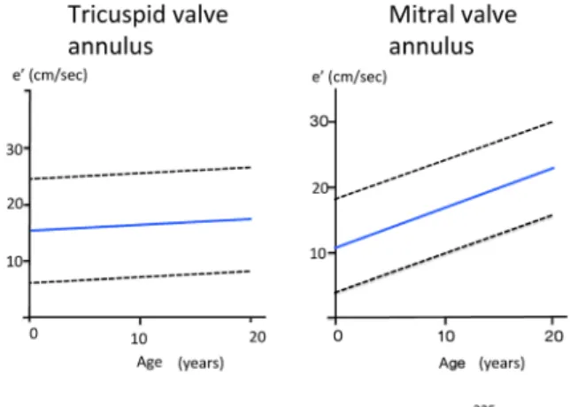
e′
も,年齢(体格)に伴い増加する(Fig. 5
).乳児 期早期の急速な増高は左室拡張能の成熟を示唆して いる.なお,右室のe
′は年齢による変化が少ない7)(
Fig. 6
).e
′は前負荷の影響を比較的受けにくく,一般に左室 拡張能の低下に伴い減高を続ける.一方,僧帽弁口拡 張早期血流速度E
は弛緩障害では減高するが,左室 コンプライアンスが極度に低下すると増高し急峻とな る.そのため,E/e
′が左室充満圧の評価に利用されて いる.小児では,出生直後の新生児ではE/e
′が高値と なる.出生直後は動脈管のため僧帽弁拡張早期血流速 度E
が増高するが,前負荷の影響を受けにくいe
′の 増高が少ないためである.その後,左室拡張能の成熟に伴い
e
′が増高するため,E/e
′は低下する8)(Fig. 7
).E/e
′は左室充満圧を推定するうえで有用とされてき たが,以下の状態では注意が必要である.i
)高心拍 出状態では,左室充満圧が上昇していても,e
′のみな らずE
も上昇するためE/e
′は増加しない9).ii
)極度 の心機能低下例では高い左房圧が弁輪を押し下げるこ とで高いe
′を形成しE/e
′は高値とならない10).e
′波の解析では僧帽弁血流E
波との時相関係にも注 目するとよい.正常では,e
′波(左室伸展)の開始時相は
E
波の開始時相とほぼ同時か若干先行する.し かし,高度の拡張機能低下例では,E
波の出現後,大 きく遅延してe
′波が出現する11)(Fig. 8
).Tei index
ICT
は左室最大陽性dP/dt
と相関し,収縮能の低 下で延長する.またIRT
は左室最大陰性dP/dt
と相 関し,拡張能低下で延長する.駆出時間(ejection
time: ET
)は1
回心拍出量が低下すると短縮する.Fig. 1 Tissue Doppler imaging (TDI) recorded from the apical 4-chamber view
Left: A sample volume is targeted on any part of myocardium. Here, the sample volume is placed on the mitral valve annulus (its lateral or septal junctions) or on the tricuspid valve annulus. Right: Recording from the lateral junction of the mitral annulus. The Doppler spectrum shows systolic waves (s1ʼ and s2ʼ), an early diastolic wave (e′), and a late diastolic wave (a′). ECG=electrocardiogram; ICT=isovolumic contraction time; IRT=isovolumic relaxation time; LA=left atrium; LV=left ventricle; RA=right atrium; RV=right ventricle; TDI=tissue Doppler imaging.
Fig. 2 Inadequate and adequate recordings of tissue Doppler imaging
The sample volume is placed on the lateral mitral valve annulus. A: Inadequate recording. Because of excess Doppler gain, the maximal velocity boundary of the spectral envelope is unclear. The vertical lineal Doppler artifacts lead to over- estimation of each velocity. B: Adequate recording. After adjustment of the Doppler gain, the spectral envelope in the Doppler recording is clearly recognized. ICT and IRT can be easily measured. s′=systolic wall motion velocity; e′=early diastolic wall motion velocity; ICT=isovolumic contraction time; IRT=isovolumic relaxation time.
用いた右室心機能評価が広く認められており,右室
s
′<10 cm/sec, Tei index
>0.55, E/e
′>6
は「右心機能 の異常」と判定される13).また,iv
)の観点から,右室機能低下の合併が左心不全患者の予後を規定する との指摘がある.
Damy
らは成人の左心不全患者で右 室s′
<9.5 cm/sec
は他の右室機能指標に比し予後を予 測しえたと報告した14).小児の検討では,術後に右 室圧負荷や容量負荷が残存すると経年的に右室s
′が低 下する15).房室弁輪収縮期移動距離 僧帽弁輪収縮期移動距離(MAPSE)
ここまで
TDI
を用いた長軸機能に関して概説し てきたが,M-mode
を用いても長軸機能の評価は Fig. 3 The relationship between the systolic wallmotion velocity (s′) and age
The sample volume is placed on the lateral mitral valve annulus. Means and 95% prediction bands are shown. r=0.73.
Fig. 4 Post-systolic shortening
A: TDI recording on the lateral mitral valve annulus from the 4-chamber view in a normal subject. B: In ischemic or fibrotic myocardium, s′ is reduced and followed by a deformation after systole, the so-called post-systolic shortening (PSS: shown by arrow). PSS is explained in part by passive recoil, which releases energy stored in the scar tissue during systolic intraventricular unloading. TDI=tissue Doppler imaging; syst=systolic period; s′=systolic wall motion velocity;
e′=early diastolic wall motion velocity.
可能である.すなわち,心尖部四腔断面で外側僧帽 弁輪にカーソルを設定し,その移動距離を測定する
(
Figs. 11, 12
).この指標はs
′波を時間積分したことに なり,最も簡単な左室長軸機能指標と言える.測定に 際しては,弁輪運動方向がM-mode
のビーム方向に 平行となるよう工夫する.Diller
らはFallot
四徴症術後413
例の検討から,MAPSE
が術後突然死や致死的不整脈出現の予測に有用であることを示した16).
Fig. 5 The relationship between the early diastolic wall motion velocity (e′) and age
The sample volume is placed on the lateral mitral valve annulus. Means and 95% prediction bands are shown. r=0.83.
Fig. 6 Comparison between early diastolic wall motion velocity of the tricuspid valve an- nulus and that of the mitral valve annulus in relation to age
Left panel: Early diastolic wall motion velocity (e′) of the tricuspid valve annulus shows a marginal increase according to age. r=0.13. Right panel:
Early diastolic wall motion velocity (e′) of the mitral valve annulus significantly increases according to age. r=0.70. Means and 95% prediction bands are shown. Arranged from reference 7 .
Fig. 7 The relationship between E/e′ ratio and age
The sample volume is placed on the lateral mitral valve annulus. The E/e′ ratio is significantly increased during the neonatal period. Thereafter, this index slowly decreases with aging. E=mitral flow velocity during early diastole; e′=early diastol- ic wall motion velocity of the mitral valve annulus.
Means and 95% prediction bands are shown. r=
−0.62.
Fig. 8 Temporal relationship between the onset of mitral flow and mitral annulus velocity during early diastole
Left: In a normal subject, the onset of the e′ wave (red line) is at the time same as or a little earlier than that of the E wave (blue line). Right: In a sub- ject with significantly increased left ventricular fill- ing pressure, the delay of the onset of the e′ wave is recognized, compared with the onset of the E wave. The dotted line shows the second heart sound. E=mitral flow velocity during early diastole;
e′=early diastolic wall motion velocity of the mitral valve annulus; ECG=electrocardiogram; IRT
=isovolumic relaxation time; PCG=phonocardio- gram; TDI=tissue Doppler imaging.
Fig. 13
は,学校心電図異常で紹介された左室緻密 化障害(15
歳男児)の1
例である.左室内径短縮率 は29
%で,軽度低下を認めた.左室長軸方向のTDI
ではs
′とe
′共に軽度低下していた.MAPSE
は著明に 低下しており,心筋障害が強く示唆された.三尖弁輪収縮期移動距離(TAPSE)
三尖弁輪の収縮期移動距離も心尖部四腔断面か
ら
M-mode
法で測定可能である.上記のように右室形態は複雑であり,
TAPSE
は簡便で再現性のよ い右室長軸機能指標として提唱されている.正常 値 はMAPSE
と 同 様 に 体 格(年 齢) と 共 に 増 加 す る17)(Fig. 14
).Forfia
らは成人の肺動脈性肺高血圧 患者で,TAPSE
<1.8 cm
が予後不良であると報告し た18).Moceri
ら はEisenmenger
症 候 群 の 心 エ コ ー 指標を検討し,TAPSE
<15 mm
,右室の収縮時間/Fig. 9 Schema for measurement of the Tei index
The Tei index is calculated as (a‒b)/b, where a is the interval between cessation of the a′ wave and onset of the subsequent e′ wave, and b is the inter- val of the s′ wave. ET=ejection time; ICT=isovo- lumic contraction time; IRT=isovolumic relaxation time; a′=late diastolic wall motion velocity; e′= early diastolic wall motion velocity; s′=systolic wall motion velocity.
Fig. 10 The relationship between the Tei index and age
The sample volume is placed on the lateral mitral valve annulus. The Tei index is significantly increased during the early neonatal period. There- after, the change in this index according to age is minimal. Means and 95% prediction bands are shown. r=−0.49.
Fig. 11 Measurement of MAPSE using the M-mode echocardiography
In the apical 4-chamber view, MAPSE is mea- sured as the longitudinal mitral annular motion from the apex using M-mode echocardiography.
It is measured as the distance between the peak and the bottom of the M-mode tracing curve. LA
=left atrium; LV=left ventricle; RA=right atrium;
RV=right ventricle; MAPSE=mitral annular plane systolic excursion.
Fig. 12 The relationship between MAPSE and age
MAPSE is significantly increased with aging.
MAPSE=mitral annular plane systolic excursion.
Means and 95% prediction bands are shown. r= 0.89.
拡張時間>
1.5
,右房面積>25 cm
2,(右房/左房面積 比>1.5
)の症例が予後不良であることを示した19).Fallot
四徴症術後症例でもTAPSE
が検討されており20),今後も多くの疾患で他の右心機能指標との対 比がなされていくであろう.
おわりに
TDI
およびMAPSE
とTAPSE
は,市販のどの心臓 超音波装置を用いても計測できる簡便な「長軸方向の 心機能指標」である.今後,種々の小児心疾患で臨床 応用されていくことを期待する.謝 辞
原稿を終えるにあたり,各種指標の正常曲線作成の ためにご尽力いただいた徳島県立中央病院小児科の井 上美紀先生,超音波検査室の技師の皆様に深謝いたし ます.
Fig. 13 MAPSE in a patient with left ventricular noncompaction
A 15-year-old boy was referred to our hospital because of ECG abnormality found during heart screening for school- children. A: ECG shows ST‒T change in the left precordial leads. B: Shortening fraction of the left ventricle is margin- ally decreased to 29%. C: Tissue Doppler imaging recorded from the lateral mitral valve annulus shows decreased velocity of systolic (s′) and early diastolic waves (e′). D: MAPSE recorded by M-mode is significantly decreased to 9.7 mm, indicating a distinct abnormality of the longitudinal systolic function of the left ventricle. MAPSE=mitral annular plane systolic excursion; ECG=electrocardiogram.
Fig. 14 The relationship between TAPSE and age
The solid line shows the mean value of TAPSE in relation to age. Sex-segregated curves are also shown. The dotted lines represent ±2 SD and ±3 SD, respectively. TAPSE=tricuspid annular plane systolic excursion. Revised with permission by Dr. Hashimoto (Ref. 17).
ular interaction as a possible mechanism for the presence of biphasic systolic velocity profile in normal left ventric- ular free walls. Heart 2008; 94: 1058‒1064
5) Oki T, Tabata T, Mishiro Y, et al: Pulsed tissue Doppler imaging of left ventricular systolic and diastolic wall mo- tion velocities to evaluate differences between long and short axes in healthy subjects. J Am Soc Echocardiogr 1999; 12: 308‒313
6) Hyler S, Pischke SE, Halvorsen PS, et al: Continuous monitoring of regional function by a miniaturized ultra- sound transducer allows early quantification of low-grade myocardial ischemia. J Am Soc Echocardiogr 2015; 48:
486‒494
7) Eidem BW, McMahon CJ, Cohen RR, et al: Impact of car- diac growth on Doppler tissue imaging velocities: A study in healthy children. J Am Soc Echocardiogr 2004; 17:
212‒221
8) 森 一博:左室拡張不全を来す疾患.小児心疾患.大木 崇,竹中 克編集:拡張期学.東京,文光堂,2010, pp 180‒185
9) Masutani S, Little WC, Hasegawa H, et al: Restrictive left ventricular filling pattern does not result from increased left atrial pressure alone. Circulation 2008; 117: 1550‒ 10) Mullens W, Borowski AG, Curtin RJ, et al: Tissue Doppler 1554
imaging in the estimation of intracardiac filling pressure in decompensated patients with advance systolic heart
right ventricular systolic echocardiographic parameters to predict adverse outcomes in chronic heart failure. Eur J Heart Fail 2009; 11: 818‒824
15) Koestenberger M, Nagel B, Ravekes W, et al: Tricuspid annular peak systolic velocity (s′) in children and young adults with pulmonary hypertension secondary to con- genital heart diseases, and in those with repaired tetralo- gy of Fallot: Echocardiography and MRI data. J Am Soc Echocardiogr 2012; 25: 1041‒1049
16) Diller GP, Kempny A, Liodakis E, et al: Left ventricular longitudinal function predicts life-threatening ventricular arrhythmia and death in adults with repaired tetralogy of Fallot. Circulation 2012; 125: 2440‒2446
17) Hashimoto I, Watanabe K, Kaneda H: Z-values of tricus- pid annular plane systolic excursion in Japanese children.
Pediatr Int 2015; 57: 199‒204
18) Forfia PR, Vachiery JL: Echocardiography in pulmonary arterial hypertension. Am J Cardiol 2012; 110 Suppl:
16S‒24S
19) Moceri P, Dimopoulos K, Liodakis E, et al: Echocardio- graphic predictors of outcome in Eisenmenger syndrome.
Circulation 2012; 126: 1461‒1468
20) Mercer-Rosa L, Parnell A, Forfia PR, et al: Tricuspid an- nular plane systolic excursion in the assessment of right ventricular function in children and adolescents after repair of tetralogy of Fallot. J Am Soc Echocardiogr 2013;
26: 1322‒1329