13 :187
<原
著>
Pure
sensory
stroke
を 呈 し た 延 髄 の lacunar
infarct
中尾
直之
久保
謙二
森脇
宏
要 旨:Pure
sensory stroke を 呈 した 延 髄 の lacunar infarct を MRI に て描 出 しえ た1例
を
報 告 す る.症 例 は58歳 男 性 で,右 半 身 の しび れ 感 を 主 訴 に て 来 院 した.神
経 学 的 検 査 で は顔 面
を除 く右 半 身 の 温 痛 覚 の 低 下 を 認 め た.そ の 他 の 神 経 脱 落 症 状 は認 め な か った.CTス
キ ャ ンで
は 明 らか な異 常 を 描 出 し え な か った が,MRIに
て延 髄 の左 辺 縁 す な わ ち 左 外 側 脊 髄 視 床 路 に ほ
ぼ一 致 す る部 位 に lacunar infarct を 確 認 した.
Key words : lacunar
infarction,
pure sensory
stroke,
MRI,
medulla
oblongata
(脳 卒 中 13 : 187-191 ,1991)
1. は じ め に
1965年,Fisherは 片 側 顔 面,上 下 肢 の 知 覚 障 害 の み
か ら な る 症 候 群 を pure sensory stroke (以 下PSS)と 称 し,そ の 原 因 を 反 対 側 の 視 床 後 腹 側 核 のlacunar
in-farctに 帰 し た1).以 来,視 床 以 外 す な わ ち thalamocor-tical sensory pathway2)3)や subthalamic sensory
pathway4)∼6)の 病 変 でPSSを 呈 し た 症 例 が 報 告 さ れ
て い る.一 方,subthalamic sensory pathway の 病 変
と し て は 中 脳 出 血4),橋 の 小 梗 塞5)や 出 血6)な ど が 報 告
され て い る が,延 髄 病 変 に つ い て の 報 告 は 筆 者 ら が 検
索 し 得 た 範 囲 で は 見 当 た ら な い.今 回,我 々 はPSSを
呈 し た 延 髄 の lacunar infarct をMRIに て 確 認 し 得 た 症 例 を 経 験 し た の で 若 干 の 文 献 的 考 察 を 加 え て 報 告 す る. II. 症 例 患者:58歳,男 性. 主 訴:右 半 身 の し び れ. 既 往 歴:5年 前 よ り高 血 圧 を 指 摘 され て い た が 降 圧 剤 は 服 用 して い な か つ た. 家族 歴:特 記 す べ き こ とな し. 現病 歴:平 成1年5月14日 起 床 時,右 半 身 の し び れ 感 に 気 付 い た.こ の しび れ 感 の た め 入 浴 時 も 右 半 身 の 温 覚 は 左 に 比 べ 鈍 か っ た.5月16日,同 し び れ 感 が 持 続 す るた め 当 院 を 受 診 した. 入 院 時 所 見 二神 経 学 的 に は 意 識 清 明 で 構 音 障 害 は 認 め られ な か っ た.嚥 下 機 能 は 正 常 で 各 脳 神 経 に 異 常 を 認 め な か っ た.四 肢 筋 力 は 正 常 で 建 反 射 に 左 右 差 を 認 め ず,病 的 反 射 も認 め な か っ た.ま た,小 脳 失 調 を 示 唆 さ せ る 所 見 も認 め ら れ な か った.顔 面 を 除 く右 半 身 の 温 痛 覚 の低 下 が 認 め ら れ た が,位 置 覚,振 動 覚 な ど の 深 部 知 覚 や 触 覚 に は 異 常 を 認 め な か っ た.理 学 的 所 見 で は170/100mmHgと 高 血 圧 を 認 め る 以 外 特 に 異 常 を 認 め な か った.ま た,一 般 血 液,生 化 学 検 査 も正 常 範 囲 内 で あ った. 神 経 放 射 線 学 的 所 見:入 院 時 の 頭 部CTで は 特 に 異 常 所 見 を 認 め な か っ た.脳 血 管 撮 影 の4-vessel study で は 頭 蓋 内 外 の 血 管 に 軽 度 の 動 脈 硬 化 を 認 め る の み で,椎 骨 脳 底 動 脈 系 に も 閉 塞 性 病 変 な ど の 異 常 所 見 は 認 め ら れ な か っ た.第40病 日に 施 行 したMRIで は,延 髄 上 部 の 左 側 辺 縁 部 に 直 径 約2mmのT1強 調 画 像 で 低 信 号(Fig.1,2),T2強 調 画 像 で 高 信 号(Fig.3)を 呈 す る 病 巣 を 認 め,同 部 位 の 梗 塞 を 思 わ せ た.一 方,入 院 後 の 一 連 のCTで はMRIで 描 出 さ れ た 延 髄 病 変 は 後 頭 蓋 窩 の 骨 の beam hardening artifact の た め 明 ら か で は な か っ た 。 入 院 後 経 過:延 髄 の lacunar infarct の 診 断 に て 高 血 圧 の コ ン トロ ー ル を 中 心 と し た 治 療 を 行 っ た.第7 病 日 頃 か ら右 半 身 の 温 痛 覚 低 下 は 軽 減 した も の の 次 第 にdysesthesiaの 要 素 が 加 わ り,約1年 後 の 現 在 も な お こ れ ら の 知 覚 障 害 は 持 続 して い る. III. 考 察
Fisher1)が pure sensory stroke (PSS)の 原 因 と し て 視 床 の 後 腹 側 核 の lacunar infarct を 提 唱 し て 以 来,
こ の 説 を 支 持 す るCT,MRIな ど の 神 経 放 射 線 学 的 所
見 や 剖 検 所 見 が 報 告 さ れ て い る3)7)∼11).し か しFisher
Fig. 1 Frontal T1-weighted magnetic resonance image showing a small area of decreased signal in the left marginal area of the upper medulla.
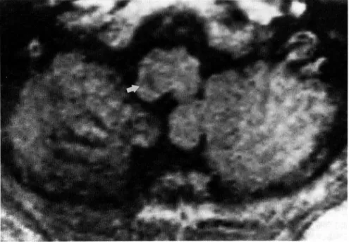
Fig. 2 Axial Ti-weighted magnetic resonance image showing a small area of decreased signal in the left margial area at the junction of the ventral and the dorsal portion of the medulla (arrow).
Pure sensory stroke を 呈 し た 延 髄 の lacunar infarct 13 :189
は最 初 にPSSを 記 述 し た 時,延 髄 か ら 大 脳 皮 質
のsen-sory pathway の い か な る 部 位 の 病 変 で もPSSは 起 こ
り うる と 述 べ て い る1).実 際,頭 頂 葉 皮 質 の 小 梗 塞12)や
thalamocortiacl sensory pathway の 病 変 と し て 内 包 後 脚2),放 射 冠3)の lacunar infarct な ど が 報 告 さ れ て
い る.さ ら に,中 脳 出 血4),橋 の 出 血5)やlacunar
infarct6)も 脳 幹 を 走 行 す る sensory pathway を 選 択 的 に障 害 し う る. 本 症 例 で の 一 連 のCTは 脳 幹 の 病 変 を 明 ら か に し え なか っ た が,MRIに て 延 髄 上 部 の 左 側 辺 縁 部 にT1強 調 画 像 で 低 信 号 域,T2強 調 画 像 で 高 信 号 域 と し て 描 出 され る直 径 約2mmの 病 巣,す な わ ち lacunar infarct を 認 め た.こ の 梗 塞 巣 の 局 在 はFig.4が 示 す よ うに 延 髄 左 側 の 外 側 脊 髄 視 床 路 に ほ ぼ 一 致 す る と 考 え ら れ る.さ らに,本 症 例 の 知 覚 障 害 は 温 痛 覚 の み に 認 め ら れ 内 側 毛 帯 が 関 与 す る深 部 知 覚 は 保 た れ て お り,MRI に 示 され た 病 巣 局 在 に 合 致 す る.Fisherが 指 摘 す る よ う に1)脳幹 病 変 で 脊 髄 視 床 路 と内 側 毛 帯 が 同 時 に 障 害 され,こ れ らの 知 覚 以 外 の 機 能 が 正 常 で あ る こ と(す な わ ちPSS)は 考 え に く い.ま た 逆 に脳 幹 か ら視 床, さ ら に 内 包 後 脚,放 線 冠 を 経 て 大 脳 皮 質 に 至 る 経 路 の 各 部 位 で のsensorymodalityの 機 能 局 在 を 考 慮 す る Fig. 3 Frontal T2-weighted magnetic resonance image showing a small area of
increased signal in the left marginal area of the upper medulla.
Fig. 4 Schematic diagram showing the topography of the lesion in the present case (oblique lines).
脈 な どか らの 内 側 延 髄 枝 な どに 分 類 され て い る13).本 症 例 で は 脳 血 管 撮 影 上,椎 骨 脳 底 動 脈 系 の 主 幹 動 脈 や そ の 分 枝 に 異 常 所 見 は な く,lacunar infarct の 原 因 と し て 従 来 か ら 言 わ れ て い る類 線 維 素 性 壊 死 や リポ ヒ ア ン症 な ど の 高 血 圧 性 変 化14)が外 側 延 髄 枝 に 生 じ た と考 え られ る. Fisher に よ る と15) lacunar infarct の 好 発 部 位 は,レ ン ズ 核41.2%,橋16.7%,視 床13.2%,尾 状 核10.3%,内 包 − 放 線 冠7.4%の 順 で あ り,延 髄 の lacunar infarct は 稀 と い え る.Rothrock らのMRIに よ る lacunar infarct の 検 討 で も10),30例 中1例 に 認 め る の み で あ る.勝 木 ら16)は脳 幹 穿 通 動 脈 の 分 枝 状 態 を 詳 細 に 観 察 し,延 髄 へ の 穿 通 枝 は 本 幹 の 血 流 に 対 し方 向 を 変 え る こ と な く鋭 角 に 分 枝 し て い るが,橋 で は 血 流 に 対 し 鈍 角 に,逆 行 性 に 分 枝 し て お り,レ ン ズ 核 線 条 体 動 脈 の そ れ と類 似 して い る と述 べ て い る.こ の よ う な事 実 か ら延 髄 の 穿 通 枝 は 橋 の そ れ や レ ン ズ 核 線 条 体 動 脈 に 比 べ,上 記 の 高 血 圧 性 変 化 を 受 け に く い の か も し れ な い. 本 症 例 で はMRIが 病 巣 診 断 に 威 力 を 発 揮 した.今 後 こ の コ ン トラ ス ト分 解 能 が 高 く,骨 の beam harden-ing artifact を 受 け な いMRIに よ り脳 幹 の lacunar infarctな どの 微 細 病 変 の 検 出 が 可 能 と な り,そ の 病 巣 局 在 と臨 床 症 状 と の 対 比 が 容 易 と な ろ う.
文
献
1) Fisher CM : Pure sensory stroke involving face, arm and leg. Neurology 15 : 76-80, 1965 2) Groothuis DR, Duncan GW, Fisher CM : The human thalamocortical sensory path in the internal capsule : Evidence from a small capsu-lar hemorrhage causing a pure sensory stroke. Ann Neurol 2: 328-331, 1977
3) Rosenberg NL, Koller R: Computed tomogra-phy and pure sensory stroke. Neurology 321 : 217-220, 1981
1986
6) Hommel M, Besson G, Pollak P, et al : Pure
sensory stroke due to a pontine lacune. Stroke
20: 406-408, 1989
7) Fisher CM : Thalamic pure sensory stroke : A
pathological
study. Neurology 28: 1141-1144,
1978
8) Landi G, Anzalone
N, Vaccari U: CT scan
evidence of posterolateral
thalamic
infarction
in pure sensory stroke. J Neurol Neurosurg
Psychiatry 47 : 570-571, 1984
9) Grosselink EL, Lodder J:
Pure sensory stroke
with lacunar infarction in the posterior ventral
thalamus on CT. Clin Neurol Neurosurg 87 : 45
−46,1985
10) Rothrock
JF, Lyden PD, Hesselink JR, et al :
Brain magnetic resonance imaging in the
evalu-ation of lacunar stroke. Stroke 18: 781-786,
1987
11) Sacco RL, Bello JA, Traub R, et al : Selective
proprioceptive
loss from a thalamic
stroke.
Stroke 18 : 1160-1163, 1987
12) Derouense C, Mas JL, Bolgert F, et al : Pure
sensory stroke caused by a small cortical
infar-ct in the middle cerebral
artery
territory.
Stroke 15 : 660-662, 1984
13) 後 藤 昇:脳 血 管 の 解 剖.診 断 と 治 療75:1738 −1743
,1987
14) Fisher CM : Lacunar strokes and infarcts : A
review. Neurology 32 : 871-876, 1982
15) Fisher CM : Lacunes ; small, deep cerebral
infarcts. Neurology 15 : 774-784, 1965
16) 勝 木 司 馬 之助,浦 本 龍 生,本 里 義 之 ら:脳 幹 穿通 動脈 分 枝 機構 の 特 異 性 に つ い て.医 学 研 究26: 3049−3052,1956
Pure sensory stroke を 呈 し た 延 髄 の lacunar infarct 13 :191
Abstract
Pure
sensory
stroke
caused
by a lacune
in the
medulla
oblongata
Naoyuki Nakao, M.D., Kenji Kubo, M.D. and Hiroshi Moriwaki, M.D.
Department of Neurological Surgery, Hidaka General Hospital