INVITED PAPER
Special Section on Information and Communication Technology for Medical, Healthcare and Welfare Applications in Conjunction with Main Topics of ISMICT 2020Cu ffl ess Blood Pressure Monitors: Principles, Standards and Approval for Medical Use
Toshiyo TAMURA†a),Nonmember
SUMMARY Cuffless blood pressure (BP) monitors are noninvasive de- vices that measure systolic and diastolic BP without an inflatable cuff. They are easy to use, safe, and relatively accurate for resting-state BP measure- ment. Although commercially available from online retailers, BP monitors must be approved or certificated by medical regulatory bodies for clinical use. Cuffless BP monitoring devices also need to be approved; however, only the Institute of Electrical and Electronics Engineers (IEEE) certify these devices. In this paper, the principles of cuffless BP monitors are described, and the current situation regarding BP monitor standards and approval for medical use is discussed.
key words: cuffless blood pressure monitor, non-invasive blood pressure monitor, regulation, standard, medical approval
1. Introduction
Blood pressure (BP) is one of the most important physiolog- ical parameters for maintaining good health. Thus, daily BP monitoring is recommended. In 2017, the American College of Cardiology and the American Heart Association intro- duced new definitions of normal BP, (<120/80 mmHg) and hypertension (>130/80 mmHg)[1]. Several cohort studies have recommended that BP be monitored either using am- bulatory blood pressure monitoring (ABPM) or home-based BP monitoring devices. The cuff-based sphygmomanometer is a noninvasive blood pressure (NIBP) monitor commonly used both in the home healthcare and clinical setting. How- ever, cuff-based sphygmomanometers are sometimes diffi- cult to handle because the cuff must be positioned at the same level as the heart. Also, cuffinflation during the night can disturb sleep, and long-term monitoring may be diffi- cult. Cuffless BP monitors have recently been introduced to monitor BP without a cuff. These monitors were devel- oped in accordance with mechanical and optical principles.
The technical performance of cuffless BP monitors has been evaluated using several methods, including machine learn- ing, deep learning, and neural networks. Medical devices have to be approved by regulatory authorities. In particular, the use of mercury in BP monitoring devices should be ap- proved only based on clinical evidence of safety. At present, there is insufficient evidence to support the clinical use of cuffless BP monitors.
In this paper, the principles of the cuffless BP moni- tor are presented and current standards and regulations are
Manuscript received October 22, 2020.
Manuscript publicized December 24, 2020.
†The author is with Future Robotics Organization, Waseda University, Tokyo, 162-0041 Japan.
a) E-mail: t.tamura3@kurenai.waseda.jp DOI: 10.1587/transcom.2020HMI0002
reviewed.
2. Principles of Cuffless BP Monitors
The basic principles of cuffless BP monitors can mainly be classified as mechanical or optical. Although the impedance cardiogram (ICG) and ballistocardiogram (BCG) are promising methods, they currently have no practical appli- cations. Cuffless NIBP devices operate according to three principles: pulse transit time (PTT), the pulse contour, and the acceleration pulse. Cuffless NIBP monitors are designed to measure trends in BP, beat-to-beat rhythms, and wave- forms.
2.1 PTT-Based Estimation
PTT-based BP devices are based either on photoplethys- mography (PPG) and electrocardiography (ECG; R wave), or on two PPGs, as shown in Fig. 1. An alternative approach to continuous NIBP measurement is based on changes in pulse wave velocity (PWV), which is the velocity of a pres- sure pulse propagating along the arterial wall. This can be calculated from the PTT, i.e., the time between two pulse waves propagating from two separate arterial sites during the same cardiac cycle, as shown in Fig. 1.
The pulse arrival time (PAT), i.e., the interval between the ECG R-wave and the starting point of the photoplethys- mographic wave (sum of PPT and the pre-ejection period [PEP]), is gaining in popularity as a method for tracking BP because it is easy to estimate[2]–[8]. PAT calculations were summarized in several review articles[6],[8]. PAT and PTT are different, in that PAT includes PEP, which is the time between electrical depolarization of the left ventricle (QRS on the ECG) and the beginning of ventricular ejection; this
Fig. 1 Estimation of BP based on the PTT, which is calculated based on the PPG waveform and ECG R wave PCG is the phonocardiograph signal.
Copyright c2021 The Institute of Electronics, Information and Communication Engineers
represents the period of left ventricular contraction during which the cardiac valves are closed. If BP increases, the du- ration of PEP sometimes increases as well, even though it is usually expected to shorten; thus, the PAT does not exactly reflect BP changes. However, several studies consider PAT equivalent to PTT. Devices based on PAT and PTT can be used as BP monitors.
PTT, including PAT, depends on the elasticity of blood vessels, calculated based on the Moens-Kortewg equation [2]and the distanceLbetween two PPG detectors.
PTT=L/
s Eh
2rρ (1)
whereρ is the blood density, r is the inner radius of the vessel, h is the vessel wall thickness, and E is the elastic modulus of the vascular wall. In this equation, the elasticity parameterEis defined as:
E=E0eaP (2)
whereαis a constant,E0 is the zero-pressure modulus of the vessel wall, andBPis the BP within the vessel. Based on the two equations of (1) and (2), the BP can be computed from PTT assuming all other parameters are held constant.
BP= 1 α
"
ln2rρL2
E0h −2linPTT
#
(3) If the changes in wall thickness, radius, and arterial elastic modulus are too small during a short period, the first term is a constant, and changes of BP are given by:
∆BP=− 2
αPTT∆PTT (4)
Thus, PTT is linearly related to the changes in BP, which in this context refers to the systolic BP (SBP). Diastolic BP (DBP) is mostly obtained from SBP, substituted from the pulse pressure (PP). PP is assumed to be linearly related to changes in SBP.
DBP=SBP−PP (5)
The PAT is determined relatively easily using ECG. It is more difficult to obtain a reliable PTT value, because of the complexity of vascular vessels. However, if PPT is mea- sured over a long distance, then the signal is more reliable such that more accurate results can be obtained.
2.2 Pulse Contour Method
The pulse contour and acceleration methods represent al- ternatives for estimating BP. Pulse demodulation analy- sis (PDA) was developed to evaluate the arterial pressure pulse. It uses ballistocardiography and invasive central artery manometers to track mechanical events, such as heart contractions and pressure pulse reflections, in the central and peripheral arteries. Studies have confirmed two major
Fig. 2 Real time (solid line) and reflected (grey line) of the arterial pres- sure pulse time signal obtained by the pulse contour method.
reflection sites in the central arteries.
Figure 2 shows the pressure waveform obtained by ap- plying the pulse contour method. The downward travelling primary pressure pulse (#1) gives rise to upward travelling pulses #2 and #3, which originate from the renal and iliac reflection sites, respectively, on which pulse #1 impinges.
The amplitude ratio of the first reflection pulse (P2) to the primary systolic pulse (P1) can be used to track changes in central beat-to-beat SBP. The time difference between the arrival of P1 and the second reflection pulse (P3) is referred to as T13, and corresponds to changes in arterial PP. T13 is the time delay between the first and third component pulses, which is correlated with the pulse pressure. T13 is directly dependent on the changes in blood pressure. For example, a T13 of about 200 ms indicates a variation of about 8 mmHg in the pulse pressure[9]. The BP was estimated from the pulse peaks and parameters included in the PDA model[9]–
[13].
For PDA, lumped parameter models of the cardiovas- cular system are commonly employed to simulate the ar- terial BP waveform and wave propagation, with resistor impedance and capacitance used to fit SBP and DBP. The BP can be measured not only by a pressure sensor, but also by reference to PPG waveforms[14],[15].
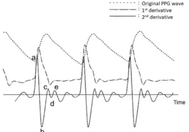
2.3 Acceleration PPG: Second Derivative Analysis The second derivative of the PPG (SDPPG) signal was an- alyzed based on the amplitudes of waves a–e, which arose in the systolic phase of the heart cycle (Fig. 3). The ampli- tudes of the waves were normalized as b/a, c/a, d/a, and e/a.
The SDPPG signal contains information on aortic compli- ance and stiffness, which is highly correlated with BP. The ratio d/a correlates with BP, along with many other physio- logical parameters. To make use of the SDPPG signal, the BP must be analyzed numerically using a neural network and/or support vector machine[17].
2.4 Tonometry
In applanation tonometry of the radial artery, when a radial artery is partially compressed or splinted against a bone, the pulsations are proportional to the intraarterial pressure, as
Fig. 3 Real time PPG signal and its first and second derivatives.
Fig. 4 Principle of applanation tonometry.
shown in Fig. 4.
However, the transducer must be positioned directly over the center of the artery; hence, the signal is highly position-sensitive. This has been dealt with by using an ar- ray of transducers placed across the artery. Although this technique was developed for beat-to-beat monitoring of the wrist BP, it requires calibration for each individual patient and is not suitable for a routine clinical setting[18]–[20].
2.5 Other Principles
Other less well-known methods for measuring PAT and PTT include electrical bio-impedance (Bimp) [21]–[24], BCG [25]–[29], and seismocardiography (SCG)[30]–[33], To use Bimp, BCG, and SCG, specialized technology are required;
no commercial medical devices are available.
For Bimp, an array of wrist-worn bio-impedance sen- sors are placed on the radial and ulnar arteries of the wrist to monitor the arterial pressure pulse resulting from blood volume changes at each sensor site. An impedance ring with spot electrodes is more suitable for wearable cuffless BP monitors than PPG sensors, in terms of noise reduction.
An ICG sensor placed on the chest for thoracic impedance measurement via radar has also been proposed[24].
BCG can measure the response of the body to blood be- ing ejected during the cardiac systole. Slight accelerations in the body caused by heart activity, mainly in the head-to- foot direction, and other relevant information were analyzed
in a previous study. The method used therein was based on ECG and BCG, and allowed efficient monitoring of central pressure[25]. Furthermore, BP was estimated from upper- limb BCG recordings, obtained using an accelerometer em- bedded in a wearable armband simultaneously with finger PPG recordings[26]–[29]. SCG measures pericardial vibra- tion during cardiac movement. It can monitor the PTT (with PEP excluded) and improve the conventional PTT analysis approach, but selection of the most appropriate measure- ment site can be difficult[30]–[33].
Ultrasound sensors are also used to measure BP and capture BP waveforms in relatively deep layers of arterial and venous sites[34]–[36]. In an NIBP sensor using ultra- sound, high frequency sound waves are bounced offa blood vessel and the echo patterns received are sent to a computer to create a representation of the vessel’s changing diameter.
When calibrated to a patient’s blood pressure, these wave- forms can be used to monitor changes in blood pressure. An ultrathin, stretchable and wearable ultrasound patch sensor that enables non-invasive, continuous, and accurate mon- itoring of cardiovascular performance has been developed [35],[36].
3. Standardization and Clinical Approval of the Cuf- fless Blood Pressure Monitor
3.1 The International Standards Organization (ISO) The ISO is an international agency that regulates industrial and medical devices. The main aim of regulation is to en- sure the high accuracy, validity, and safety of devices. ISO 81060-1:2007 and ISO 81060-2:2018, 09 have been pub- lished as standards for BP devices. Also, the ISO in co- operation with the IEC partly uses IEC 60601-1:2005, and IEC 60601-2-30:20. ISO 81060-2:2018 pertains to cuff- based sphygmomanometers. Recently, the ISO Technical Committee (TC) 121/Subcommittee (SC) 3/Joint Working Group (JWG) 7, which is concerned with NIBP monitors, discussed continuous BP monitors and proposed the follow- ing standard: “ISO DIS 81060-3: Noninvasive sphygmo- manometers - Part 3: Clinical investigation of continuous automated measurement type”. Most national ISO commit- tees approved this proposed standard, but the Comit´e Eu- rop´een de Normalisation (CEN) did not. The standard per- tains mainly to continuous automated BP monitors, as ex- emplified by the Finapres (Finapres Medical Systems, En- schede, The Netherlands), CNAP (CNSystems, Graz, Aus- tria) and Nexfin (Edwards Lifesciences, Draper, UT, USA) devices. These devices use a voltage clamp method based on a continuous PPG waveform and passive control. ISO JWG 7 is scheduled to discuss the cuffless NIBP monitor in April 2021 following publication of ISO 81060-3.
3.2 The Institute of Electrical and Electronics Engineers (IEEE)
The IEEE Standards Association (SA) has long been con-
cerned with wearable BP monitors. In 2014, the 1708- 2014 IEEE standard for wearable cuffless BP measuring devices was approved by the IEEE SA. The standard is applicable to all types of wearable BP measurement de- vices, including wearable and unobtrusive BP devices hav- ing different modes of operation (e.g., short- vs. long-term measurement, discrete vs. continuous readings, beat-to-beat BP, BP variability measurement, etc.). The content of the 1708-2104 IEEE standard is in accordance with that of ISO 81060-2:2009. With regard to static accuracy evaluation, ISO81060-2 requires 85 subjects, but the IEEE standard in- troduced a paired t-test and minimum number of subjects is 45 persons. Thus, the IEEE standard has a static accuracy of±5±8 mmHg with 45 subjects instead of 85 subjects.
For the purposes of the US Food and Drug Adminis- tration (FDA), an amendment was proposed for the stan- dard, published in October 2019 as IEEE 1708A1-2019.
The main amendments of the revision were that the num- bers of subjects are 45 to 85, and that two clauses should be included concerning the estimation of noise reduction, and measurement site during rest. The IEEE submitted a combined IEEE 1708-2014 and 1708A-2019 to the FDA to request the recognized consensus standards. The FDA database provides an up-to-date list of voluntary standards, for which a supplier can make a declaration of confor- mity. The FDA partly recognizes IEEE standards for wear- able cuffless BP monitoring devices excluding the follow- ing items[37]. No recognized clauses for observed mea- surements, which is in conflict with the ISO 81060-2 defini- tion of special patient populations, detailed requirement for testing BP change, accuracy of dynamic changes in BP lev- els with no statistical justification for the proposed criteria, and risk management of wireless technologies used for com- munication. The IEEE standard association working group (P1708) will try to revise the comments mentioned.
3.3 The FDA
The FDA is responsible for protecting public health in the US by ensuring the safety and efficacy of human and veteri- nary drugs, biological products, and medical devices. The FDA operates in association with other regulatory bodies, such as the Association for the Advancement of Medical Instrumentation (AAMI), the American National Standards Institute (ANSI), the IEEE, and the ISO, and adheres to in- ternational standards.
Based on ISO 81060-2, the FDA approved the com- mercially available BP monitors shown in Fig. 5 and Ta- ble 1. ViSi Mobile sensor is based on PTT with PPG and ECG, Caretaker uses a pulse contour method with a pres- sure sensor, Biobeat displays BP from calculated from the PTT signal, and Bro is a continuous tonometric BP moni- tor. However, the clinical utility of these devices is difficult to evaluate, although evidence of the clinical efficacy of the Caretaker device (Caretaker Medical LLC, Charlottesville, VA, USA) has been provided. Cuffless BP monitors could be used on a large scale to assess cardiovascular function
Fig. 5 FDA-approved cuffless BP monitors. (a) ViSi, https://www.
soterawireless.com/(b) Caretaker, https://www.caretakermedical.net/ (c) Biobeat, https://www.bio-beat.com/and (d) BPro. https://www.medtach.
com
Table 1 FDA-approved cuffless blood pressure monitors.
during the ongoing Covid-19 pandemic, but to date there have been no reports of its use in this capacity.
3.4 The Pharmaceuticals and Medical Devices Agency (PMDA)
BP monitors should ideally be cuff-based according to Japanese Standard JIST1115. Several manufacturers have attempted to obtain class II certification by third parties, but none has been successful. The main purpose of cuffless BP devices is for screening and health check-ups, so clinical ap- proval is mandatory.
3.5 Trends in Medical Device Approval in Japan
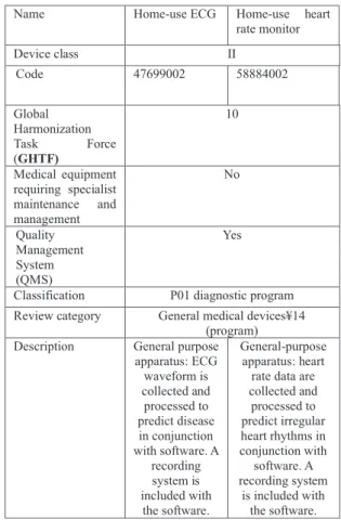
Medical devices not intended to aid physicians in making a diagnosis still require medical approval. A new trend in device approval has emerged in Japan. On July 20, 2020, the Ministry of Health and Welfare added two new items to the Japanese Medical Device Nomenclature (JMDN), as shown in Table 2. On September 4, 2020, the Apple Watch, which has ECG and heart rate monitoring capabilities, was approved by the PMDA. The definition of “device” now in- cludes “the information obtained from general-purpose dig- ital apparatus such as portable watch, specially Apple watch can be used to analyze ECG waveform and to support the
Table 2 Japanese Medical Device Nomenclature (JMDN) newly intro- duced on July 20, 2020.
detection of diagnosis or find irregular heart rhythm noti- fications”. Here, the terms “general digital apparatus” and
“support” are especially interesting. The Apple Watch can be purchased through the Apple Store, as opposed to from medical equipment specialists providing maintenance and management services.
The FDA approved Apple Electrocardiograph software for over-the-counter purchase following a de novo classifi- cation request. To obtain this classification, medical devices must demonstrate safety and efficacy with respect to the in- tended use.
4. The Market for Cuffless BP Monitors and Their Clinical Utility
Cuffless BP monitors are actively being sought. The clin- ical use of cuffless BP monitors has been described [46], but has not been approved by the PMDA or the Ministry of Health, Labor and Welfare of Japan. Third-party certifica- tion has been obtained only for the Somnotouch NIBP mon- itor (Somnomedics, Randersacker, Germany)[47]. Several inexpensive smart watches with embedded BP monitors are commercially available from online retailers. Based on per- sonal communications, inexpensive smart watches do not meet the accuracy and validation requirements of cuff-based sphygmomanometers.
To be approved by regulatory authorities, clinical util- ity, safety, and accuracy must be demonstrated. No clinical protocol for cuffless BP monitors has been established, but this is important for clinical application.
5. Conclusion
The principles of cuffless BP monitors, which show promise for healthcare applications, were described. However, more detailed and precise evaluation of these devices is needed to confirm their clinical efficacy.
Acknowledgments
This work was supported in part by grants-in-aid from the Japanese Ministry of Education, Culture, Sports, Sci- ence and Technology, Scientific Research (C) (Kakenhi) (#17K01440), and the Ministry of Economy, Trade and In- dustry (project proposing a new ISO/IEC standard).
References
[1] P.K. Whelton, R.M. Carey, W.S. Aronow, et al., “2017 ACC/AHA/
AAPA/ABC/ACPM/AGS/APhA/ASH/ASPC/NMA/PCNA guide- line for the prevention, detection, evaluation, and management of high blood pressure in adults,” J. Am. Coll. Cardiol., vol.71, no.19, pp.e127–e248, 2018.
[2] W. Chen, T. Kobayashi, S. Ichikawa, Y. Takeuchi, and T. Togawa,
“Continuous estimation of systolic blood pressure using the pulse arrival time and intermittent calibration,” Med. Biol. Eng. Comput., vol.38, no.5, pp.569–574, 2000.
[3] C.C.Y. Poon and Y.T. Zhang, “Cuff-less and noninvasive measure- ments of arterial blood pressure by pulse transit time,” Conference
Proceedings of the IEEE Engineering and Medical Biology Society, vol.6, pp.5877–5880, 2005.
[4] A.-G. Pielmus, M. Pflugradt, T. Tigges, M. Klum, A. Feldheiser, O.
Hunsicker, and R. Orglmeister, “Novel computation of pulse transit time from multi-channel PPG signals by wavelet transform,” Cur- rent Directions in Biomedical Engineering, vol.2, no.1, pp.209–213, 2016. doi: 10.1515/cdbme-2016-0047
[5] Z. Tang, T. Tamura, M. Sekine, M. Huang, W. Chen, M. Yoshida, K. Sakatani, H. Kobayashi, and S. Kanaya, “A chair-based unobtru- sive cuffless blood pressure monitoring system based on pulse arrival time,” IEEE J. Biomed. Health Inform., vol.21, no.5, pp.1194–1205, 2017. doi: 10.1109/JBHI.2016.2614962
[6] X. Ding, B.P. Yan, Y.T. Zhang, J. Liu, N. Zhao, and H.K. Tsang,
“Pulse transit time based continuous cuffless blood pressure estima- tion: A new extension and a comprehensive evaluation,” Sci. Rep., vol.7, 11554, 2017. https://doi.org/10.1038/s41598-017-11507-3 [7] M. Kachuee, M.M. Kiani, H. Mohammadzade, and M. Shabany,
“Cuffless blood pressure estimation algorithms for continuous health-care monitoring,” IEEE J. Biomed. Health Inform., vol.64, no.4, pp.859–869, April 2017. doi: 10.1109/TBME.2016.2580904 [8] X. Ding and Y.T. Zhang, “Pulse transit time technique for cuf-
fless unobtrusive blood pressure measurement: From theory to al- gorithm,” Biomed. Eng. Lett., vol.9, pp.37–52, 2019. doi: 10.1007/
s13534-019-00096-x
[9] M.C. Baruch, D.E. Warburton, S.S. Bredin, A. Cote, D.W. Gerdt, and C.M. Adkins, “Pulse decomposition analysis of the digital arte- rial pulse during hemorrhage simulation,” Nonlinear Biomed. Phys., vol.5, no.1, p.1, 2011.
[10] M. Elgendi, “On the analysis of fingertip photoplethysmogram sig- nals,” Current Cardiology Reviews, vol.8, no.1, pp.14–25, 2012.
[11] S. Epstein, M. Willemet, P.J. Chowienczyk, and J. Alastruey, “Re- ducing the number of parameters in 1D arterial blood flow modeling:
Less is more for patient-specific simulations,” Am. J. Physiol. Heart Circ. Physiol., vol.309, no.1, pp.H222–H234, 2015.
[12] H. Shin and S.D. Min, “Feasibility study for the non-invasive blood pressure estimation based on PPG morphology: Normotensive sub- ject study,” BioMed. Eng. Online, vol.16, p.10, 2017.
[13] I. Gratz, E. Deal, F. Spitz, M. Baruch, I.E. Allen, J.E. Seaman, E. Pukenas, and S. Jean, “Continuous non-invasive finger cuff CareTakerR comparable to invasive intra-arterial pressure in pa- tients undergoing major intra-abdominal surgery,” BMC Anesthe- siology, vol.17, no.1, p.48, 2017.
[14] F. Rundo, A. Ortis, S Battiato, and S. Conoci, “Advanced bio- inspired system for noninvasive cuff-less blood pressure estimation from physiological signal analysis,” Computation, vol.6, no.3, 46, 2018.
[15] M. Proenc¸a, P. Renevey, and F. Braun, “Pulse wave analysis tech- niques,” J. Sol`a and R. Delgado-Gonzalo, eds., The Handbook of Cuffless Blood Pressure Monitoring, pp.107–137, Springer, Cham, 2019. https://doi.org/10.1007/978-3-030-24701-0 8
[16] M.H. Chowdhury, M.N.I. Shuzan, M.E. Chowdhury, Z.B. Mahbub, M.M. Uddin, A. Khandakar, and M.B.I. Reaz, “Estimating blood pressure from the photoplethysmogram signal and demographic fea- tures using machine learning techniques,” Sensors, vol.20, no.11, 3127, 2020.
[17] M. Liu, L.-M. Po, and H. Fu, “Cuffless blood pressure estimation based on photoplethysmography signal and its second derivative,”
International Journal of Computer Theory and Engineering, vol.9, no.3, pp.202–206 2017.
[18] G.M. Drzewiecki, J. Melbin, and A. Noordergraaf, “Arterial tonom- etry: Review and analysis,” J. Biomech., vol.16, no.2, pp.141–152, 1983.
[19] T. Sato, M. Nishinaga, A. Kawamoto, T. Ozawa, and H. Takatsuji,
“Accuracy of a continuous blood pressure monitor based on arterial tonometry,” Hypertension, vol.21, no.6, pp.866–874, 1993. https://
doi.org/10.1161/01.HYP.21.6.866
[20] H. Smulyan, D.S. Siddiqui, R.J. Carlson, G.M. London, and M.E.
Safar, “Clinical utility of aortic pulses and pressures calculated from applanated radial-artery pulses,” Hypertension, vol.42, no.2, pp.150–155, 2003.
[21] S.H. Liu, D.C. Cheng, and C.H. Su, “A cuffless blood pressure mea- surement based on the impedance plethysmography technique,” Sen- sors, vol.17, no.5, 1176, 2017. https://doi.org/10.3390/s17051176 [22] T.H. Huynh, R. Jafari, and W.Y. Chung, “Noninvasive cuffless blood
pressure estimation using pulse transit time and impedance plethys- mography,” IEEE Trans. Biomed. Eng., vol.66, no.4, pp.967–976, 2019. doi: 10.1109/TBME.2018.2865751. Epub 2018 Aug. 17.
PMID: 30130167.
[23] B. Ibrahim and R. Jafari, “Cuffless blood pressure monitoring from an array of wrist bio-impedance sensors using subject-specific re- gression models: Proof of concept,” IEEE Trans. Biomed. Circuits Syst., vol.13, no.6, pp.1723–1735, 2019. doi: 10.1109/TBCAS.
2019.2946661
[24] D. Buxi, J.M. Redout, and M.R. Yuce, “Blood pressure estimation using pulse transit time from bioimpedance and continuous wave radar,” IEEE Trans. Biomed. Eng., vol.64, no.4, pp.917–927, 2017.
DOI: 10.1109/TBME.2016.2582472
[25] G. Fierro, F. Silveira, and R. Armentano, “Central blood pressure monitoring method oriented to wearable devices,” Health Technol., vol.6, no.3, pp.197–204, 2016.
[26] C-S. Kim, A.M. Carek, R. Mukkamala, O.T. Inan, and J.-O. Hahn,
“Ballistocardiogram as proximal timing reference for pulse tran- sit time measurement: Potential for cuffless blood pressure moni- toring,” IEEE Trans. Biomed. Eng., vol.62, no.11, pp.2657–2664, 2015, doi: 10.1109/TBME.2015.2440291
[27] C.-S. Kim, A.M. Carek, O.T. Inan, R. Mukkamala, and J.-O. Hahn,
“Ballistocardiogram-based approach to cuffless blood pressure mon- itoring: Proof of concept and potential challenges,” IEEE Trans.
Biomed. Eng., vol.65, no.11, pp.2384–2391, 2018, doi: 10.1109/
TBME.2018.2797239
[28] Y. Yao, S. Shin, A. Mousavi, C.-S. Kim, L. Xu, R. Mukkamala, and J.-O. Hahn, “Unobtrusive estimation of cardiovascular parameters with limb ballistocardiography,” Sensors, vol.19, no.13, 2922, 2019.
[29] P. Yousefian, S. Shin, A. Mousavi, C.-S. Kim, R. Mukkamala, D.- G. Jang, B.-H. Ko, J. Lee, U.K. Kwon, Y.H. Kim, and J.-O. Hahn,
“The potential of wearable limb ballistocardiogram in blood pres- sure monitoring via pulse transit time,” Sci Rep, vol.9, 10666, 2019.
https://doi.org/10.1038/s41598-019-46936-9
[30] A.K. Verma, R. Fazel-Rezai, A. Blaber, and K. Tavakolian, “Pulse transit time extraction from Seismocardiogram and its relationship with pulse pressure,” Comput. Cardiol., pp.37–40, 2015.
[31] C. Yang and N. Tavassolian, “Pulse transit time measurement us- ing seismocardiogram, photoplethysmogram, and acoustic record- ings: Evaluation and comparison,” IEEE J. Biomed. Health Inform., vol.22, no.3, pp.733–740, 2018.
[32] A.M. Carek, J. Conant, A. Joshi, H. Kang, and O.T. Inan, “Seis- moWatch: Wearable cuffless blood pressure monitoring using pulse transit time,” Proc. ACM Interact. Mob. Wearable Ubiquitous Tech- nol., vol.1, no.3, 40, 2017. doi: 10.1145/3130905
[33] J. Park, S. Yang, J. Sohn, J. Lee, S. Lee, Y. Ku, and H.C. Kim,
“Cuffless and continuous blood pressure monitoring using a single chest-worn device,” IEEE Access, vol.7, pp.135231–135246, 2019, doi: 10.1109/ACCESS.2019.2942184
[34] S. Weber, P. Scharfschwerdt, T. Schauer, T. Seel, U. Kertzscher, and K. Affeld, “Continuous wrist blood pressure measurement with ultrasound,” Biomedical Engineering/Biomedizinische Tech- nik, vol.58, suppl 1, (SI-1-Track-E), 2013. 000010151520134124.
doi: https://doi.org/10.1515/bmt-2013-4124
[35] S.R. Steinhubl and E.J. Topol, “A skin patch for sensing blood pres- sures,” Nat. Biomed. Eng., vol.2, pp.633–634, 2018. https://doi.org/ 10.1038/s41551-018-0296-9
[36] C. Wang, X. Li, H. Hu, L. Zhang, Z. Huang, M. Lin, Z. Zhang, Z. Yin, B. Huang, H. Gong, S. Bhaskaran, Y. Gu, M. Makihata, Y. Guo, Y. Lei, Y. Chen, C. Wang, Y. Li, T. Zhang, Z. Chen, A.P.
Pisano, L. Zhang, Q. Zhou, and S. Xu, “Monitoring of the central blood pressure waveform via a conformal ultrasonic device,” Nat.
Biomed. Eng., vol.2,, pp.687–695, 2018. https://doi.org/10.1038/ s41551-018-0287-x
[37] FDA recognized consensus standards, https://www.accessdata.fda.
gov/scripts/cdrh/cfdocs/cfStandards/detail.cfm?standard identificat ion no=41190 accessed on 1 Sept. 2020
[38] Sotera wireless Inc., https://www.soterawireless.com/accessed on 7 Oct. 2020.
[39] Caretaker Medical LLC, https://www.caretakermedical.net/ ac- cessed on 7 Oct. 2020.
[40] Biobeat Technologies Ltd., https://www.bio-beat.com/accessed on 7 Oct. 2020.
[41] Med Tach Inc., https://www.medtach.com/accessed on 7 Oct. 2020.
[42] M.C. Baruch, K. Kalantari, D.W. Gerdt, and C.M. Adkins, “Vali- dation of the pulse decomposition analysis algorithm using central arterial blood pressure,” BioMed. Eng. OnLine, vol.13, 96. 2014.
https://doi.org/10.1186/1475-925X-13-96
[43] D. Nachman, Y. Gepner, N. Goldstein, E. Kabakov, A.B. Ishay, R.
Littman, Y. Azmon, E. Jaffe, and A. Eisenkraft, “Comparing blood pressure measurements between a photoplethysmography-based and a standard cuff-based manometry device,” Sci. Rep., vol.10, 16116, 2020. https://doi.org/10.1038/s41598-020-73172-3
[44] D. Nair, S.-Y. Tan, H.-W. Gan, S.-F. Lim, J. Tan, M. Zhu, H.
Gao, N.-H. Chua, W.-L. Peh, and K.-H. Mak, “The use of ambu- latory tonometric radial arterial wave capture to measure ambula- tory blood pressure: The validation of a novel wrist-bound device in adults,” J. Hum. Hypertens, vol.22, pp.220–222, 2008. doi: 10.1038/ sj.jhh.1002306
[45] T. Komori, K. Eguchi, S. Hoshide, B. Williams, and K. Kario,
“Comparison of wrist type and arm-type 24-h blood pressure mon- itoring devices for ambulatory use,” Blood Press. Monit., vol.18, no.1, pp.57–62, 2013. doi: 10.1097/MBP.0b013e32835d124f [46] D.M. Bard., J.I. Joseph, and N. van Helmond, “Cuff-less methods
for blood pressure telemonitoring,” Front. Cardiovasc. Med., 6, 40, 2019. https://www.frontiersin.org/article/10.3389/fcvm.2019.00040 [47] G. Bilo, C. Zorzi, M. Ochoa, E. Juan, C. Torlasco, V. Giuli, and G. Parati, “Validation of the Somnotouch-NIBP noninvasive con- tinuous blood pressure monitor according to the European Soci- ety of Hypertension International Protocol revision 2010,” Blood Press. Monit., vol.20, no.5, pp.291–294, 2015. doi: 10.1097/MBP.
0000000000000124
[48] Device classification under Section 513(f)(2)(De Novo), https://
www.accessdata.fda.gov/scripts/cdrh/cfdocs/cfpmn/denovo.cfm?id= DEN180044 accessed on 10 Oct. 2020.
Toshiyo Tamura received his Ph.D.
from Tokyo Medical and Dental University in 1980. He is currently a visiting profes- sor, Future Robotics Organization, Waseda Uni- versity, Japan. His research interests include biomedical instrumentation, biosignal process- ing, telemedicine telecare, home care technol- ogy and rehabilitation engineering. He has served as a chair of IEEE/EMBS Tokyo Chapter in 1996–2000, and the Asian Pacific represen- tative for the EMBS from 2000 to 2004. He is fellows of IAMBE, Japanese Society of Medical Electronics and Biological Engineering, Japanese Society of Life Support Technology, and Japanese Society for Nursing Science and Engineering.