Title
Functional Histology of Endocrine Cells in Bovine Intestine at
Different Developmental Stages( 本文(FULLTEXT) )
Author(s)
PYAROKHIL, Asadullah Hamid
Report No.(Doctoral
Degree)
博士(獣医学) 甲第346号
Issue Date
2012-03-13
Type
博士論文
Version
publisher
URL
http://hdl.handle.net/20.500.12099/42930
※この資料の著作権は、各資料の著者・学協会・出版社等に帰属します。
Functional
Histology
of Endocrine
Cells in Bovine
Intestine
at
Different
Developnental
Stages
(BtR50
#Q)BB
tTLB
81
8
Fq3ji&haFn&Q)i&eEgAnSiB9#*
)
-:''.:I:.I;I
L _ .i 2011(?
7u
5p
The Uhited Graduate School of Veterinary Sciences, Gifu Uhiversity
(Obihiro
Uhiversity of Agriculture and VeterinaryMedicine)
Table
of
Contents
Chapter 1
General introduction
1.1. Overview...
...1 1.2. Chemical Signals including Regulatory Peptides of the Gut...2
1.3. GeneralMaterials and methods...6
Chapter 2
Immunohistochemical study on the ontogenetic development of the regional
distribution of peptide W, pancreatic polypeptide, and glungon-like peptide I
endocrine cells in bovine gastrointestinal tract
2.1. Introduction...ll
2.2. Materials and methods...
...12
2.3. Results..."...13
2.4. Discussion...-...18
Chapter 3
Quantitative
Immunohistochemical Study of Endocrine Cell Distribution in theBovine Large Intestine
3.1.Introduction...
...38 3.2. Materials andmethods...39
3.4. I)iscussion...41
Chapter 4 The developmental plasticity of colocalization pattern of peptide W and glucagon-like peptide- 1 in the endocrine cens of bovine rectum 4.1. Introduction... ...56 4.2. Materials andmethods...57 4.3. Results..."...58 4.4. Discussion...59 Chapter 5 Generaldiscussion...65 Abstract... ...70 AbstractinJapanese...74 Acknowledgements.. References...-...80
Chapter
IGeneral
introduction
1. I. Overview
Endocrine cells distributed in the gastrointestinal tract produce various
kinds of signal materials to regulate diverse functions ofgut such as secretion,
Secretion and motility
(65,78).
Many studies have elucidated the distributionof various endocrine cells in the gastrointestinal tract of different mammals including domestic animals such as cat
(40),
cattle(39),
sheep(12),
horse(38),
pig
(36),
water buffalo(8, 52),
and camel(16).
These studies have aimed todemonstrate their distribution and relative frequencies in different parts of
the gastrointestinal tract and to understand their functional roles in the
digestive system. Several peptides produced by endocrine cells were newly
identified in recent decades. In addition, Some kinds ofgut peptides including
PW and GLP-1 were distinguished to participate in the control mechanism
within the gut as wen as outside it such aS nutritional and food intake
regulation
(30)
which hashigh
significance in the domestic animal husbandry. The ruminant has drastic changes twice in nutrition, from fetus topostnatal, and suckling to herbivorous. IIowever, no study has been focusing
relative frequency of endocrine cens in the gastrointestinal tract of cattle from early fetus to adult cow. Furthermore, it is intended to emphasize the
regulatory role of the large intestine because ruminants are usually noticed
on the stomach.
The present study investigated the distribution of endocrine cells in the
gastrointestinal tract of cattle at pre- and post-natal stages with different developmental stages
(Chapter 2).
It was focusing on the large intestine bythe precise investigations on the detailed segments of the convoluted gut
(Chapter 3).
Furthermore, the detail study was done on thedevelopmental
plasticity of the colocahzation pattern of gut hormones
(Chapter 4).
Thepresent study would play a key role to understand the control mechanism of
the bovine digestive tract from the view points of the developmental plasticity
of the regulatory systems.
1.2. Chemical Signals including Reguhtory Peptides of the Gut
In this section, brief descriptions are made on the characteristics of
chemical signal materials which are dealt in the present study.
Peptide W
(PYY)
is a member of PP family along with pancreaticpolypeptide
(PP)
and neuropeptide Y(WY)
(24).
These three peptides sharesimilar chemical structure based on the number of amino acids
(36-amino-acid residue
peptides).
The regional distribution of PW has been studied in aradioimmunoassy of tissue extracts. Lundberg et a1.
(53)
first localized PYYto a population of endocrine cells in the intestinal mucosa of a variety of
species, including man. PW immunoreactive cells are present in the distal
small intestine, colon and rectum, but were rare and absent in the stomach
and duodenum. Immunohistochemical and tissue extraction studies
subsequently confirmed the distal pattern of distribution of PW in a number
of species
(I,
21, 22, 23,81).
Recent studies have demonstrated that PW1.36and PW3.36 are the
major
molecular forms of the peptide in tissue and in thecirculation
(15).
PYY is present inhigh
concentration in mucosal extracts of the human ileum, colon and rectum(1).
PW is a multifunctional hormone;the main function is to inhibit gastric emptying and mediates the effects of
deal fat on gastric motility as indicated in rodents. PW has been identified
as a peptide involved in the ileal brake
(50,64).
The temporal pattern ofPYYrelease is different from that of other gastrointestinal peptides with
suggested roles in feeding control. Plasma levels of PW gradually increase
aaer meal initiation, reaching a peak at about 60 minutes, and
high
levelsare maintained for a number of hours after a meal
(61).
PP is a structurally related member of the PP family. PP isa 36 amino
acid peptide predominantly expressed in the endocrine pancreas. However,
rare PP-immunoreactive endocrine cells have been described in some but not
PP celk are also present in the intestinal mucosa, especially in the ileum and
colon
(10,
47,49).
PP secretion is stimulated by food ingestion and exercise,and vagal tone is an important determinant regulating PP secretion in
rodents and human
subjects (14).
The main function of PP is to inhibitpancreatic exocrine secretion and relax the gallbladder
(54).
PP is consideredas food intake regulator
(14)
and also plays an important role in negative feedback control of the pancreas, inhibiting netually mediated stimulation ofthis
gland (54).
Glucagon-like peptide- I
(GLP- I)
belongs to a large family of glucagon(34, 37),
which is secreted from L-cells of the gastrointestinal mucosa(34,
37,79).
GLP-1-producing L-cells have been identified in thejejunum,
neum andcolon, with the
highest
cell densities reported in the ileum and colon(34,37,).
GLP- 1 is secreted after nutrient ingestion and stimulates insulin secretion ina glucose dependent manner
(42,
56,63).
GLP-I functions as a gut hormone that helps regulate blood glucose and feeding behavior in mammals. GLP-I isinvolved in the ileal brake mechanism. Gastric inhibitory and intestinal
motility actions of ileal nutrients are partiany mediated
through
GLP-1release. On the other hand, GLP-1 increase insulin release and reduce
appetite in human
(30).
GLP-1 is considered to be abiologicany
and therapeutically interesting hormone, because of its potentialChromogranin
(CG)
is an acidic protein widely distributed inentero-endocrine cells and in other members of the paraneuron family. Therefore,
CG has been claimed as a common "marker" of an neuro-endocrine cens
(13,
25,
28).
As to the gastro-entero-pancreatic(GEP)
endocrine system, previousstudies localized CG in a variety ofendocrine cells
(ll,
13, 17, 25, 66, 85,90).
Evidence increases that CG in this location may serve as a precursor forother hormones to be identified
(18,
19, 35, 41, 71, 70, 73,81).
The mainphysiological
function of CG is not yet clearly understood, but studiessuggested that CG and other related proteins
might
function in theorganization of the granule matrix
(e.g.
binding ofcalcium),
in the processingor
packaging
of peptide hormones or their precursors, and possibly in theregulatory mechanisms after secretion
(41,
72, 91).
Serotonin
(Ser)
is a regulatory amine of mucosal enterochromaffin(EC)
cells. EC cells constitute the largest endocrine cell population in the
gastrointestinal tract and produce over 90% of an the Ser synthesized in the body
(4).
Ser plays an important role in the control of gastrointestinal smoothmuscle contraction and epithelial secretion
(26).
Somatostatin
(Son)
is a peptide hormone symihesized and secreted byendocrine D cells. Son cells are widely distributed in the gastrointestinal
mucosa, pancreatic islets and in the autonomic and central nervous system
biological activities in different parts of the body such as the secretion of
pituitary hormones, release of a wide variety of peptides hormones from all
regions of the gut and endocrine pancreas, gastric acid Secretion, pancreatic
exocrine secretion, intestinal absorption, proliferation and gastrointestinal
motility
(51, 57).
1.3. General Materials and methods
The present experiments on animals were carried out in accordance
with the guideLnes of the Committee of Animal Exp.eriments of Obihiro University of Agriculture and Veterinary Medicine
(No.
17-51, 20-95, 21-lO8,23-46).
Totanyforty-eight
Holstein cattle in different pre- and postnatalstages were used in this study. Prenatal animals were obtained at the
autopsy of their euthanized mothers for pathological inspection. The prenatal
animals
(fetuses)
were divided into 3 developmental stages according to theircrown-rump length
(CRL):
early fetus(CRL:
20 - 40cm),
mid fetus(CRL:
41-70
cm),
and late fetus(CRL:
71-100
cm).
The postnatal animals were dividedinto four developmenta1 Stages according to their age: suckling calf
(1
-
2-week-old and
5-7-week-old),
2. weaning calf(2-month-old),
3. weaned calf(7-month-old and
10-month-old),
and 4. adult cattle(1
-8-year-old)(Table
1.1).
The postnatal animals were exsanguinated from the carotid artery under the
anesthesia
(xylazine
hydrochloride 0.3mgnig and thiopenta17mgnig)
and dissected.Tissue samples were taken from the whole gastrointestinal tract;
esophagus
(upper
and lowerpart),
rumen,r6ticulum,
omasum, oxyntic(body)
and pyloric parts of abomasum
(glandular stomach),
duodenum(cranialpart),
jejunum
(mid part),
ileum(terminal part),
cecum(body),
ascending colon(proximal
loop, centripetal turns, central nexure, centrifugal turns, dista1loop),
transverse colon, descending colon, sigmoid colon(cranial
to the rectalampulla)
and rectum(ampulla
andjust
cranial to the anorectallme).
Thesamples were then fixed in the Bouin's solution at room temperature
(RT)
for24 hours
(h).
The obtained samples were trimmed into small pieces and were
dehydrated in ascending concentration of ethano1, cleared by xylene and
embedded in parafBn
(Paraplast
Plus@, Kendall, MA,USA).
The sampleswere then cut at 1-4pm thickness used sliding microtome
(Leica
SM 2000 R,Germany, with blades S35 Feather,
Japan),
collected on gelatin coated slidesand kept
overnight
in the warming cupboard(40oC).
Hematoxylin and eosin
(HE)
staining was performed to investigate thegeneral histological inspection of the gastrointestinal tract.
Immunohistochemical staining was applied for the detection of targeted
endocrine cells with specific antibodies. The sections were deparaffinized and
rehydrated. The endogenous peroxidase activity was blocked with 0.3% H202 in methanol, and washed three times by phosphate buffer saline
(PBS,
pHnormal goat serum
(I:50,
S-1000, Vector Laboratories Inc., Burlingame, CA,USA)
f.r 30 min at RT. The secti.ns were again washed by PBS yandincubated with targeted primary antisera and kept
overnight
in refrigerator(approximately
5oC).
The dilution tests of prlmary antiserum wereperformed to get the optimal dilution fTor the best condition of specific immunoreactivity before applying. In the second day, the sections were
washed by PBS and incubated with secondary anti-rabbit IgG raised in goat
(1:200,
BA-1000, Vector LaboratoriesInc.)
for 30 min at RT. Aaer washingagain three times by PBS, the sections were further incubated with the
avidin-biotin peroxidasae complex
hBC)
method using Vectastain EEte ABCkit
(Vector
Laboratories,Inc.).
Immunoreaction sites were revealed by Trisbuffer
(pH7.4)
containing 0.02% 3, 3'-diaminobenzidine tetrahydrochlorideand 0.06% H202. The Sections were
lightly
counterstained with hematoxy1inand dehydrated in ascending concentration of ethanol, cleared by xylene and then mounted.
Each ofimmunoreative cellsin different gastrointestinal portions were
observed under the conventional
light
microscope and photomicrographswere taken by a digital camera
(DS-5M,
Nikon, Tokyo,Japan).
For the cellcounting, special counting chamber with the counting grid was &ed in the
eyepiece of the microscope and the number of immunoreactive cells was
estimated in ten random unit areas per section of each gastrointestinal part.
only with clear and identifiable nucleus were counted and recorded for further analyzing.
The obtained data were arranged and calculated by Microsoa Excel
2010, and the values were statistically analyzed by GraphPad Prism
(version
5.00, GraphPad SoRware, San Diego, Cahfornia,USA)
usingone-way ANOVA followed by Tukey's post hoe multiple comparison tests. The
values were expressed as Mean i SEM and differences were considered
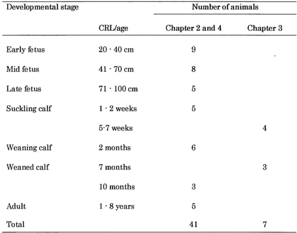
Table 1. I.The detail information of cattle used in the present study
Develop mental stage Number of animals
CRL/age Chapter 2 and 4 Chapter 3
Early Betus Mid fetus Late fetus Suckling calf Weaning calf Weaned calf 20-40cm 41-70cm 71 -lOOcm 1.2weeks 5-7 weeks 2 months 7 months 10 months I -8years 9 8 5 5 CRL: Cmwn-rump length
Chapter
2
Immunohistochemical
study
onthe
ontogenetic
development
of
the
regional
distribution
of peptide
W,
pancreatic
polypeptide,
and
glucagon-like
peptide-
Iendocrine
cen8
in bovine
gastrointestinal
tract2. 1. Introduction
Endocrine cells dispersed in the gastrointestinal tract comprise the
largest endocrine organ of the body. It is composed ofmore than 20 different
cell populations
(78).
These gastrointestinal endocrine cells synthesize andrelease various types of gastrointestinal hormones to regulate the digestive
system and the body
(78).
In recent decades, peptide W(PW)
andglucagon-like peptide-1
(GLP- 1)
have been proven to influence feeding mechanisms inrodents and human
(24,
77,93).
PW is a member of the pancreatic polypeptide
&P)
family(83),
whichincludes neuropeptide Y
(NPY)
and PP, all consisting of 36 amino acids.NPY is found only within the brain and peripheral nervous system, while
PW and PP are found mostly within endocrine cens of the
gastro-entero-pancreatic system. PW is Synthesized and released from the endocrine cells
populations of cells located at the periphery of pancreatic islets
(58,76).
PPcens are aho distributed at a low level in the exocrine pancreas and in the
gastrointestinal tract, mainly in the colon and rectum
(39,
78,83).
GLP-1 is one of the members that are produced from proglucagon
sequence
(37)
Large numbers of GLP-1 endocrine cells have been identified injejunum,
ileum, and colon(77,93).
It has been reported that PW and GLP-1have important roles in the regulation of appetite and feeding, and have an
additive effect on feeding control in rodents and human
(59,
77,93).
However,the
physiological
importance of these three peptides in ruminants is yet to bedetermined
(61, 62).
There have been no studies on the ontogeneticdevelopment of these three kinds of endocrine celk in domestic animals. The
present study was conducted to reveal the
regional
distribution and relativefrequencies of PW, PP, and GLP-1 endocrine ceb in the gastrointestinal
tract of pre- and postnatal cattle.
2.2. Materials and methods
In this study, twenty-one Hohtein cattle in the fonowing seven
ontogenetic stages were examined. The detail of animals and method of
sampling were described in the section of general materials in Chapter I. Brief explanations are fonows: early fetus: 20-40 cm in crown-rump length
(CRL, n=3),
mid-fetus: CRL 41-70 cm(n=3),
late fetus: CRL 71-loo cm(n=3),
calf
(10-month-old, n=3),
and adult(I-8-year-old,n=3).
Tissue samples were obtained from esophagus, rumen, reticulum, omasum, abomasum, duodenum(cranialpart), jejunum (mid-portion),
ileum(terminal portion),
cecum(body),
colon
(central flexure),
and rectum(terminal
portion,just
cranial to theanorecta1
line).
The detail method of paraffin section and immunohistochemistry were
explained in the Chapter 1. The primary antisera used in this chapter were
raised in rabbit against porcine PW
(I:10,000,
IHC7173, Peninsula Lab. Inc., Belmont,USA),
human PP(1:10,000,
YO80, Yanaihara Institute, Inc.,Shizuoka,
Japan)
and human GLP-1(1:10,000,
Y-320, YanaiharaInstitute).
2.3. Results
The present study demonstrated the characteristic distribution ofPW-,
PP-, and GLP- 1-immunoreactive endocrine cells, whereas immunoreactivity
for these peptides in the nervous system was not revealed. Immunoreactive
endocrine cells were not found in the esophagus, rumen, reticulum, omasum,
or abomasum of all seven ontogenetic groups. They were detected in the
sman and large intestines of all groups at different frequencies. The
mentioned immunoreactive cells were mostly detected in the basal
region
of the intestinal crypt glands and a few numbers were also detected in the vinuswith the presence of cytoplasmic processes ending with the lumen. Round and
spherical shaped cells were also found rarely in different
regions.
The frequencies of PW-, PP- and GLP-1-immunoreactive cells were varydepending on intestinal
regions
and perhaps food habits of animals atdifferent developmental ages.
Dispersed PW-immunoreactive cells were detected mostly in the large
intestine, especiany in the rectum, but only a few cens were detected in the
small intestine at allstages. PW-immunoreactive cells were detected in the basal
regions
of intestinal crypt glands and a few numbers were also detectedin the villus glands. PYY-immunoreactive cens were mostly spindle shape
with long cytoplaSmic processes reached the intestinal 1umen. Few cens with the oval, round and spherical shapes were also found in different regions.
PYY-lmmunoreactive celh were immunohistochemicany detected
abundantly in the prenatal
(early,
mid andearly)
stages. In early fetal stage,PW-immunoreactive cenS were not detected in the duodenum portion of the
small intestine. They were detected from
jejunum
to rectum parts. The mostabundant immunoreactivities were observed in the ileum
(Fig. 2.1A)
andrectum
(Fig.
2.1B)
parts. In mid fetal stage, PW-immunoreactive cells weredistributed almost similar to that of early stage, but their frequencies were
higher
in mid fetal stage. Very few PW-immunoreactive cells were detectedin the
jejunum
of mid fetal stage. PW-immunoreactive cens were observed(Fig. 2.2C)
and rectum(Fig. 2.2D)
parts of the mid feta1 Stage.PYY-lmmunoreactive cells were also detected in the late fetal stage; their
frequencies in the late iTetalstage were also similar to that of early and
mid-fetal stages. However, in the late fetal stage, PYY-immunoreactive cens were
also detected with a very low frequency in the duodenum portion.
The frequency and distribution of PW-immunoreactive cens were low
in the postnatal
(sucklmg,
weaning, weaned andadult)
stages.PYY-immunoreactive cells were not detected in the duodenum of suckling,
weaning, weaned and adult stages. Very rare PW-immunoreactive cens were
observed in the
jejunum
of suckling, weaning and weaned stages. However,PW-immunoreactive cells were detected from the ileum to the rectum
portions of all postnatal
(suckling,
weaning, weaned andadult)
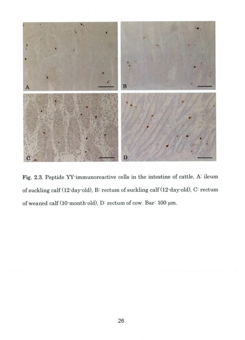
stages. Insuckling stage, the distribution of PW-immunoreactive cens was
higher
in the ileum(Fig. 2.3A)
and rectum(Fig. 2.3B)
portions. The distribution ofPW-immunoreactive cens was almost same in the
jejunum,
ileum, cecumand rectum of weaning, weaned and adult stages. However,
high
number ofPW-immunoreactive cells was observed in the rectum portions of weaned
(Fig.2.3C)
and adult(Fig.2.3D)
stages.The
regional
distribution of PW-immunoreactive celh wassignificantly
(P
<0.05) higher
in therectum at mid fetus
(16.27
i1.7l),
differences among the intestinal segments were observed in early and late
fetusland weaning calf stages
(Table
2.1, Fig.2.10-ll).
With regard to ontogenetic stage, on the other hand, the frequency of
pw-immunoreactive cells was significantly
(P
<0.05) higher
in the ileumof the early fetus
(7.60
i0.25)
and the rectum of the mid fetus(16.27
i1.7l)
than at other stages, while in other
regions
of the gut, such a markedtendency was not observed
(Table
2-1, Fig. 2.12-13).
Open-type PP-immunoreactive cens were detected in the intestinal
crypt
gland
with the spindle, oval and spherical shapes. In the smallintestine, PP-immunoreactive cells were not detected in the duodenum part.
However, very few PP-immunoreactive cens were detected in the
jejunum
ofprenatal
(early,
mid and latefeta1)
and postnatal(sucklmg
calf and weanedcalf)
stages. pp-immunoreactive cells were not detected in thejejunum
ofweaning calf and adult stages. PP-immunoreactive cell were mostly detected from the ileum to the rectum portions of allpre- and postnatal stages.
In the prenatal stages, the distribution of PP-immunoreactive cens was
higher
in the mid and late fetal stages. However, in the postnatal stages, theywere detected with very low number from the ileum to the rectum portions.
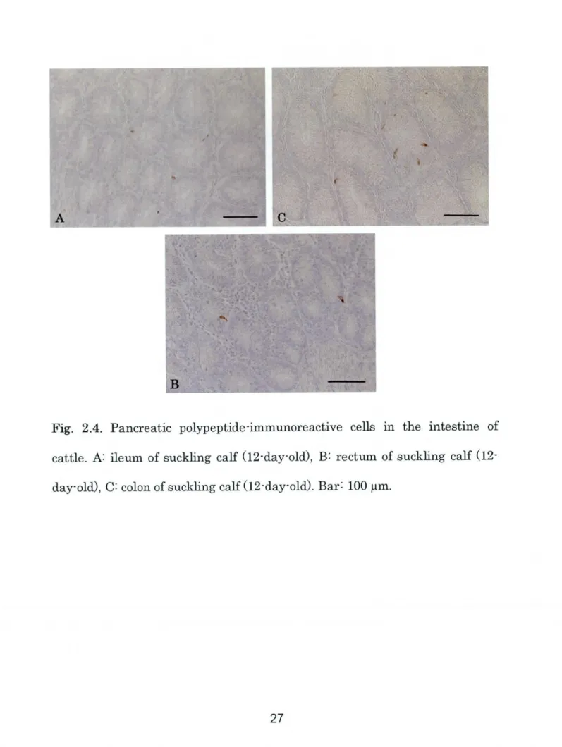
In the suckling stage, PP-immunoreactive cells were clearly observed in the
ileum, colon and rectum portions
(Fig. 2.4A-C).
PP-immunoreactive cells, inthe weaned and adult stages were mostly observed in the colon and rectum
PP-immunoreactive cens were observed between any intestinal segments at
different developmental stages
(Table
2. 1, Fig. 2.10-13).
Relatively abundant GLP- 1-immunoreactive cens were mostly detected
in the crypt
glands
of small and large intestines at all developmental stages.GLP-I-immunoreactive cells were all open-type with the Spherical and
spindle shapes, which had long cytoplasmic processes ended with the lumen
similar to PYY-immunoreactive cells.
The localization of GLP-1-immunoreactive cens was
higher
in theprenatal
(early,
mid and latefetus)
stages than in that of postnatal(suckling,
weaning, weaned and
adult)
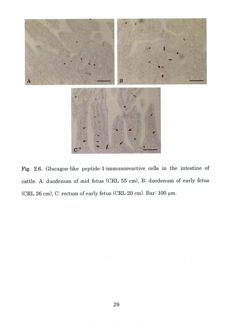
stages. GLP-1-lmmunoreactive ceh, in the earlyand mid-fetal stages, were more numerous in the duodenum
(Fig. 2.6A-B)
and rectum
(Fig.2.6C)
portions. GLP- I-immunoreactive cells in the late fetalstage were detected in all portions. However, they were more numerous in
the small intestine
(Fig.2.7A-B).
GLP-I-immunoreactive cells were also moderately detected in all
portions of the postnatal
(sucklmg,
weaning, weaned andadult)
stages. Theirimmunohistochemical distribution was almost slmilar in the suckling and
weaning stages. However, GLP- 1-lmmunoreactive cens were observed
higher
in the duodenum of the suckling calf stage(Fig.2.8A).
In the weaned stage,GLP-I-immunoreactive cens were
higher
in the small intestine(Fig.2.8B-D).
The
regional
distribution of GLP-1-immunoreactive cells wassignificantly
(P
<0.05)
increased in the duodenum(6.00
i0.70)
and rectum(5.66
iI.25)
of sucklmg calf,rectum(5.56
i2.01)
of weaning calf, duodenum(3.80
i0.96),
ileum(3.63
i0.53),
and rectum
(3.77
i:0.50)
of weaned calf, andrectum
(5.63
i1.67)
of adult compared with those of other intestinalregions
(Table
2.1, Fig. 2.10-ll).
The distribution of GLP- 1-immunoreactive cens wasalso significantly
(P<0.05)
increased ontogenetically at mid fetus stage(13.33
i
1.77)
in the duodenum, mid and late fetus stages(8.26
i 1.33, 9.06 i2.03)
in the
jejunum,
mid fetus stage(ll.6
i2.03)
in the neum, late fetus stage(7.73
i2.36)
in the cecum, mid and late fetus stages(9.46
i 1.75, 8.93 i2.46)
in the colon, and mid fetus stage
(15.73
iI.54)
in the rectum(Table
2.1, Fig.2.12-13).
2.4. Discussion
In the present study, the
regional
distributions of PW-, PP-, andGLP-1-immunoreactive cells were for the first time investigated in the
gastrointeStinal tract of cattle at different ontogenetic Stages. The present
results indicate that PW, PP, and GLP-I endocrine cells were absent in the
esophagus and stomach. However, they were widely distributed in the small
and large intestines of all cattle with different relative frequencies depending
Different endocrine cells have been studied in the gastrointestinal tract
of various mammals including human
(74),
cat(40),
cattle(39),
sheep(12),
horse
(38),
pig(36),
water buffalo(8,52)
and camel(16)
to demonstrate theirdistribution and relative frequencies in different parts of the gastrointestinal
tract and to understand their functional roles in the digestive system.
However, no detail studies on distribution of PW, PP and GLP-1 endocrine
cells in different pre and postnatal stages of neither human and rodents nor
ruminants we reported yet. Thus, the present study could be a key paper for
the distributions of mentioned endocrine cens in the gastrointestinal tract of
cattle. The distribution and frequencies of PW, PP and GLP-1 endocrine
cells will be individuany discussed with the available previous studies on
ruminant species.
In the present study, PYY-immunoreactive cens were detected in an
portions of the intestine with different regional frequencies; they were more
numerous in the large intestine. Previous studies demonstrated
PW-immunoreactive cens mostly in the distal portions of the small intestine and
in the large intestine
(2,
3, 8, 52,62).
In cattle, PW-immunoreactive ceuswere much less abundant in the ileum than in the large intestine, such as in
colon and rectum, aS has been reported in the intestine of babiruSa
(2),
water buffalo(8, 52)
and sheep(62).
PW-immunoreactive cells were not detected(philippine
waterbuffalo) (8),
while they were numerous in the largeintestine, especially in the rectum of carabao
(8).
The role of PYY peptide in the gastrointestinal tract has been widely
investigated in monogastric species, especially in human
(7).
The nutrientinfusion into the ileum inhibits
jejunal
motnity(75, 87)
and decreases antraland duodenal peristaltic pressure wave
(27),
the so-called "ileal brake". Moreover, it has been reported that colonic food infusion reduced ilealmotility
(88)
and pancreatic secretion(32)
with increasing PYY and GLP-1 in the colon(88).
In rats, cecal nutrient infusion reduced food intake more thanileal infusion
(55),
suggesting the importance of the large intestine for theregulation of food intake. Onaga et a1.
(62)
reported controversial findings for the ileal brake in sheep. At the same time, however, they reported theincreasing abundance of PW content in the dista1 large intestine. In cattle, it
is assumed that abundant PW-immunoreactive cells in the distal portions of
Small intestine and in the large intestine
might
be involved, at least in part,in the dista1-to-proximal intestinal feedback. However, in ruminants, foregut
fermenters, actual regulatory mechanisms are stillunclear
(6
1,62).
In the bovine ontogenetic results, PW-immunoreactive cens were
decreased in postnatal stages. This decreasing tendency in the postnatal
period is consistent with results of a previous study on the ontogeny of gut
endocrine cens in water buffalo
(52).
They found that the frequency ofbuffalo than in the 5-month and 5-yearold buffalo. It is suggested that other
intestinal epithelial cens such as goblet and absorptive epithelial cens show a
greater increase in number with intestinal development after birth. The
decreasing tendency of PW-immunoreactive ceus was prominent at the
weaning period as wen as nursing. The adaptation of digestive system to the herbivorous nature may also have drastic influence in the regulatory system
of the gut.
At all developmental stages of the cattle, PP-immunoreactive cells were
sparsely distributed in different intestinal portions. Similar to PYY-immunoreactive cen distribution, PP-immunoreactive cells also tended to be
increased in the large intestine. However, no significant differences were
observed among any intestinal regions of the seven stages examined in the
present study. Similar to the present study, small numbers of
PP-immunoreactive cells in the distal small intestine and relatively large
numbers in the large intestine were reported in calf and cow
(39),
as well assheep
(12).
However, intestinal PP-lmmunoreactive cens seem to differ depending on the anlmal species. In human colon and rectum, fewPP-immunoreactive cells were detected
(74).
Moreover, rat PP-immunoreactivecells were transiently expressed for a short time in the endocrine cens of
colon at the postnatal stage
(20).
It is thus assumed that the functional rolesto reduce food intake and may induce long term suppression of appetite
(74).
However, the main role of PP in ruminants including cattle remains unclear.
In the present study, numerous GLP-I-immunoreactive cells were
detected in all parts of the small and
_large
intestines at all developmental
stages. GLP-1-immunoreactive cells were more numerous at the prenatal
stages than at calf and adult stages. The relative frequencies of
GLP-I-immunoreactive cells tended to be
higher
in the duodenum and rectum of theintestine. The general tendency of the distribution of GLP- 1-lmmunoreactive
cells in the large intestine was basicany Similar to those of
ghcentin-lmmunoreactive cells in calf and cow
(39),
as well as sheep(12).
This may berelated to the fact that GLP-1 and glicentin are derived from the same
precursor molecule
(37).
GLP- 1 is secreted in the distal small intestine and colon by stimulation
of carbohydrate and fat, and inhibits gastroduodenal motility
(69)
and gastricacid secretion
(89).
Therefore, it is considered that GLP-1 also has animportant role in ileal brake and/or dista1-to-proxlmal feedback, like PYY,
and the two peptides may act cooperatively
(59, 89).
The reasons for the decreased frequency of GLP-1-immunoreactive cells with development maybe the same aS those for PW-immunoreactive cells.
The present study demonstrated the regional distribution and relative
frequencies of PW-, PP-, and GLP- I-immunoreactive cens in the intestine of
frequency of these endocrine cells vary among different intestinal segments
and at different developmental stages in cattle. These differences of PYY-, PP-, and GLP-1-immunoreactive cell distribution
might
be due to thestage-dependent changes in intestinal growth, secretion, motility, and diet. Further
studies on the
physiological
functions of these three kinds of hormones - PW,PP, and GLP-I - in
ruminants are needed. The present results on the
ontogenetic distribution of three types of endocrine cells in the
gastrointestinal tract of cattle should provide the basis for future extensive
∼.
-
'L-I
∼__i&_
∼Fig. 2.I. Peptide YY-immunoreactive cells in the ileum
W
of early fetus(CRL
26
Jl i:. =itL3)..I:..7
y-'u--- ----Wti3r I.gi8B3ZF1/A
Fig. 2.2. Peptide W-inmunoreactive cens in the ileum
W,
cecum(B),
colon(c)
and rectum(D)
of mid fetus(CRL
55Fig. 2.3. Peptide W-immunoreactive ceus in the intestine of cattle, A: ileum
of suckling calf
(12-day-old),
B: rectum ofSuck1ing calf(12-day-old),
C= rectum_i!Ei
-_ij@;iL
∼TFig. 2.4. Pancreatic polypeptide-immunoreactive cells in the intestine of
cattle. A: ileum of suckhg calf
(12-day-old),
B: rectum of Suckling calfrib Tll i I
.I.S::
i iFig. 2,5. Pancreatic polypeptide-immunoreactive cells in the intestine of
cattle. A: colon of cow, B: rectum ofcow, C: rectum of weaned calf
Fig. 2.6. Glucagon-like peptide-1-immunoreactive cells in the intestine Of
cattle. A: duodenum of mid fetus
(CRL
55cm),
B: duodenum of early fetus- -' _I I. : I iA I I I l I I I Rttt. LL'_ t'.
'Tt5
a,:k
Fig. 2.7. Glucagon-Eke peptide-1-lmmunoreactive endocrine cells in the
intestine of cattle. A: duodenum of late fetus
(CRL
96cm),
B:jejunum
of latefetus
(CRL
90Fig. 2.8. Glucagon-like peptide-I-immunoreactive cells in the intestine Of
cattle. A: duodenum of suckling calf
(12-day-old),
B: duodenum of weaned calf(10-month-old),
C=jejunum
of weaned calf(10-month-old),
D= ileum of weanedI?r- :Tif7PL-&.7E::.T;:a-TTTEVk7f7'W.1:.;.qT_5F
ty
.yT.:.{ ( iI"Ill....A. .
I
Fig. 2.9. Glucagon-like peptide-I-immunoreactive cells in the intestine of
DtIO Jej ne Ccc Col Rcc (c) 1S 16 ∼ 14
S12
l!
":
5:
2 0Duo Jcj ne eec Col Rcc htestine
t= PYY = PP II GLP-1
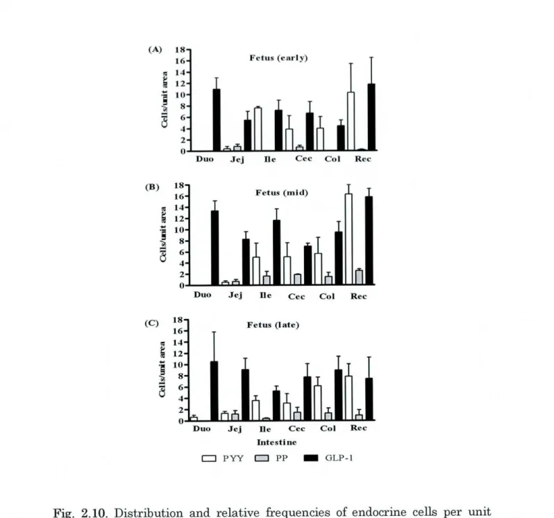
Fig. 2.10. Distribution and relative frequencies of endocrine cells per unit
area
(625 Llm2)
in the intestine of prenatalA:
early fetus, B: mid fetus, andc: late
fetus).
PW: peptide W, PP: pancreatic polypeptide, GIJP-1:glucagon-hke peptide-1. Duo: duodenum,
Jej: jejunum,
Iie: ileum, Cec: cecum, Col:a I,I cl
I-g
U cI L< cq 'a 5 UDuo Jej []c eec Cot Rec 18 16 14 12 10 l8 16 14 12 10 8 CaJ f(w eancd)
Duo Jej Ile eec Col Rec
(a) 18 16 cl l4 4J 512 ・-a10
f
∼ 5 6 U 4 (D) l8 16 d14 V 512I-i
I:
% 6 U 4Cat f(yveani ng)
Duo Jej ne eec Col Ref
DtLO Jej ne eec Col Rec htesti ne
t= Pry a PP )GLP-1
Fig. 2.ll. Distribution and relative frequencies of endocrine cells per unit
area
(625 pm2)
in the intestine of postnatalh=
suckling calf,B: weaning calf,c: weaned calf, D:
adult).
PYY: peptide YY, PP: pancreatic polypeptide,GIJP-1: glucagon-like peptide-1. I)uo: duodenum,
Jej: jejunum,
Ile: ileum, Cec:・B,
::
a::
31:
5
6 4 2 0 18 (C) l6{L
I.:.
.i.I:
5
6 4 2 0 4 5 6 7 Developmental stiLgeS D PYY D PP - GLP-1Fig. 2.12. Distribution and relative frequencies of endocrine cells per unit
area
(625 pm2)
in the small intestineA:
duodenum, B:Jejunum,
and C:ileum)
of seven developmental Stages of cattle(l:
early fetus, 2: mid fetus, 3:late fetus, 4: suckling calf,5: weaning calf, 6: weaned calf and 7=
adult).
PW:18 (J)) l6
i
:I:I
'U:
a
6 4 2 0 4 5 6 7 Develop-ental stages E= PYY Q PP Ill GLP-IFig. 2.13. Distribution and relative frequencies of endocrine cells per unit
area
(625 pm2)
in the large intestineh=
cecum, B: colon, and C:rectum)
ofseven developmental Stages Of cattle
(1:
early fetus, 2: mid fetus, 3: late fetus,4: suckling
calf;
5: weaning calf, 6= weaned calf and 7=adult).
PW: peptideTable 2. 1.Distribution and relative frequency ofPW-, PP-, and GLP- 1-immunoreactive cells
in the bovine intestine at different developmental stages.
Small intestine Large intestine
Duo Jej Ile Ce c Col Re c
Fetus (early) PYY ND PP ND GLP-1 10.12 j=2.04 Fetus (mid) PYY ND PP ND 0.40iOAO 7.60iO.25 0.80 iO.41 ND
5A6i: 1.54 7.20i: I.75
0.53 iO.26 5.00i2.54 0.66iO.35 1.60iO.85 GLP-1 13.33i1.771 8.26j=1.332 ll.6i2,035 Fetus (late) PYY 0.75 j=0.35 PP ND GLP-I 10.53 i 5.27 Calf (su&1ing) PYY ND PP ND
I.33 iO.33 3.56iO.81 I.20iO.60 0.40iO.05 9.06 i2.033 5.23 j=0.89 0.03 iO,03 2.36i 1,07 0.03 iO.03 0A6iOA6 GLP-1 6.00 i 0.70c 2.80 i 0.49 2.43 j=0.95 Calf (weiming) PYY ND 0.06 i 0.06 0.80 j=0.56 PP 0.95iO.03 ND 0.16iO.12 GLP-1 3.73 i 1.25 2.80 j=0.70 I.70 j=0.46 Calf (weane d) PYY ND 0.06 i 0.06 0.76 iO.37 PP 0.33 i 0.17 0.03 i 0.03 0.33 iO.24
GLP-I 3.80iO.96g 2.57i1.18 3.63iO.53h
Adult, PYY ND ND 0.56 i 0.29 PP ND ND 0.06 iO.03 GLP-1 4.96 i 0.98 2.60 j=0.05 3.20 j=0.23 3.86i2.37 4,00i2.06 0.66iO.32 ND 6.66j=2.01 4.40i I.04 5.06i2.53 5.6 i2.91 1.86iO,12 1A6j=0.75 10.27i5.14 0.13 j=0.13 ll.73 j=4.72 16,27 j=1.71a, 9 2.53 iO.31 6.93iO.55 9.46i1.757 15.73j=l.5410 3.06i 1.68 6.13 j=1.52 1A6iO.86 I.33 j=0.88 7.73i2.366 8.93j=2.468 1.30 i 0.73 2.30 j=0.45 0.13 iO.13 0,90iO.32 0.93 iO.32 2.63 iO.20 1.33 i 0.52 1.93 i=0.36 0.10iO.10 0.SOLO.05 0.60iO.45 I.06iO.61 0.96 i 0.29 1.87 j=0.43 0.03 iO.03 0A6iO.08 0.96 i 0.38 2.33 iO.28 2.46iO.29 1.36iO.75 0.36 i 0.08 0.50 iO.40 1,33 iO.08 2.20iO.91 7.80 i=2.27 0.93 i 0,93 7.40 i3.81 3.76 iO.53b 1.00 iO.57 5.66i 1.25d 3.26 iO.14 0.90 iO.58 5.56i2.01e 5.03 i:0.33f 0.83 iO.18 3.77 i 0.50i 4.20 i 2.10j 0.73 iOAO 5.63 i 1.67k
Data represent mean i SEM; Duo: duodenum, Jej:jejunum, Ile: ileum, Col: colon, bc: rectum.
PW: peptide YY, PP: pancreatic polypeptide, GLP- 1:glucagon-like peptide-1, ND: not detected.
Chapter
3
Quantitative
Immunohistochemical
Study
of Endocrine
Cell
Distribution
in the Bovine
Large
Intestine
3.1.Introduction
The gastrointestinal tract is the largest endocrine organ in the body
(65).
Gut hormones are chemical messengers which are localized in theendocrine cells distributed
throughout
the mucosa of glandular stomach andintestine
(65,
68,59).
They are playing important roles in entire digestivetract to regulate many digestive functions, Such as secretion, absorption and
motility. Many studies have elucidated the regional distribution and relative frequencies of various kinds of endocrine cells in the gastrointestinal tract of
different animals. However, detail studies on the distribution of endocrine
cells in different parts of the large intestine especially in domestic animals have not been reported yet. The large intestine, as wen as stomach, has
important physiological function in the digestive process of ruminants
(33).
Previous reports on the endocrine cells in the ruminant large intestine
examined only few portions
(3,
8, 12,39).
The discriminative portions, whichare characteristic to the spiral colon and rectum, were not examined at so
clear the detail of endocrine cell distributions in eleven different portions of large intestine of cattle.
3.2. Materials and methods
six calves in two groups
(suckling
and'weaned)
were used in this study.The age of the suckling calves were between 5
-7 weeks, and weaned calves
were 7 months. Tissue samples were taken from eleven parts of the large intestine detailed in Chapter 1 and processed for paraffin sections as
described before. The primary antisera used in this chapter, in addition to
those used in Chapter 2, were raised in rabbit against chromogranin
(CG:
1:15,000, Code 20085, INCSTAR, Stillwater, MN,
USA),
serotonin(Ser:
15,000, Code sero-23, donated by Dr.
Nishiitsutsuji-Uwo, Kyoto),
andsomatostatin
(son:
I:10,000, Code 20H2T,INCSTAR).
Immunohistochemicalprocedures were mentioned in Chapter I.
3.3. Results
Six types of endocrine cells were detected with the antisera against CG,
Ser, GLP- 1, PW, PP and Son.in eleven different portions oflarge intestine of
suckling and weaned calves. The
regional
distribution and relative frequencies of these endocrine cells were different in each parts of largebetween two groups, suckling and weaned. The distribution and relative
frequencies of each immunoreactive endocrine cen will be discussed
individually.
CG-immunoreactive cens were abundantly detected in an segments of
the large intestine. The general distribution of CG-immunoreactive cells was
higher
in the most distal parts of the colon and two parts of the rectum(Fig
3.1A-C and
3.2A-C).
The frequency of CG-immunoreactive cells weresignificantly
higher
in the descending colon of both suckling and weanedgroups
(sucklmg:
19.03 i 0.61, weaned: 18.53 i3.06),
sigmoid colon of bothgroups
(suckling:
22.90 i 0.87, weaned: 18.37 i2.39),
ampulla of rectum ofboth groups
(suckling:
27.90 i 1.55, weaned: 23.37 i5.79)
and rectumjust
cranial to the anorecta1 lme of both groups
(suckling:
21.60 j= 0.86, weaned:17.40 i
0.78) (Fig.
3.10A, Table3.I)
The general distribution of Ser-immunoreactive cens was almost
similar to that of CG;
higher
in the most distal parts of the colon and twoparts of the rectum
(Fig
3.3A-C, and Fig.3.4A-C).
Significant differences(P
<o.o5).f
Ser-immun.reactivetells
were.bserved in sign.id c.1.n.f sucklmggroup
(19.33
i0.87),
ampuna ofrectum of suckling group(23.37
i0.81)
andrectum
just
cranila to the anorecta1 line of both groups(suckling:
18.77 j= I.03,weaned: 10.37 i
2.60) (Fig
3. 10B, Table3.1).
GLP- 1-immunoreactive cens were rarely detected in the cecum and the
distal parts of the colon and two parts of the rectum
(Fig.
3.5A-C and Fig.3.6A-C).
No significant differences of GLP-1-immunoreactive cells wereobserved between any regions of the large intestine as well as two different developmental stages, suckling and weaned
(Fig
3. 10C, Table 3.1).
The frequencies of PW-immunoreactive cells were varied among
regions (Fig.
3.7A-C,3.8A-C).
They were few in the cecum and the proximalportions of the colon, but were significantly
higher
in the sigmoid colon(4.
10i
0.40)
and ampuna ofrectum(3.60
i0.43)
of the suckling group(Fig.
3.llAand Table
3.1).
PP-immunoreactive cens were detected rarely in all portions of the
large intestine.
(Fig 3.9A-C).
The PP-immunoreactive cells were very few inthe cecum and proximal parts of the colon, increased in the middle portions,
descending c.1.n, Sigm.id c.1.n and ampulla.frectum
(Fig.
3.llB, Table6.
I).
Son-immunoreactive cells were only detected with a very few numberin the transverse colon of the suckling stage. They were not detected in any
other portions of both Sucklmg and weaned groups
(Fig.
3. llC, Table 3.1).
3.4. Discussion
In chapter 1,the regional distribution and relative frequencies of three
types of endocrine cens, PW-, GLP-I- and PP-immunoreactive cells were
numerous in the large intestine of an developmental groups. This fact leads
the present study to investigate the distribution in more detailed topography
of bovine large intestine. The relative frequencies of CG-, Ser-, GLP-1-, PW-,
PP- and Son-immunoreactive endocrine cells were evaluated in eleven
different portions of large intestine in order to understand the characteristics
of the tortuous intestine of the ruminant. The distinguishing differences in
distributions of these six types of endocrine cens were found in each portions
of large intestine.
General distribution of endocrine cells represented by
CG-lmmunoreactive cens indicates the distal abundance in the bovine gut. In
addition, each type of endocrine ceus immunoreactive for Ser, GLP-1, PW,
and PP also showed similar tendency to be more abundant in the distal
portions. Son-immunoreactive cells were too low in frequency to discuss here.
The present study demonstrated the detail distribution of endocrine
cells along with the tortuous large intestine of ruminant. Previous studies on
the ruminant have so far examined only three portions in the large intestine
(cecum,
central flexure of the spiral colon, terminalrectum)
of cattle(39),
sheep
(12),
water buffalo(8),
and mouse deer(3).Although
these resultsshowed the tendency of the abundance ofendocrine cells in the rectum, itwas
not compared in detail with the rest of the large intestine. The eleven
portions of the intestine examined in the present study are the peculiar
distinguished the distal distribution of endocrine cells in the bovine large
intestine. It is suggesting that the huge numbers of endocrine cells in the
large intestine especially distal portions of colon and rectum
might
be relatedto the
physiological
importance of those portions of the digestive system inruminants. This emphasize again that the dista1-to-proximal feedback
regulation
(59, 89)
may play important roles in the digestive tract of theruminant. It iS necessary to make detail
physiological
studies on the lmkagebetween different portions of the large intestine of ruminant and gut
J[ i I > I , I A Q
Fig- 3.I. Chromogranin-immunoreactive cells in the large intestine of
Suckling and weaned calves.
W
cecum of the weaned caw,(B)
centripetalturns of colon of the suckling
call;
(C)
transvel.Se COlon of the weaned calf.BarI 4 JQ. i ∼ I . I r V I i QL /- +-I I I . 1 II ・ IJ I I I 1
i.
(I t4 I I ・4 .1I I I 1 ・L1 I I L -'I I ∼jt∼ I I Jl. fr tJ r .;I ,. F iFig. 3.2. Chromogranin-immunoreactive cells in the large intestine of
suckling and weaned calves.
W
descending colon in the sucklingcalf
(B)
ampuna of rectum in the Suckhg
calf (C)
rectumjust
cranial to the. I 8 . I I I f I :-piH1
Fig. 3,3. Serotonin-immunoreactive cells in the large intestine of 8uCklmg and
weaned calves.
W
cecum of the Sucklmg calf,0)
proximal loop ofcolon in theweaned
calf (C)
centripetal turns ofcolon in the weaned calf.Bar = 100I
L. -.I. i . -I/ ;:_:.T.-8
'4 i IA + I JrA
I . 4I I A o I I 1 IFig. 3.4. Serotonin-imnunoreactive cens in the large intestine of suckling and
weaned calves.
W
descending colon of the suckling calf,0)
ampul1a ofrectum in the Suckling calf,
(C)
rectumjust
cranial to the anorectal he in therT-^T{ f,I--- AAIIT--I
:LL: l.'l
i/.A.;/ i
;'ht..i;
'1-_._.__ _._____
Fig. 3.5. Glucagon-like peptide- 1-immunoreactive cells in the large intestine
of suckling and weaned calves.
W
proximal loop of colon in the suckling calf,0)
transverse colon in the weaned calf,(C)
descending colon in the sucklingrI +
tI:
i.'Jli3-_if,I,;A;ti;
:i..
;i.
Fig. 3.6. Glucagon-Eke peptide-I-immunoreactive Gens in the large intestine
of suckhg and weaned calves.
W
sigmoid colon in the suckhg calf,(B)
ampul1a of rectum in the suckhng
calf (C)
rectumjust
cranial to theFig. 3.7. Peptide W-inmunoreactive cens in the large intestine Of suckling
and weaned calves.
W
distal loop of colon in the weanedcall;
(B)
transversecolon in the suckling calf,
(C)
descending colon in the suckling calf.Bar = looT ・/-IF:--i:#i Ill, ∼y ILL1{ I (;.:jb A I ∼ ;:T -_i-?TdW>'7Tl ∼ .LL -r..i ・t :,lJ.Y1:-(.:i , L,/.;I i,I .-JL i:. I. .I. - -...--Yt .
i
--.r ;.I -.'(. -I J 1.L 1-. .-.)jp';.; :. I I.:...-.::,.:..
,:::;.:i"I.33f:
.1L -J! ..)I -T<. A-( I.I.ti..:
.T. [t.'r'S.
L '*:li.
i::;..-il.
I:Li_
_-,,_ir!ii
4._'./ Tit .I.; > -tl ∼'. ; _:rL,"_..iL_Fig. 3.8. Peptide YY-inmunoreactive cells in the large intestine of suckling
and weaned calves.
W
signoid colon in the Suckling calf,(B)
ampuna ofrectum in the suckling
calf
(C)
rectumjust
cranial to the anorectal lme in theweaned calf.Bar = 100
/tti 1 1
LB
y. I''l
l4?;.=if3
i.I lTpY8 I -
-'l-:=:>
_. .∼_'1 .)-L :.= ,? ,I-7;Fig. 3.9. Pancreatic polypeptide-inmunoreactive cells in the large intestine of
suckhg and weaned calves.
W
centrifugal turns Of colon in the weaned calf,Q})
transverse colon of the suckling calf, Bar = loow
40g30
I-ho
7BguE
0 1S
・i
Taa
I 2 3 4 5 6 7 8 9 10 ll Large intestinal portionsd Suckling D Weaned
Fig. 3.10. Distribution and relative frequencies of endocrine cells per unit area
(625 Ltm2)
in the large intestine of suckling and weaned calvesL CG:chromogranin, Ser: serotonin, GLP-1:
glucagon-like
peptide-1, 1: cecum, 2:proximal loop of colon, 3: centripetal turns of colon, 4: central flexure of colon, 5: centrifugal turns of colon, 6: diStal loop of colon, 7: transverse colon, 8: descending colon, 9: Sigmoid colon, 10: ampulla of rectum, ll: rectum
just
(A) 5 4 a =3 1= E} i2 y U I I 2 3 4 5 6 7 8 9 10 I ) 2 3 4 5 6 7 8 9 10 1L Large intestinal porLioTLS
( Sucklitlg a Weaned
Fig. 3.ll. Distribution and relative &equencies of endocrine cells per unit
area
(625 pm2)
in the large intestine Of suckling and weaned calves. PYY: peptide W, PP: pancreatic polypeptide, Son: somatostatin, 1= cecum, 2:proximal loop of colon, 3: centripetal turns of colon, 4: central nexure of colon,
5: centrifugal turns of colon, 6: distal loop of colon, 7: transverse colon, 8:
descending colon, 9: sigmoid colon, 10'. ampulla of rectum, ll: rectum