Effects of Kampo Formulas on the Progression of Hypercholesterolemia and Fatty
Liver Induced by High-Cholesterol Diet in Rats
Weibin Qian,* Junichi Hasegawa,* Satoshi Tsuno,* Yusuke Endo,* Akiko Matsuda† and Norimasa Miura*
*Division of Pharmacotherapeutics, Department of Pathophysiological and Therapeutic Science, School of Medicine, Tottori University Faculty of Medicine, Yonago 683-8503, Japan and †Department of Fundamental Nursing, School of Health Science, Tottori University Faculty of Medicine, Yonago 683-8503, Japan
ABSTRACT
Background
Bofutsushosan is a well known Kampo,
traditional Japanese medicine, based on ancient Chinese
medicine mainly used in the treatment of
hypercholes-terolemia in Japan. We selected two Kampo formulas,
Boiogito and Keishibukuryogan mainly used in the
treatment of hypercholesterolemia in China to compare
with Bofutsushosan and cholesterol absorption inhibitor
ezetimibe.
Methods
Hypercholesterolemia and fatty liver were
induced by high cholesterol (containing 2% cholesterol
and 0.5% cholic acid) diet in male Wistar rats for 6 and
12 weeks. Kampo formulas Boiogito, Bofutsushosan,
Keishibukuryogan and ezetimibe were added to the
high-cholesterol diet, respectively. After 6 and 12 weeks,
body and liver weights, blood chemistry, cholesterol
concentrations, fat-related and inflammatory-related
fac-tors were examined.
Results
High-cholesterol diet increased body and
liver weights, and serum cholesterol concentrations.
Boiogito and ezetimibe improved them. Serum
ICAM-1 and RBP4 were increased in the high cholesterol diet
group. Boiogito and ezetimibe improved them too. In
the histological examinations of liver and adipose
tis-sues, we observed a significant improvement after
treat-ment. Immunostaining expression of ICAM-1 in aorta
was improved by Boiogito, Bofutsushosan,
Keishibuku-ryogan and ezetimibe. The mRNA expression of RBP4,
Corresponding author: Junichi Hasegawa, MDhasegawa@med.tottori-u.ac.jp Received 2014 October 14 Accepted 2014 November 10
Abbreviations: ALP, alkaline phosphatase; ALT, alanine amino-transferase; BOT, Boiogito; BTS, Bofutsushosan; C, control group; CFABP, cutaneous fatty acid-binding protein; CVD, cardiovascu-lar disease; H, high-cholesterol diet group; HA, high-cholesterol diet with Boiogito group; HB, high-cholesterol diet with Bofutsu-shosan group; HC, high-cholesterol diet with Keishibukuryogan group; HDL-C, density lipoprotein cholesterol; HE, high-cholesterol diet with ezetimibe group; HFABP, heart fatty acid-binding protein; ICAM-1, intercellular adhesion molecule-1; KBG, Keishibukuryogan; LDL-C, low-density lipoprotein choles-terol; MCP1, monocyte chemoattractant protein-1; NAFLD, non-alcoholic fatty liver disease; RBP4, retinol-binding protein 4; TC, total cholesterol; VEC, vascular endothelial cells
HFABP, CFABP, MCP1 and CCR2 in liver and adipose
tissue were decreased by Boiogito and ezetimibe.
ConclusionBoiogito has a protective effect on the
progression of hypercholesterolemia and fatty liver
in-duced by high-cholesterol diet in rats and more effective
than Bofutsushosan and Keishibukuryogan. The
lipid-lowering effect of Boiogito is not stronger than
ezeti-mibe. But the anti-inflammatory (MCP1, CCR2) and
anti-arteriosclerotic (ICAM-1) effects of Boiogito are
more potent than ezetimibe.
Key words
Bofutsushosan; Boiogito;
Keishibukuryo-gan; fatty liver; hypercholesterolemia
Hypercholesterolemia is one of the major risk factors
for many cardiovascular diseases, such as
atheroscle-rosis, hypertension and myocardial infarction.
1, 2Non-alcoholic fatty liver disease (NAFLD) is common in the
general population, and it occurs even more frequently
in patients with hypercholesterolemia.
3, 4Patients with
NAFLD have a high risk of cardiovascular disease
(CVD).
5In addition, NAFLD is often associated with
atherosclerotic signs including the presence of carotid
plaques
6and coronary arterial calcification.
7, 8It is generally believed that the occurrence and
de-velopment of hypercholesterolemia have significantly
correlation to lipid metabolism-related genes, such
as retinol-binding protein 4 (RBP4), heart fatty
acid-binding protein (HFABP), cutaneous fatty acid-acid-binding
protein (CFABP).
9, 10RBP4, a protein secreted by
hepa-tocytes and adipose tissue, is closely related to
hyper-cholesterolemia and NAFLD.
11HFABP and CFABP
belongs to FABPs family which may play a broad role in
cellular fatty acid metabolism.
12In addition to lipid
me-tabolism-related genes, inflammatory cytokines are
as-sociated with hypercholesterolemia. Engström et al. have
supported the view that inflammation could be a risk
factor for developing hypercholesterolemia.
13Monocyte
chemoattractant protein-1 (MCP-1) and its receptor CC
chemokine receptor 2 (CCR2) are important
inflamma-tory chemokines linked with hypercholesterolemia.
14, 15Intercellular adhesion molecule-1 (ICAM-1) can promote
the development and progression of atherosclerosis.
16Therefore, it is a simple and convenient method to detect
hypercholesterolemia by monitoring RBP4, C-FABP,
H-FABP, MCP1, CCR2, ICAM-1 expression.
Bofutsushosan (BTS), Boiogito (BOT) and
Keishi-bukuryogan (KBG) are well-known Japanese Kampo
and Chinese traditional herbal medicines which are used
to improve obesity.
17, 18BTS
has been reported to inhibit
atherosclerosis,
19obesity,
17, 18, 20hypertension
17and
hy-perglycemia.
21BOT and KBG are also Kampo
prepara-tions which have been used in patients with obesity.
22, 23However, pharmacological evidence for the effects of
treatment of NAFLD of BOT and KBG still remains
obscure. Using the well-established experimental model
of high-cholesterol diet in rats, we compared the
phar-macological efficacies of BOT, BTS and KBG.
MATERIALS AND METHODS
Rats and feeding method
Sixty male Wistar rats aged 8-weeks (purchased from
Shimizu Laboratory Supplies, Kyoto, Japan) were kept
in an air-conditioned room at 25 ˚C with 55% humidity
and given standard chow. After 3 days of acclimation,
the rats were divided into 6 groups: control group (C,
n
= 10), cholesterol diet group (H, n = 10),
high-cholesterol diet with Kampo formula BOT group (HA,
n
= 10), high-cholesterol diet with Kampo formula BTS
group (HB, n = 10), high-cholesterol diet with Kampo
formula KBG group (HC, n = 10), high-cholesterol diet
with ezetimibe group (HE, n = 10). The rats in each
group were numbered from 1 to 10. Group C was fed
a standard rat diet (CE-2; Japan Clea, Tokyo, Japan). A
high-cholesterol diet
11, 24was supplied for Group H; it
was prepared by adding 2% cholesterol and 0.5% cholic
acid to the standard diet. The high-cholesterol diets with
BOT, BTS or KBG were made by adding 1% extract
for-mulations of BOT, BTS or KBG to the high-cholesterol
diet, respectively. These Kampo extract formulations
were generous gift from Tsumura (Tokyo, Japan). The
high-cholesterol diet with ezetimibe was made by
add-ing 0.0006% of ezetimibe (LKT laboratories, St Paul,
MN). The amount of feed for each rat was regulated
to 25 g/day and water was supplied ad libitum. Body
weights, systolic and diastolic blood pressure and heart
rate were measured weekly. Blood pressure and heart
rate were measured by a noninvasive computerized
tail-cuff method (BP-98A; Softron, Tokyo).
Sample collection
On days 42 and 84, 5 rats of each group in the order of
how they were numbered were sacrificed by collecting
blood from the heart under pentobarbital anesthesia
af-ter fasting for 12 h. Liver tissue, adipose tissue around
the left kidney and abdominal aorta were removed,
and then portions of the samples were stored in a 10%
formalin solution for hematoxylin-eosin staining and
oil red O staining.
25The remaining samples were
im-mediately transferred into EP tubes containing 500 μL
of RNA later (Ambion, Austin, TX), quickly frozen in
liquid nitrogen, and stored at –80 ˚C. Serum levels of
to-tal cholesterol (TC), high-density lipoprotein cholesterol
(HDL-C), low-density lipoprotein cholesterol (LDL-C),
triglyceride, blood sugar, creatinine, total bilirubin,
ala-nine aminotransferase (ALT) and alkaline phosphatase
(ALP) were analyzed for rats using an auto analyzer at
an accredited clinical laboratory (SRL, Tokyo).
ICAM-1 and RBP4
Serum samples were applied for an enzyme-linked
immunosorbent assay (ELISA) of ICAM-1 (R & D,
Minneapolis, MN) and RBP4 (Aviscera Bioscience, CA)
according to the manufacturer’s instructions.
RT-PCR
Total RNA was extracted from the liver and adipose
tissue around the left kidney using TRIzol reagent
ac-cording to the manufacturer’s instructions (Promega,
Carlsbad, CA). A semiquantitative real-time PCR
(RT-PCR) was performed with Line-Gene (Toyobo, Tokyo)
and SYBR Green I (Roche, Basel, Switzerland). The
detection was executed at the extension reaction stage in
each cycle.
The primer sets for RBP4, HFABP, CFABP, MCP1,
CCR2 and beta-actin mRNA were all synthesized by
Hokkaido System Science (Sapporo, Japan). The
se-quence of each primer is listed in Table 1. Using the
2
-ΔΔCTmethod, mRNA expression was
semi-quantita-tively measured as a relative amount of each target RNA
to a known housekeeping gene (beta-actin) expression
level.
26, 27Table 1. The sequence of each PCR primer
Gene Primer
RBP Forward 5'-gacaaggctcgtttctctgg-3'
Reverse 5'-gactcgtcccttggctgtag-3'
H-FABP Forward 5'-ctagcatgagggaagcaagg-3' Reverse 5'-tgcttcatccagacaagtgg-3' C-FABP Forward 5'-gggctggctcttaggaagat-3'
Reverse 5'-aaaacacggtcgtcttcacc-3'
MCP1 Forward 5'-ctgtagcatccacgtgctgt-3' Reverse 5'-tgctgctggtgattctcttg-3' CCR2 Forward 5'-gatcctgcccctacttgtca-3' Reverse 5'-agatgagcctcacagcccta-3' Beta-actin Forward 5'-gtagccatccaggctgtgtt-3'
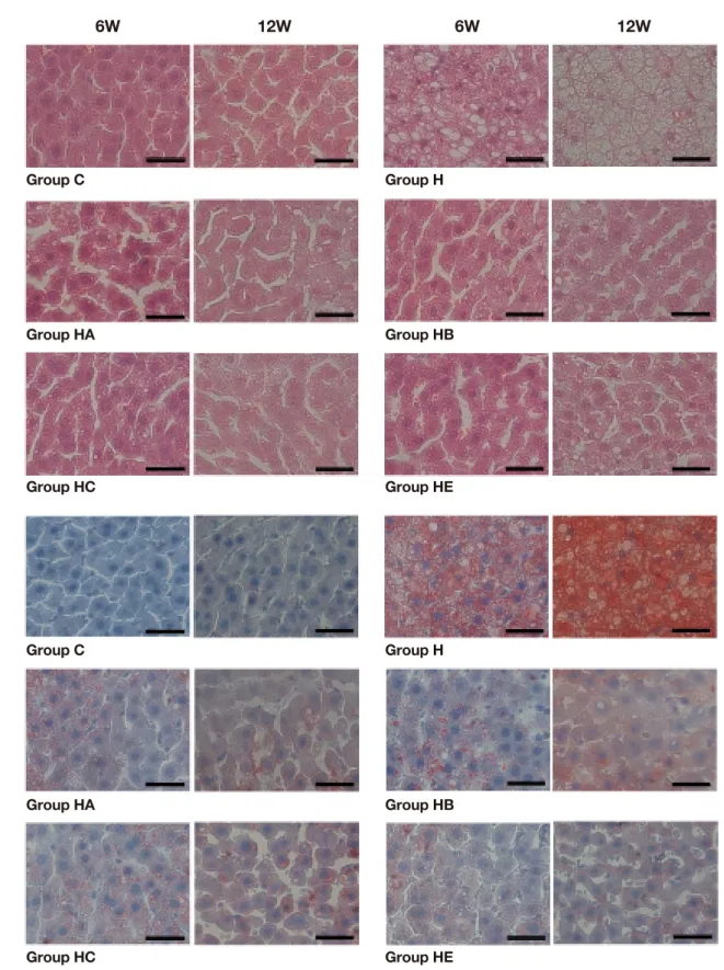
Fig. 1. Histopathological examination of liver.
A: Fatty degeneration (steatosis) of the liver is observed in the high-cholesterol diet-fed (H, HA, HB, HC and HE) groups, but not in
Group C in hematoxylin-eosin stained tissues. Fatty changes increase with time to a greater extent in Group H than Groups HA, HB, HC and HE.
B: Oil red O staining reveals more lipid droplets (stained red) to be accumulated in vacuoles in Group H than other groups.
6W, 6 weeks; 12W, 12 weeks. C, control: standard diet for 6 and 12 weeks (n = 5). H: high-cholesterol diet for 6 and 12 weeks (n = 5). HA: high-cholesterol diet with Boiogito for 6 and 12 weeks (n = 5). HB: high-cholesterol diet with Bofutsushosan for 6 and 12 weeks (n = 5). HC: high-cholesterol diet with Keishibukuryogan for 6 and 12 weeks (n = 5). HE: high-cholesterol diet with ezetimibe for 6 and 12 weeks (n = 5). Bars express 25 μm.
Immunohistochemical studies
During the immunohistochemical analyses, 4%
form-aldehyde-fixed aorta tissue specimens were processed.
The following monoclonal antibody was used:
anti-ICAM-1 (Abcam, Cambridge, United Kingdom). As a
negative control, tissues were stained without the
prima-ry antibody. The optical densities were measured by
Im-age-Pro Plus version 6.0 software (Media Cybernetics,
Rockville, MD).
Statistical analyses
The data are expressed as the mean ± SEM. For the
continuous variables, differences in responses among
groups were compared using Mann-Whitney’s U test for
non-parametric variables by SPSS 11.0 J (SPSS Japan,
Tokyo). P < 0.05 was considered statistically significant.
RESULTS
Histological examination of liver tissues
As Fig. 1 shows, the fatty degeneration (steatosis) of liver
was observed in the high-cholesterol diet-fed (H, HA,
HB, HC and HE) groups, but not in Group C. These
changes comprising tiny and large vacuoles as well as
pleomorphic nuclei were more prominent in Groups
HA, HB, HC and HE than in Group H (Fig. 1A). Oil red
O staining revealed that the livers in all high-cholesterol
diet supplemented groups were filled with
microvesicu-lar or macrovesicumicrovesicu-lar fat deposits; they were depicted as
reddish deposits (Fig. 1B). Overall, fatty liver changes
were more prominent in Group H than Groups HA, HB,
HC and HE (× 400).
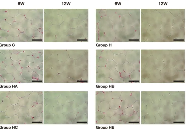
Histological examination of adipose tissue
As Fig. 2 shows, the larger fat cells were observed in
the high-cholesterol diet-fed (H, HA, HB, HC and HE)
groups, but not in Group C. It was more prominent in
Group H than Groups HA, HB, HC and HE (× 400).
Body and liver weightsAs Table 2 shows, there were no significant differences
in the baseline of body weights at the beginning of the
experiment. The body weights had no significant
dif-ferences during the experiment too. Liver weights were
significantly lower in Group HE than Group H after 6
and 12 weeks (P < 0.05).
Fig. 2. Histopathological examination of adipose tissue.
The larger fat cells are observed in the high-cholesterol diet-fed (H, HA, HB, HC and HE) groups, but not in Group C (56.13 ± 11.51 and 64.88 ± 13.96 μm after 6 and 12 weeks, respectively) in hematoxylin-eosin stained tissues. Fat cells change with time to a greater extent in Group H (97.00 ± 20.14 and 104.63 ± 22.56 μm after 6 and 12 weeks, respectively) than Groups HA, HB, HC and HE (55.63 ± 5.85 and 78.88 ± 16.34 μm, 75.63 ± 11.53 and 78.13 ± 16.47 μm, 74.63 ± 13.94 and 88.13 ± 8.58 μm, 74.38 ± 13.21 and 83.13 ± 16.63 μm after 6 and 12 weeks, respectively). Bars express 50 μm.
Table 2. Body and liver weights
Group C Group H Group HA Group HB Group HC Group HE
Body weight (g) 0W 233.1 ± 6.6 233.5 ± 6.6 235.9 ± 6.7 237.2 ± 9.4 237.1 ± 7.8 236.1 ± 6.7 6W 402.8 ± 15.9 398.5 ± 23.2 390.0 ± 21.9 392.4 ± 8.6 395.9 ± 23.9 385.8 ± 24.6 12W 448.6 ± 14.0 477.1 ± 15.5* 461.6 ± 26.1 457.6 ± 32.7 470.2 ± 32.9 453.3 ± 29.2 Liver weight (g) 6W 10.1 ± 0.9 15.0 ± 0.8* 15.5 ± 2.6 15.8 ± 1.4 14.9 ± 1.1 10.8 ± 1.0† 12W 12.5 ± 3.5 19.0 ± 3.1* 18.9 ± 1.5 19.9 ± 2.4 19.9 ± 2.2 12.1 ± 1.1†
0W, 0 weeks; 6W, 6 weeks; 12W, 12 weeks. C, control: standard diet for 6 and 12 weeks (n = 5). H: high-cholesterol diet for 6 and 12 weeks (n = 5). HA: high-cholesterol diet with Boiogito for 6 and 12 weeks (n = 5). HB: high-cholesterol diet with Bofutsushosan for 6 and 12 weeks (n = 5). HC: high-cholesterol diet with Keishibukuryogan for 6 and 12 weeks (n = 5). HE: high-cholesterol diet with ezetimibe for 6 and 12 weeks (n = 5). *P < 0.05 versus Group C. †P < 0.05 versus Group H. Data are expressed as the mean ± SEM.
Table 3. Blood chemistry and cholesterol concentrations
Group C Group H Group HA Group HB Group HC Group HE
TC (mg/dL) 6W 55.5 ± 15.2 68.2 ± 6.8 67.4 ± 13.7 65.4 ± 9.8 68.6 ± 13.8 78.5 ± 12.4 12W 56.6 ± 13.0 78.8 ± 5.4* 73.7 ± 10.4 75.2 ± 12.0 70.5 ± 12.0 73.2 ± 3.5 HDL-C (mg/dL) 6W 18.0 ± 2.6 21.0 ± 2.2 20.4 ± 4.5 19.4 ± 2.9 20.0 ± 3.2 17.5 ± 2.7 12W 16.0 ± 2.6 22.2 ± 0.8* 22.7 ± 3.2 23.8 ± 3.1 20.0 ± 3.4 19.4 ± 1.1 LDL-C (mg/dL) 6W 5.6 ± 1.1 8.4 ± 1.7* 8.6 ± 2.3 8.0 ± 1.9 8.8 ± 2.6 8.8 ± 2.2 12W 6.0 ± 1.4 11.8 ± 0.8* 9.0 ± 1.7† 9.8 ± 2.7 10.5 ± 4.4 8.0 ± 1.2† TG (mg/dL) 6W 31.5 ± 7.5 34.4 ± 11.2 28.0 ± 17.1 18.2 ± 3.9† 21.8 ± 8.8 25.0 ± 8.0 12W 29.2 ± 9.2 26.4 ± 4.7 22.7 ± 6.9 24.0 ± 7.9 19.8 ± 5.4 16.4 ± 7.0† BS (mg/dL) 6W 109.4 ± 7.7 99.6 ± 7.2 112.8 ± 8.8† 105.4 ± 6.6 110.8 ± 12.0 112.6 ± 10.5 12W 102.8 ± 6.1 112.0 ± 11.0 111.3 ± 20.0 114.8 ± 17.8 99.2 ± 7.6 101.2 ± 9.2 Cr (mg/dL) 6W 0.30 ± 0.03 0.30 ± 0.04 0.35 ± 0.06 0.35 ± 0.05 0.34 ± 0.04 0.34 ± 0.07 12W 0.32 ± 0.04 0.35 ± 0.03 0.34 ± 0.02 0.36 ± 0.11 0.34 ± 0.03 0.32 ± 0.03 TBil (μmol/L) 6W 0.03 ± 0.01 0.03 ± 0.01 0.03 ± 0.01 0.04 ± 0.01 0.03 ± 0.01 0.03 ± 0.02 12W 0.04 ± 0.02 0.03 ± 0.02 0.03 ± 0.01 0.03 ± 0.01 0.03 ± 0.01 0.04 ± 0.01 ALT (U/L) 6W 60.0 ± 11.7 72.2 ± 11.4* 71.8 ± 18.2 61.3 ± 10.2 93.6 ± 17.1 111.0 ± 11.1† 12W 53.4 ± 3.0 148.7 ± 25.7* 79.0 ± 17.4† 104.3 ± 26.1† 79.2 ± 18.5† 109.7 ± 25.8 ALP (U/L) 6W 413.0 ± 22.6 497.8 ± 24.8 377.5 ± 31.2† 399.0 ± 20.1† 450.0 ± 35.6 519.0 ± 34.9 12W 331.0 ± 28.5 432.7 ± 31.7* 334.3 ± 32.1† 337.0 ± 25.5† 283.3 ± 27.1† 514.8 ± 17.8† 6W, 6 weeks; 12W, 12 weeks. ALP, alkaline phosphatase; ALT, alanine aminotransferase; BS, blood sugar; Cr, creatinine; HDL-C, high-density lipoprotein cholesterol; LDL-C, low-high-density lipoprotein cholesterol; TBil, total bilirubin; TC, total cholesterol; TG, triglyceride. *P < 0.05 versus Group C. †P < 0.05 versus Group H. Data are expressed as the mean ± SEM.
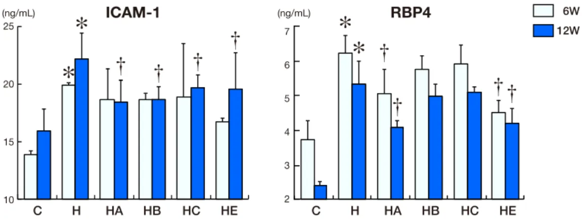
Fig. 3. Intercellular adhesion molecule-1 (ICAM-1) and retinol-binding protein 4 (RBP4) concentration in serum. ICAM-1 and RBP4
concentrations in control, standard diet group (C, n = 5), high-cholesterol diet group (H, n = 5), high-cholesterol diet with BOT group (HA, n = 5), high-cholesterol diet with BTS group (HB, n = 5), high-cholesterol diet with KBG group (HC, n = 5) and high-cholesterol diet with ezetimibe group (HE, n = 5) for 6 and 12 weeks. *P < 0.05 versus Group C. †P < 0.05 versus Group H.
*
ICAM-1
RBP4
C H HA HB HC HE 6W 12W C H HA HB HC HE 25 20 15 10 7 6 5 4 3 2 (ng/mL) (ng/mL)*
†
†
†
†
†
†
††
*
*
Blood chemistry and cholesterol concentrations
After 6 weeks of the experiment, the concentration of
LDL-C in Group H (8.4 ± 1.7 mg/dL) was significantly
elevated compared to the concentration in Group C (5.6
± 1.1 mg/dL) (P < 0.05). After 12 weeks, the
concentra-tion of TC in Group H (78.8 ± 5.4 mg/dL) was
signifi-cantly elevated compared with it in Group C (56.6 ±
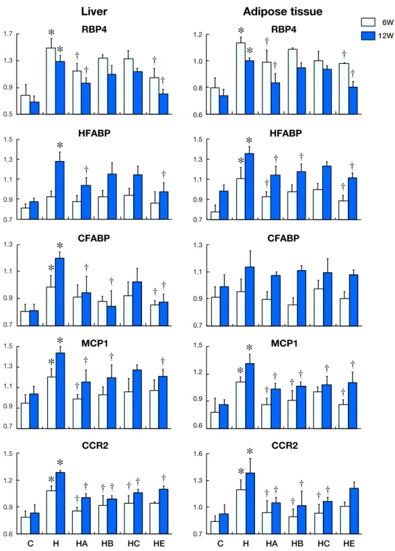
Fig. 4. Changes in mRNA expression with time in liver and adipose tissue. Levels of RBP4, HFABP, CFABP, MCP1 and CCR2 against
beta-actin mRNA expression are shown in the above histograms. The left-side graphs show the expression in liver. The right-side graphs show the expression in adipose tissue around the left kidney.
13.0 mg/dL) (P < 0.05). The concentration of HDL-C in
Group H (22.2 ± 0.8 mg/dL) was significantly elevated
compared with it in Group C (16.0 ± 2.6 mg/dL) (P < 0.05).
The concentration of LDL-C elevated to a greater extent
in Group H (11.8 ± 0.8 mg/dL) than that in Groups HA
and HE (9.0 ± 1.7 and 8.0 ± 1.2 mg/dL, respectively) (P
< 0.05). The concentration of ALT in Group H (148.7 ±
*
*
†
†
†
†
*
*
†
†
†
†
*
*
†
†
†
†
†
*
*
†
†
*
†
†
†
†
*
*
†
†
†
†
†
†
*
*
†
†
†
†
†
†
†
*
*
†
†
†
†
†
†
†
*
*
†
†
†
†
Liver
Adipose tissue
C H HA HB HC HE C H HA HB HC HE 1.7 1.3 0.9 0.5 1.2 1.0 0.8 0.6 1.3 1.1 0.9 0.7 1.3 1.1 0.9 0.7 1.5 1.2 0.9 0.6 1.5 1.2 0.9 0.6 1.6 1.3 1.0 0.7 1.5 1.3 1.1 0.9 0.7 1.5 1.3 1.1 0.9 0.7 1.5 1.3 1.1 0.9 0.7 HFABP RBP4 MCP1 CFABP CCR2 HFABP RBP4 MCP1 CFABP CCR2 6W 12W
Group C Group H Group HA
Group HB Group HC Group HE
C H HA HB HC HE 0.026 0.022 0.018 0.014 0.010 Optical density †
*
† † †25.7 U/L) was significantly elevated compared with it in
Group C (53.4 ± 3.0 U/L) (P < 0.05). It was decreased in
Groups HA, HB and HC (79.0 ± 17.4, 104.3 ± 26.1, 79.2
± 18.5 U/L, respectively) than in Group H (P < 0.05).
The concentration of ALP in Group H (432.7 ± 31.7 U/
L) was significantly elevated compared with it in Group
C (331.0 ± 28.5 U/L) (P < 0.05). It was lower in Groups
HA, HB and HC (334.3 ± 32.1, 337.0 ± 25.5, 283.3 ± 27.1
U/L, respectively) than in Group H (P < 0.05) (Table 3).
ICAM-1 and RBP4 in serumICAM-1 concentrations were increased in Group H
compared to Groups C, HA, HB and HE after 12 weeks
(Fig. 4, P < 0.05). RBP4 concentrations were increased
in Group H compared to the other groups
includ-ing Groups C, H, HA and HE after 6 and 12 weeks
(Fig. 3, P < 0.05).
mRNA expression in liver and adipose tissue
Figure 4 demonstrates the changes of mRNA expression
in liver and adipose tissue round the left kidney of rats
Fig. 5. Immunohistochemistry expression of ICAM-1 in aorta.
A: Seldom ICAM-1 immunostaining (red-brown deposits indicate positive staining) is found on the whole layers of abdominal aortas in
Group C. Bars express 25 μm.
B: Mean optical density values of ICAM-1. The photographs generated were quantitatively analyzed the optical density of ICAM-1 with
Image-Pro Plus version 6.0 software. *P < 0.05 versus Group C. †P < 0.05 versus Group H.
for 6 and 12 weeks.
RBP4 mRNA expression in liver in Group H was
significantly increased compared to that in Groups C,
HA and HE after 6 and 12 weeks (P < 0.05). HFABP
mRNA expression in Group H was significantly
in-creased compared to that in Groups C, HA and HE after
12 weeks (P < 0.05). Compared with the other groups,
the mRNA expression of CFABP in liver in Group H
were higher than that in Groups C, HA, HB and HE at
12 weeks (P < 0.05), whereas they were not obvious at
6 weeks. Compared with the other groups, the mRNA
expression of MCP1 in liver in Group H remained high
during the entire experiment, especially after 12 weeks
(P < 0.05). The expression of CCR2 in liver in Groups
HA, HB, HC (at 6 and 12 weeks) and HE (at 12 weeks)
were lower than that in Group H (P < 0.05).
A down-regulation of RBP4 mRNA expression in
Group H appeared in adipose tissue around the left
kid-ney after 12 weeks; even it was higher in Group H than
that in Groups C, HA and HE (P < 0.05). The mRNA
expression of HFABP was higher in Group H than that
in Groups C, HA, HE (at 6 and 12 weeks) and HB (at
12 weeks) (P < 0.05). CFABP mRNA expression had
no significant difference during the whole experiment
among all the groups. The expression of MCP1 in
adi-pose tissue in Groups HA, HB, HE (at 6 and 12 weeks)
and HC (at 12 weeks) was lower than that in Group H (P
< 0.05). The CCR2 mRNA expression in Group H was
higher than that in Groups HA, HB and HC (P < 0.05).
ICAM-1immunostaining expression in aortaAs shown in Fig. 5, after 12 weeks, little ICAM-1
immunostaining (red-brown deposits indicate
posi-tive staining) was found on the whole layers of
ab-dominal aortas in Group C. Significantly positive
ICAM-1 immunostaining was observed in Group H
and mainly localized on the endothelial layers.
Posi-tive ICAM-1 immunostaining was also observed in
Group
s
HA, HB, HC and HE, but less than Group H.
While ICAM-1 immunoreactivities were very weak
in Group
s
HA, HB, HC and HE, the positive area and
the strength of ICAM-1 stainings was markedly lower
than those in Group H (× 400).
Blood pressure and heart rate
No significant changes in systolic or diastolic blood
pressure and heart rates were observed during the
ex-periment.
DISCUSSION
Hypercholesterolemia is one of major risk factors
contributing to cardiovascular diseases.
3, 4Patients
with NAFLD have a high risk of CVD, too.
5Elevated
concentrations of plasma TC and LDL-C as well as
reduced concentration of HDL-C are negative risk
fac-tors for CVD is well documented.
28–30Lipid-lowering
drugs, such as statins,
hydroxyl-methylglutaryl-coen-zyme A reductase inhibitors can well reduce LDL-C
and increase HDL-C. Adverse reactions such as
rhab-domyolysis and hepatitis were sometimes caused by
them. Therefore, minimizing the side effects of
lipid-lowering drugs is also very important. The mechanism
of Kampo formula is to restore the balance of the body.
It is a safe way to achieve the balance between LDL-C
and HDL-C concentrations. In this study, we observed
the effect of Kampo formulas (BOT, BTS, KBG) on
the progression of hypercholesterolemia and fatty
liver induced by high-cholesterol diet in rats. LDL-C,
ALT and ALP concentrations in the serum of rats fed
high-cholesterol diet with BOT decreased. ALT (at 6
weeks) and ALP (at 12 weeks) concentrations in Group
HE was significantly elevated compared with that in
Group H. The reason is still unknown. The steatosis
of liver and hypertrophy of fat cells caused by
high-cholesterol diet could be alleviated by BOT, BTS and
KBG as shown by histopathological examinations.
Retinol-binding protein 4 (RBP4), a protein
secret-ed by hepatocytes (80%) and adipose tissue (20%), is a
21-kDa protein that facilitates the transport of retinol
through the circulation to peripheral tissues.
11It plays
a key role in the control of metabolic and proliferative
cell functions,
31including steatogenesis.
32Recently,
the role of adipokines, specifically RBP4 in the
patho-genesis of obesity-related diseases is widely being
discussed.
33–36Many studies have reported the
rela-tionship between RBP4 and obesity as well as its
re-lated complications.
35–37Recent studies demonstrated
that RBP4 levels were increased in obese and
insulin-resistant humans and mouse models.
38–41Stefan et al.
found a direct relation between hepatic fat content and
blood levels of RBP4 in healthy subjects, too.
42It has
been reported that RBP4 mRNA expression can be
up-regulated in liver and adipose tissue of rats with
high cholesterol diet for 12 weeks.
11In our experiment,
RBP4 expression in liver and adipose tissue of rats
fed a high-cholesterol diet as well as the expression in
serum were up-regulated. BOT and ezetimibe shows
greater effect to decrease the level of RBP4 mRNA
expression in liver and adipose tissue, the serum
ex-pression of RBP4 can also be decreased by BOT and
ezetimibe.
HFABP is a member of a family of binding
pro-teins with distinct tissue distributions and diverse
roles in fatty acid metabolism, trafficking, and
signal-ing.
43It is a low molecular-weight cytoplasmic protein
that is abundant in the myocardium
44and produced
by skeletal muscle,
45cardiomyocytes, kidney distal
tubular cells
46and specific parts of the brain.
47, 48HFABP mRNA expression has been reported to be
up-regulated in livers of rats fed high-cholesterol diet
for 28 days.
11In our study, this phenomenon was also
demonstrated. HFABP mRNA expression in the liver
and adipose tissue of high-cholesterol diet groups was
significantly increased compared to the control group
after 12 weeks.
CFABP was originally identified as being
over-expressed in the psoriatic skin.
49, 50It is typical of the
FABP family and binds long-chain fatty acids with
high affinity
49, 51and were thought to play a important
role in the storage and transport of fatty acids.
50, 52, 53CFABP was reported to be up-regulated after 12
weeks in liver and adipose tissue of high-cholesterol
diet rats.
11Here, the mRNA expression of CFABP in
liver of high-cholesterol diet rats were higher after 12
weeks while there were no significant differences
be-tween groups in adipose tissue. BOT, BTS and
ezeti-mibe can down-regulate the CFABP mRNA
expres-sion in liver.
BOT, BTS and KBG are all used as anti-obesity
medications in Japan.
54BOT is used for people with a
solid build and thick abdominal subcutaneous fat.
54It
could decrease the mouse body weight, fat
accumula-tion, TC and triglyceride level, but cannot influence
fasting blood glucose levels or insulin levels.
22BTS is
used for patients with flabby constitution and
prone-ness to fatigue.
54It can decrease food intake, body
weight, blood pressure, white adipose tissue weight
and ameliorate the adipocytokine dysregulation in
white adipose tissue.
55KBG is commonly used for
women with sudden weight gain in menopause and
could led to a reduction in blood cholesterol.
54, 56Ac-cording to these reports, even BOT, BTS and KBG are
all used in the treatment of obesity, they are suitable
for different situations. In our study, BOT has a
bet-ter effect on the progression of hypercholesbet-terolemia
and fatty liver induced by high-cholesterol diet in rats.
Ougi and Ginger are the key components of BOT,
and it has been reported that Ougi and Ginger of BOT
contribute greatly to the beneficial effects on abnormal
lipid metabolism.
22Matsuda et al. also found that
Gin-ger tends to improve lipid metabolism.
24Wang et al.
reported that Ougi could significantly reduced plasma
levels of TC and LDL-C and improved the
atheroscle-rosis profile.
57These results partly explain the present
findings. We speculate that inhibition of lipid
absorp-tion might contribute to the effect of BOT.
22However,
the mechanisms of BTS and KBG are still unknown.
Inflammatory factors play important roles in
NAFLD and atherosclerosis progression.
8MCP1 is
an important inflammatory chemokine that can be
produced by a variety of cells including vascular
endo-thelial cells, vascular smooth muscle cells, monocytes
and other cells. It is a member of the CC chemokine
family.
58, 59Plasma concentration of MCP1 increases
with obesity.
60, 61CCR2 is a receptor of MCP1 and
can help MCP1 to accomplish its effect.
62Dietary
cho-lesterol can induce the MCP1 gene expression,
14and
CCR2 expression was increased in
hypercholesterol-emic patients compared with normocholesterolhypercholesterol-emic
controls.
15, 63In our experiment, compared with the
other groups, the mRNA expression of MCP1 and
CCR2 in both liver and adipose tissue in Group H
re-mained high level during the entire experiment,
espe-cially after 12 weeks. BOT, BTS, KBG and ezetimibe
can down-regulate the MCP1 and CCR2 expression.
This effect of BOT, BTS and KBG is direct or indirect
through inhibition of cholesterol metabolism is still
unknown.
ICAM-1, a member of the immunoglobulin
super-family, is one of the markers of endothelial cell
activa-tion. It plays an important role in neutrophil migration
and adhesion of endothelial cells
64and is partly involved
in the whole process of monocyte adhesion, migration
and transformation.
16This migration is one of the earliest
events in the atherosclerotic process.
65ICAM-1 was
up-regulated in neointimal and medial smooth muscle cells
after vascular injury.
66Sekiya et al. reported that KBG
could prevent the progression of atheromatous plaque,
67but no literature could prove the effect of BOT and BTS
on preventing atherosclerosis. In our experiment, serum
ICAM-1 concentration and immunostaining expression
were increased in Group H after 12 weeks. BOT, BTS,
KBG and ezetimibe can down-regulate it. We speculate
that down-regulated ICAM-1 may be one of the
mecha-nisms of BOT, BTS and KBG to prevent atherosclerosis.
Overall, BOT has a protective effect on the
progres-sion of hypercholesterolemia and fatty liver induced by
high-cholesterol diet in rats and more effective than BTS
and KBG. Ezetimibe has the similar effect on lipid
relat-ed factors, such as LDL-C, RBP4, HFABP and CFABP.
However, the anti-inflammatory (MCP1, CCR2) and
anti-arteriosclerotic (ICAM-1) effects of BOT are more
potent than ezetimibe. This may be a guide on clinical
use. The mechanism is still uncertain and further studies
should be designed.
Acknowledgments: We would like to express our sincere apprecia-tion for the fellowship and research grant from the Japan Research Foundation of Clinical Pharmacology (JRFCP).
A preliminary report has appeared in abstract form in Japanese.68
The authors declare no conflict of interest.
REFERENCES
1 Roffi M, Brandle M, Robbins MA, Mukherjee D. Current per-spectives on coronary revascularization in the diabetic patient. Indian Heart J. 2007;59:124-36. PMID: 19122245.
2 Mohamed AR, El-Hadidy WF, Mannaa HF. Assessment of the Prophylactic Role of Aspirin and/or Clopidogrel on Ex-perimentally Induced Acute Myocardial Infarction in Hyper-cholesterolemic Rats. Drugs R D. 2014 Sep 18. [Epub ahead of print]. PMID: 25231707.
3 Angulo P, Lindor KD. Non-alcoholic fatty liver disease. J Gas-troenterol Hepatol. 2002;17 Suppl:S186-90. PMID: 12000605. 4 Marchesini G, Moscatiello S, Di Domizio S, Forlani G.
Obesity-associated liver disease. J Clin Endocrinol Metab. 2008;93(11 Suppl 1):S74-80. PMID: 18987273.
5 Targher G, Marra F, Marchesini G. Increased risk of cardio-vascular disease in non-alcoholic fatty liver disease: causal effect or epiphenomenon? Diabetologia. 2008;51:1947-53. PMID: 18762907.
6 Dick TJ, Lesser IA, Leipsic JA, Mancini GB, Lear SA. The effect of obesity on the association between liver fat and
ca-rotid atherosclerosis in a multi-ethnic cohort. Atherosclerosis. 2013;226:208-13. PMID: 23141586.
7 Sung KC, Wild SH, Kwag HJ, Byrne CD. Fatty liver, insulin resistance, and features of metabolic syndrome: relationships with coronary artery calcium in 10,153 people. Diabetes Care. 2012;35:2359-64. PMID: 22829522; PMCID: PMC3476919. 8 Kim EJ, Kim BH, Seo HS, Lee YJ, Kim HH, Son HH, et al.
Cholesterol-induced non-alcoholic fatty liver disease and ath-erosclerosis aggravated by systemic inflammation. PLoS One. 2014;9:e97841. PMID: 24901254; PMCID: PMC4046981. 9 Margareto J, Gómez-Ambrosi J, Marti A, Martínez JA.
Time-dependent effects of a high-energy-yielding diet on the regula-tion of specific white adipose tissue genes. Biochem Biophys Res Commun. 2001;283:6-11. PMID: 11322759.
10 Kushiro M, Takahashi Y, Ide T. Modulation of cutaneous fatty acid-binding protein mRNA expression in rat adipose tissues by hereditary obesity and dietary fats. J Oleo Sci. 2007;56:533-41. PMID: 17898460.
11 Wang X, Hasegawa J, Kitamura Y, Wang Z, Matsuda A, Shinoda W, et al. Effects of hesperidin on the progression of hypercholesterolemia and fatty liver induced by high-choles-terol diet in rats. J Pharmacol Sci. 2011;117:129-38. PMID: 21979313.
12 Bass NM. Function and regulation of hepatic and intestinal fatty acid binding proteins. Chem Phys Lipids. 1985;38:95-114. PMID: 4064226.
13 Engström G, Hedblad B, Janzon L, Lindgärde F. Long-term change in cholesterol in relation to inflammation-sensitive plasma proteins: a longitudinal study. Ann Epidemiol. 2007;17:57-63. PMID: 17178329.
14 Tous M, Ferré N, Rull A, Marsillach J, Coll B, Alonso-Villaverde C, et al. Dietary cholesterol and differential mono-cyte chemoattractant protein-1 gene expression in aorta and liver of apo E-deficient mice. Biochem Biophys Res Commun. 2006;340:1078-84. PMID: 16403442.
15 Han KH, Tangirala RK, Green SR, Quehenberger O. Che-mokine receptor CCR2 expression and monocyte chemoat-tractant protein-1-mediated chemotaxis in human monocytes. A regulatory role for plasma LDL. Arterioscler Thromb Vasc Biol. 1998;18:1983-91. PMID: 9848893.
16 Nighoghossian N, Derex L, Douek P. The vulnerable carotid artery plaque: current imaging methods and new perspectives. Stroke. 2005;36:2764-72. PMID: 16282537.
17 Hioki C, Yoshimoto K, Yoshida T. Efficacy of bofu-tsusho-san, an oriental herbal medicine, in obese Japanese women with impaired glucose tolerance. Clin Exp Pharmacol Physi-ol. 2004;31:614-9. PMID: 15479169.
18 Ono M, Ogasawara M, Hirose A, Mogami S, Ootake N, Aritake K, et al. Bofutsushosan, a Japanese herbal (Kampo) medicine, attenuates progression of nonalcoholic steato-hepatitis in mice. J Gastroenterol. 2014;49:1065-73. PMID: 23800945; PMCID: PMC4048468.
19 Ohno K, Chung HJ, Maruyama I, Tani T. Bofutsushosan, a traditional Chinese formulation, prevents intimal thicken-ing and vascular smooth muscle cell proliferation induced by balloon endothelial denudation in rats. Biol Pharm Bull. 2005;28:2162-5. PMID: 16272712.
20 Yoshida T, Sakane N, Wakabayashi Y, Umekawa T, Kondo M. Thermogenic, anti-obesity effects of bofu-tsusho-san in MSG-obese mice. Int J Obes Relat Metab Disord. 1995;19:717-22. PMID: 8589765.
21 Morimoto Y, Sakata M, Ohno A, Maegawa T, Tajima S. [Ef-fects of Byakko-ka-ninjin-to, Bofu-tsusho-san and
Gorei-san on blood glucose level, water intake and urine volume in KKAy mice]. Yakugaku Zasshi. 2002;122:163-8. Japanese. PMID: 11857957.
22 Shimada T, Akase T, Kosugi M, Aburada M. Preventive Ef-fect of Boiogito on Metabolic Disorders in the TSOD Mouse, a Model of Spontaneous Obese Type II Diabetes Mellitus. Evid Based Complement Alternat Med. 2011;2011:931073. PMID: 19208721; PMCID: PMC3139392.
23 Nagata Y, Goto H, Hikiami H, Nogami T, Fujimoto M, Shibahara N, et al. Effect of keishibukuryogan on endothelial function in patients with at least one component of the diag-nostic criteria for metabolic syndrome: a controlled clinical trial with crossover design. Evid Based Complement Alternat Med. 2012;2012:359282. PMID: 22675380.
24 Matsuda A, Wang Z, Takahashi S, Tokuda T, Miura N, Hasegawa J. Upregulation of mRNA of retinoid binding pro-tein and fatty acid binding propro-tein by cholesterol enriched-diet and effect of ginger on lipid metabolism. Life Sci. 2009;84:903-7. PMID: 19379761.
25 Koopman R, Schaart G, Hesselink MK. Optimisation of oil red O staining permits combination with immunofluorescence and automated quantification of lipids. Histochem Cell Biol. 2001;116:63-8. PMID: 11479724.
26 Livak KJ, Schmittgen TD. Analysis of relative gene expression data using real-time quantitative PCR and the 2(-Delta Delta C(T)) Method. Methods. 2001;25:402-8. PMID: 11846609. 27 Schmittgen TD, Zakrajsek BA, Mills AG, Gorn V, Singer
MJ, Reed MW. Quantitative reverse transcription-polymerase chain reaction to study mRNA decay: comparison of endpoint and real-time methods. Anal Biochem. 2000;285:194-204. PMID: 11017702.
28 Daniels TF, Killinger KM, Michal JJ, Wright RW Jr, Jiang Z. Lipoproteins, cholesterol homeostasis and cardiac health. Int J Biol Sci. 2009;5:474-88. PMID: 19584955; PMCID: PMC2706428.
29 Sharrett AR, Ballantyne CM, Coady SA, Heiss G, Sorlie PD, Catellier D, et al.; Atherosclerosis Risk in Communities Study Group. Coronary heart disease prediction from lipoprotein cholesterol levels, triglycerides, lipoprotein(a), apolipopro-teins A-I and B, and HDL density subfractions: The Athero-sclerosis Risk in Communities (ARIC) Study. Circulation. 2001;104:1108-13. PMID: 11535564.
30 Liu Y, Lei L, Wang X, Ma KY, Li YM, Wang L, et al. Plasma cholesterol-raising potency of dietary free cholesterol ver-sus cholesteryl ester and effect of b-sitosterol. Food Chem. 2015;169:277-82. PMID: 25236227.
31 Desvergne B. RXR: from partnership to leadership in metabolic regulations. Vitam Horm. 2007;75:1-32. PMID: 17368310.
32 Larter CZ, Farrell GC. Insulin resistance, adiponectin, cyto-kines in NASH: Which is the best target to treat? J Hepatol. 2006;44:253-61. PMID: 16364488.
33 Lebensztejn DM, Wojtkowska M, Skiba E, Werpachowska I, Tobolczyk J, Kaczmarski M. Serum concentration of adiopo-nectin, leptin and resistin in obese children with non-alcoholic fatty liver disease. Adv Med Sci. 2009;54:177-82. PMID: 20022856.
34 Louthan MV, Barve S, McClain CJ, Joshi-Barve S. Decreased serum adiponectin: an early event in pediatric nonalco-holic fatty liver disease. J Pediatr. 2005;147:835-8. PMID: 16356442.
35 Christou GA, Tselepis AD, Kiortsis DN. The metabolic role of retinol binding protein 4: an update. Horm Metab Res.
2012;44:6-14. PMID: 22205567.
36 Kotnik P, Fischer-Posovszky P, Wabitsch M. RBP4: a contro-versial adipokine. Eur J Endoc rinol. 2011;165:703-11. PMID: 21835764.
37 Saki F, Karamizadeh Z, Honar N, Moravej H, Ashkani-Esfahani S, Namvar Shooshtarian MH. Association of Plasma Retinol Binding Protein-4 (RBP4) and Sonographic Grad-ing of Fatty Liver in Obese Iranian Children. Hepat Mon. 2012;12:e7103. PMID: 23423766; PMCID: PMC3575548. 38 Graham TE, Yang Q, Blüher M, Hammarstedt A, Ciaraldi
TP, Henry RR, et al. Retinol-binding protein 4 and insulin resistance in lean, obese, and diabetic subjects. N Engl J Med. 2006;354:2552-63. PMID: 16775236.
39 Norseen J, Hosooka T, Hammarstedt A, Yore MM, Kant S, Aryal P, et al. Retinol-binding protein 4 inhibits insulin signal-ing in adipocytes by inducsignal-ing proinflammatory cytokines in macrophages through a c-Jun N-terminal kinase- and toll-like receptor 4-dependent and retinol-independent mechanism. Mol Cell Biol. 2012;32:2010-9. PMID: 22431523; PMCID: PMC3347417.
40 Klöting N, Graham TE, Berndt J, Kralisch S, Kovacs P, Wason CJ, et al. Serum retinol-binding protein is more highly expressed in visceral than in subcutaneous adipose tissue and is a marker of intra-abdominal fat mass. Cell Metab. 2007;6:79-87. PMID: 17618858.
41 Liu Y, Wang D, Li D, Sun R, Xia M. Associations of retinol-binding protein 4 with oxidative stress, inflammatory mark-ers, and metabolic syndrome in a middle-aged and elderly Chinese population. Diabetol Metab Syndr. 2014;6:25. PMID: 24559154; PMCID: PMC3938900.
42 Stefan N, Hennige AM, Staiger H, Machann J, Schick F, Schleicher E, et al. High circulating retinol-binding protein 4 is associated with elevated liver fat but not with total, subcu-taneous, visceral, or intramyocellular fat in humans. Diabetes Care. 2007;30:1173-8. PMID: 17259477.
43 Qian Q, Kuo L, Yu YT, Rottman JN. A concise promoter region of the heart fatty acid-binding protein gene dictates tis-sue-appropriate expression. Circ Res. 1999;84:276-89. PMID: 10024301.
44 Glatz JF, van Bilsen M, Paulussen RJ, Veerkamp JH, van der Vusse GJ, Reneman RS. Release of fatty acid-binding protein from isolated rat heart subjected to ischemia and reperfu-sion or to the calcium paradox. Biochim Biophys Acta. 1988; 961:148-52. PMID: 3260112.
45 Zschiesche W, Kleine AH, Spitzer E, Veerkamp JH, Glatz JF. Histochemical localization of heart-type fatty-acid binding protein in human and murine tissues. Histochem Cell Biol. 1995;103:147-56. PMID: 7634154.
46 Maatman RG, van de Westerlo EM, van Kuppevelt TH, Veerkamp JH. Molecular identification of the liver- and the heart-type fatty acid-binding proteins in human and rat kidney. Use of the reverse transcriptase polymerase chain reaction. Biochem J. 1992;288(Pt 1):285-90. PMID: 1280113; PMCID: PMC1132111.
47 Pelsers MM, Hanhoff T, Van der Voort D, Arts B, Peters M, Ponds R, et al. Brain- and heart-type fatty acid-binding pro-teins in the brain: tissue distribution and clinical utility. Clin Chem. 2004;50:1568-75. PMID: 15217991.
48 Cakir E, Ozbek M, Sahin M, Cakal E, Gungunes A, Ginis Z, et al. Heart type fatty acid binding protein response and subsequent development of atherosclerosis in insulin resistant polycystic ovary syndrome patients. J Ovarian Res. 2012;5:45. PMID: 23249450; PMCID: PMC3574048.
49 Madsen P, Rasmussen HH, Leffers H, Honoré B, Celis JE. Molecular cloning and expression of a novel keratinocyte protein (psoriasis-associated fatty acid-binding protein [PA-FABP]) that is highly up-regulated in psoriatic skin and that shares similarity to fatty acid-binding proteins. J Invest Der-matol. 1992;99:299-305. PMID: 1512466.
50 Jing C, Beesley C, Foster CS, Chen H, Rudland PS, West DC, et al. Human cutaneous fatty acid-binding protein induces me-tastasis by up-regulating the expression of vascular endothelial growth factor gene in rat Rama 37 model cells. Cancer Res. 2001;61:4357-64. PMID: 11389060.
51 Adamson J, Morgan EA, Beesley C, Mei Y, Foster CS, Fujii H, et al. High-level expression of cutaneous fatty acid-binding protein in prostatic carcinomas and its effect on tumorigenic-ity. Oncogene. 2003;22:2739-49. PMID: 12743598
52 Bass NM. The cellular fatty acid binding proteins: aspects of structure, regulation, and function. Int Rev Cytol. 1988;111: 143-84. PMID: 3074959.
53 Horrobin DF. Essential fatty acids in clinical dermatology. J Am Acad Dermatol. 1989;20:1045-53. PMID: 2526823. 54 Yamakawa J, Moriya J, Takeuchi K, Nakatou M, Motoo Y,
Kobayashi J. Significance of Kampo, Japanese traditional medicine, in the treatment of obesity: basic and clinical evi-dence. Evid Based Complement Alternat Med. 2013;2013: 943075. PMID: 23662155; PMCID: PMC3639634.
55 Azushima K, Tamura K, Wakui H, Maeda A, Ohsawa M, Uneda K, et al. Bofu-tsu-shosan, an oriental herbal medicine, exerts a combinatorial favorable metabolic modulation in-cluding antihypertensive effect on a mouse model of human metabolic disorders with visceral obesity. PLoS One. 2013;8: e75560. PMID: 24130717. PMCID: PMC3794018.
56 Fujimoto M, Tsuneyama K, Kinoshita H, Goto H, Takano Y, Selmi C, et al. The traditional Japanese formula keishibuku-ryogan reduces liver injury and inflammation in patients with nonalcoholic fatty liver disease. Ann N Y Acad Sci. 2010;1190: 151-8. PMID: 20388146.
57 Wang D, Zhuang Y, Tian Y, Thomas GN, Ying M, Tomlinson B. Study of the effects of total flavonoids of Astragalus on ath-erosclerosis formation and potential mechanisms. Oxid Med Cell Longev. 2012;2012:282383. PMID: 22496932. PMCID: PMC3306992.
58 Yang X, Coriolan D, Murthy V, Schultz K, Golenbock DT, Beasley D. Proinflammatory phenotype of vascular smooth muscle cells: role of efficient Toll-like receptor 4 signaling. Am J Physiol Heart Circ Physiol. 2005;289:H1069-76. PMID: 15863460.
59 Anand AR, Bradley R, Ganju RK. LPS-induced MCP-1 ex-pression in human microvascular endothelial cells is mediated by the tyrosine kinase, Pyk2 via the p38 MAPK/NF-kappaB-dependent pathway. Mol Immunol. 2009;46:962-8. PMID: 18954908; PMCID: PMC2680789.
60 Panee J. Monocyte Chemoattractant Protein 1 (MCP-1) in obesity and diabetes. Cytokine. 2012;60:1-12. PMID: 22766373; PMCID: PMC3437929.
61 Heber D, Zhang Y, Yang J, Ma JE, Henning SM, Li Z. Green tea, black tea, and oolong tea polyphenols reduce visceral fat and inflammation in mice fed high-fat, high-sucrose obeso-genic diets. J Nutr. 2014;144:1385-93. PMID: 25031332. 62 Siebert H, Sachse A, Kuziel WA, Maeda N, Brück W.
The chemokine receptor CCR2 is involved in macrophage recruitment to the injured peripheral nervous system. J Neuro-immunol. 2000;110:177-85. PMID: 11024548.
high-fat and high-cholesterol diet. Am J Physiol Gastrointest Liver Physiol. 2011;301: G1031-43. PMID: 21885686.
64 Shimizu N, Suzuki H, Wakabayashi K, Iso Y, Shibata M, Yorozuya M, et al. [Expression of intercellular adhesion molecule-1 and vascular cell adhesion molecule-1 in the pig coronary artery injury model: comparison of plain old balloon angioplasty and stent implantation]. J Cardiol. 2004;43:131-9. Japanese. PMID: 15067801.
65 Amran AA, Zakaria Z, Othman F, Das S, Al-Mekhlafi HM, Nordin NA. Changes in the vascular cell adhesion molecule-1, intercellular adhesion molecule-1 and c-reactive protein following administration of aqueous extract of piper sarmentosum on experimental rabbits fed with cholesterol diet. Lipids Health Dis. 2011;10:2. PMID: 21214952. PMCID: PMC3024236.
intima formation and expression of inflammatory cytokines in rats after carotid artery balloon injury. Indian J Pharmacol. 2014;46:510-4. PMID: 25298580. PMCID: PMC4175887. 67 Sekiya N, Kainuma M, Hikiami H, Nakagawa T, Kouta K,
Shibahara N, et al. Oren-gedoku-to and Keishi-bukuryo-gan-ryo inhibit the progression of atherosclerosis in diet-induced hypercholesterolemic rabbits. Biol Pharm Bull. 2005;28:294-8. PMID: 15684487.
68 Qian W, Tsuno S, Endo Y, Matsuda A, Miura N, Hasegawa J. [Effects of Kampo recipes on the progression of fatty liver induced by high-cholesterol diet in rats]. Jpn J Clin Pharmacol Ther. 2013;44(Suppl):S308. Japanese.