Abbreviations: CH, chronic hepatitis; DNMT, DNA methyltransferase; HCC, hepatocellular carcinoma; LC, liver
cir-Expression of DNA Methyltransferase (DNMT) 1, 3a and 3b
Proteins in Human Hepatocellular Carcinoma
Takanori Miyake, Kanenori Endo, Soichiro Honjo, Yasuaki Hirooka and Masahide Ikeguchi
Division of Surgical Oncology, Department of Surgery, School of Medicine, Tottori University Fac-ulty of Medicine, Yonago 683-8504, Japan
Alteration of aberrant DNA methylation is one of the most consistent epigenetic changes found in human cancers. DNA methylation is catalyzed by DNA methyltransferase (DNMT). In this study, we examined DNMT protein expression by immunohistochemis-try in surgically resected hepatocellular carcinomas (HCCs). Sections of paraffin-embed-ded specimens were obtained from 95 patients with HCC between 1989 and 2002. The specimens were stained with anti-DNMTs (DNMT1, DNMT3a and DNMT3b) antibodies. There were statistically significant associations between DNMT protein expression and tumor differentiation (P < 0.05) and intrahepatic metastasis (P < 0.05). DNMT3a protein expression was significantly correlated with portal vein involvement of tumors (P < 0.05). The overall survival rates of patients with DNMT3a-positive HCCs and DNMT3b-positive HCCs were significantly lower than those of patients negative for these proteins (P < 0.005, respectively). To further evaluate the correlation between DNMT protein expression and patient survival, we classified patients into 3 groups: Group 1, DNMT1(+), 3a(–) and 3b(–); Group 2) DNMT1(+), 3a or 3b(+); and Group 3) DNMT1(+), 3a(+) and 3b(+). The over-all survival rate of patients in Group 3 was significantly lower than those of patients in Groups 1 and 2 (P = 0.0009). In conclusion, the results of this study suggest that DNMT1, DNMT3a and DNMT3b are cooperatively involved in determining the extent of HCCs, and that DNMT protein overexpression in HCCs may be a predictive factor for poor sur-vival.
Key words: DNA methyltransferase; hepatocellular carcinoma; immunohistochemistry; prognosis
DNA methylation plays an important role in the regulation of gene expression, and gene silencing by aberrant hypermethylation of CpG islands in gene promoter regions frequently occurs in human cancers. DNA methyltransferases are involved in the transfer of methyl groups to promoter CpG islands, and many tumor suppressor genes can be silenced by promoter CpG island methylation. Alteration of aberrant DNA methylation is one of the most consistent epigenetic changes found in
hu-man cancers (Girault et al., 2003; Ding et al., 2008; Nosho et al., 2009).
DNA methyltransferases have been reported to be encoded by 3 distinct families of DNMT genes: DNMT1, DNMT2, and DNMT3. DNMT1 is the best known DNMT involved in the mainte-nance of methylation, while DNMT3 is involved in de novo methylation and is encoded by 2 dis-tinct genes, DNMT3a and DNMT3b (Łuczak and Jagodzinski, 2006). The function of DNMT2
remains to be determined. Interactions among DNMTs and methyl CpG-binding domain proteins should also be considered as a mechanism for maintaining DNA methylation patterns using the proofreading or remethylation function (Hattori et al., 2004).
Aberrant hypermethylation of CpG islands has recently been implicated in hepatocarcinogen-esis, and it has been reported that tumor suppressor genes in hepatocellular carcinomas (HCCs) are affected by such silencing (Saito et al., 2001; Yang et al., 2003). To study the significance of aberrant DNMT (DNMT1, 3a and 3b) expression in human HCC, we evaluated the correlation between DNMT protein expression and clinicopathologic features and prognosis in patients with HCC.
Materials and Methods
Patients and samples
Stored HCC and corresponding non-cancerous tis-sue specimens from our laboratory were obtained for 95 patients from 1989 to 2002. The proto-col was approved by the ethics committee of the School of Medicine, Tottori University (1040). In-formed consent was obtained from all patients.
The clinicopathologic features of the 95 pa-tients are shown in Table 1. A mean age was 61 ± 10 years (range, 17–80 years). For comparison, normal liver tissues were also obtained from 3 patients who had undergone surgical resection for hepaticolithiasis.
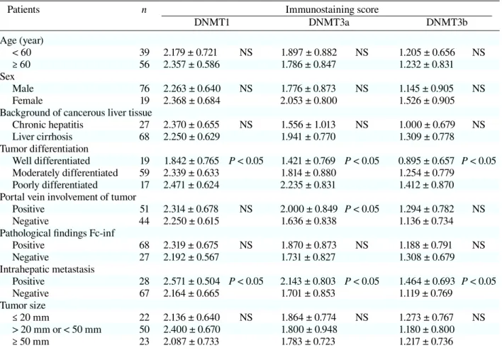
Table 1. Relationship between DNMTs and tissue specimens
Patients n Immunostaining score
DNMT1 DNMT3a DNMT3b Age (year) < 60 39 2.179 ± 0.721 NS 1.897 ± 0.882 NS 1.205 ± 0.656 NS ≥ 60 56 2.357 ± 0.586 1.786 ± 0.847 1.232 ± 0.831 Sex Male 76 2.263 ± 0.640 NS 1.776 ± 0.873 NS 1.145 ± 0.905 NS Female 19 2.368 ± 0.684 2.053 ± 0.800 1.526 ± 0.905
Background of cancerous liver tissue
Chronic hepatitis 27 2.370 ± 0.655 NS 1.556 ± 1.013 NS 1.000 ± 0.679 NS Liver cirrhosis 68 2.250 ± 0.629 1.941 ± 0.770 1.309 ± 0.778 Tumor differentiation Well differentiated 19 1.842 ± 0.765 P < 0.05 1.421 ± 0.769 P < 0.05 0.895 ± 0.657 P < 0.05 Moderately differentiated 59 2.339 ± 0.633 1.814 ± 0.880 1.254 ± 0.779 Poorly differentiated 17 2.471 ± 0.624 2.235 ± 0.831 1.412 ± 0.870 Portal vein involvement of tumor
Positive 51 2.314 ± 0.678 NS 2.000 ± 0.849 P < 0.05 1.294 ± 0.782 NS
Negative 44 2.250 ± 0.615 1.636 ± 0.838 1.136 ± 0.734
Pathological findings Fc-inf
Positive 68 2.319 ± 0.675 NS 1.870 ± 0.873 NS 1.188 ± 0.791 NS Negative 27 2.192 ± 0.567 1.731 ± 0.827 1.308 ± 0.679 Intrahepatic metastasis Positive 28 2.571 ± 0.504 P < 0.05 2.143 ± 0.803 P < 0.05 1.464 ± 0.693 P < 0.05 Negative 67 2.164 ± 0.665 1.701 ± 0.853 1.119 ± 0.769 Tumor size ≤ 20 mm 22 2.136 ± 0.640 NS 1.864 ± 0.774 NS 1.273 ± 0.767 NS > 20 mm or < 50 mm 50 2.400 ± 0.670 1.800 ± 0.948 1.180 ± 0.800 ≥ 50 mm 23 2.087 ± 0.733 1.783 ± 0.723 1.217 ± 0.736 n, number of patients.
Immunohistochemistry
Formalin-fixed, paraffin-embedded tissues were cut into 4-μm-thick continuous sections and mounted on poly-L-lysine-coated glass slides. Slides were deparaffinized in xylene, rehydrated in graded al-cohols and washed in tap water. Endogenous per-oxidase activity was blocked by incubating sections in 3% H2O2. Slides were heated in a microwave at
600 W for 15 min with 10 mM sodium citrate buf-fer (pH 6.0) and treated with polyoxyethylene sorbi-tan monolaurate (Tween 20) for 30 min for antigen retrieval. Rabbit polyclonal antibodies for DNMT1 (H-300, sc-20701, dilution 1:200), DNMT3a (H-295, sc-20703, dilution 1:200) and DNMT3b (H-230, sc-20704, dilution 1:200) (Santa Cruz Biotechnol-ogy, Santa Cruz, CA) were applied overnight, fol-lowed by incubation with biotinylated secondary antibodies [anti-rabbit IgG, N-Histofine, Simple Stain MAX PO (MULTI)] at room temperature for 30 min. Antigen-antibody complexes were visual-ized using a streptavidin-horseradish peroxidase conjugate (LSAB kit; Dako, Carpinteria, CA) with diaminobenzidine as a chromogen. Slides were counterstained with Harris’ hematoxylin and meth-ylgreen for 3 to 5 s.
Scoring of immunoreactivity
Two independent observers (T.M. and K.E.) inde-pendently evaluated the immunostaining without knowledge of the clinical outcomes; the concor-dance ratio was > 90%. When differences in scor-ing were encountered, a consensus of two was used to obtain a final decision.
Nuclear positivities for the 3 proteins were evaluated using the scoring system previously re-ported by Choi et al. (2003), according to staining intensity (0, negative; 1, weak; 2, moderate; and 3, severe) and proportion of positive cells (0, negative; 1, positive in ≤ 10%; 2, positive in > 10% and ≤ 33%; 3, positive in > 33% and ≤ 66%; 4, positive in > 66% of cells). The 2 scores were added in each case, and the expression was graded as – (0), 1+ (1 or 2), 2+ (3 to 5) and 3+ (6 or 7).
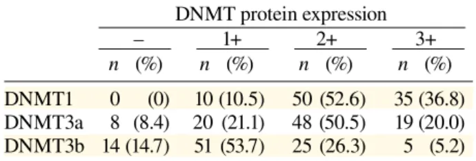
Table 2. Expression of DNMT 1, 3a and 3b pro-teins in patients with hepatocellular carcinoma
DNMT protein expression – 1+ 2+ 3+ n (%) n (%) n (%) n (%) DNMT1 0 (0) 10 (10.5) 50 (52.6) 35 (36.8) DNMT3a 8 (8.4) 20 (21.1) 48 (50.5) 19 (20.0) DNMT3b 14 (14.7) 51 (53.7) 25 (26.3) 5 (5.2) n, number of patients: total number, 95.
Nuclear positivities for the 3 proteins were evaluated using the scoring system, according to staining intensity (0, nega-tive; 1, weak; 2, moderate; and 3, severe) and proportion of positive cells (0, negative; 1, positive in ≤ 10%; 2, positive in > 10% and ≤ 33%; 3, positive in > 33% and ≤ 66%; 4, posi-tive in > 66% of cells). The 2 scores are added in each case, and the expression is graded as – (0), 1+ (1 or 2), 2+ (3 to 5) and 3+ (6 or 7).
Statistical analysis
Statistical analysis was performed using Prism soft-ware (GraphPad Softsoft-ware, La Jolla, CA). Spear-man’s rank correlation test and Student’s t-test were used to study the relationships between DNMT im-munoreactivities and various clinicopathologic pa-rameters. Overall survival rates were determined by the Kaplan-Meier method, and the significance of differences between survival rates was studied using the log-rank test. Differences or correlations with P values < 0.05 were considered significant. Positive DNMT expression was defined as moder-ate (2+) or strong (3+), and negative DNMT expres-sion as absent (–) or weak (1+) expresexpres-sion. To fur-ther evaluate the correlation between DNMT pro-tein expression and patient survival, we subdivided the patients into 3 groups: Group 1) DNMT1(+), 3a(–) and 3b(–); Group 2) DNMT1(+), 3a or 3b(+); and Group 3) DNMT1(+), 3a(+) and3b (+).
Results
The immunohistochemical staining results are shown in Table 2 and Fig. 1. The non-cancerous liver tissues showed no expression of nuclear DNMT proteins by immunohistochemistry. There were no differences in cytoplasmic DNMT
expres-Fig. 1. Immunohistochemical staining results for (a) DNMT1,
(b) DNMT3a and (c) DNMT3b proteins in hepatocellular carcinomas. Strong nuclear expression and faint cytoplasmic expression are seen in these 3 cases. a and b: counterstained with Harris’ hematoxylin. c: conterstained with methylgreen. Bar = 100 µm.
Fig. 2. Kaplan-Meier survival curves for patients
with hepatocellular carcinomas according to DNMT protein expression levels. [ ], number of patients. a: DNMT 1. b: DNMT 3a. c: DNMT 3b.
a
b
c
sion between patients with CH and LC (data not shown). None of the 3 normal control livers dem-onstrated nuclear expression of DNMTs.
We indicated the immunostaining scores according to the clinicopathological findings in Table 1. There were significant associations be-tween DNMT protein expression and tumor dif-ferentiation or intrahepatic metastasis. DNMT3a protein expression was significantly correlated with portal vein involvement in the tumor. On the other
hand, no significant correlations were noted be-tween DNMT1 or 3b and portal vein involvement. There were no significant associations between DNMT expression and age, sex, tumor size, infil-tration to the capsule or underlying liver disease.
Follow-up information was available for all 95 patients, covering periods ranging from 0 to 183 months (mean, 63.7 months). Kaplan-Meier surviv-al curves were plotted for the 95 patients according to expression of each DNMT.
The overall survival rates of DNMT3a-positive and DNMT3b-DNMT3a-positive HCC patients were significantly lower than those of patients negative for these proteins (Fig. 2b, P = 0.003, log-rank test; Fig. 2c, P = 0.0034, log-rank test). However, the overall survival rate of DNMT1-positive HCC patients was not significantly different from that of DNMT1-negative HCC cases (Fig. 2a, P = 0.1038, log-rank test).
The overall survival rate of patients in Group 3 [DNMT1(+), 3a(+) and 3b(+)] was significantly lower than those of patients in Groups 1 and 2 (Fig. 3, P = 0.0009, log-rank test).
Discussion
DNA methylation is well known to be involved in the early developmental stages of HCC with a background of chronic liver disease, including CH and LC. It has recently been suggested that aber-rant DNA methylation of CpG islands is an early
Fig. 3. Kaplan-Meier survival curves
were plotted for 94 patients accord-ing to the correlation with expression of the DNMT protein complex. [ ], number of patients.
Group 1: DNMT1(+), 3a(–) and 3b(–). Group 2: DNMT1(+), 3a or 3b(+). Group 3: DNMT1(+), 3a(+) and 3b(+).
and frequent event, and that stepwise progression of methylating events contributes to multistep hepa-tocarcinogenesis (Roncalli et al., 2002; Lee et al., 2003; Yang et al., 2003). The levels of DNMT1, DNMT3a and DNMT3b mRNA in HCCs are sig-nificantly higher than in noncancerous liver tissues, including normal, chronic CH and LC tissues (Saito et al., 2001; Oh et al., 2007).
In the present study, we used an immunohis-tochemical technique to directly examine the pro-tein expression of 3 DNMTs in HCCs. The results demonstrated that DNMT protein expression in HCCs was significantly higher than in noncancer-ous liver tissues including normal, CH and LC tis-sues. In addition, there was no significant relation-ship between DNMT expression, CH and LC.
Immunohistochemical staining showed very strong nuclear expression and faint cytoplasmic expression of DNMT1, and strong nuclear and faint cytoplasmic expression of DNMT3a and DNMT3b. Some immunohistochemical staining of these DNMTs has previously been performed in a vari-ety of cancers (Girault et al., 2003; Lin et al., 2007; Ding et al., 2008) and HCC (Saito et al., 2003; Oh et al., 2007). Although nuclear and cytoplasmic DNMT3b expression has been detected in other cancers, no results of immunohistochemical stain-ing of DNMT3b have previously been reported in HCC.
The present study showed a significant re-lationship between DNMT3a protein expression and portal vein involvement, and significant
rela-tionships between DNMT protein expression and tumor differentiation or intrahepatic metastasis. However, a previous report found no significant relationship between DNMT mRNA levels and portal vein involvement and tumor differentiation in HCCs (Saito et al., 2001), but the same research group reported a significant relationship between DNMT1 protein expression and portal vein in-volvement or tumor differentiation (Saito et al., 2003). Reverse transcriptase-polymerase chain reaction is sufficiently sensitive to detect small el-evations in DNMT mRNA levels, while immuno-histochemistry is only able to detect elevations in protein expression when they reach a certain level in more malignant HCCs. We suggest that DNMT protein expression may be more strongly correlated with progression of the cancerous stage of HCCs, rather than the precancerous stage. To the best of our knowledge, there have been no reports of any significant relationship between DNMT protein expression and intrahepatic metastasis, and there have been some reports of a lack of any significant relationship between DNMT protein expression and lymphogenous metastasis in other cancers (Ding et al., 2008; Nosho et al., 2009). However, the relationship between DNMT protein expres-sion and hematogenous metastasis has not been previously reported. E-cadherin, an intercellular adhesion-related protein, is coded for by a tumor suppressor gene. E-cadherin gene methylation has been associated with reduced expression of the pro-tein, resulting in a loss of intercellular adhesion and tissue destruction in HCCs (Kanai et al., 1997). In a recent study, E-cadherin expression was reported to be significantly increased after concomitant siRNA-mediated depletion of DNMT1, 3a and 3b in endometrial epithelial carcinoma cells (Rahnama et al., 2009). DNMTs are thought to be involved in E-cadherin methylation. In addition, reduction of E-cadherin may also be related to the acquisition of cell motility, tumor cell invasion and vascular inva-sion. E-cadherin gene methylation in HCCs has been shown to be associated with a poor prognosis (Kozyraki et al., 1996; Lee et al., 2003). DNMT
protein expression may therefore contribute to he-matogenous metastasis in HCC.
We investigated the relationship between expression of the 3 DNMTs and overall patient survival. This is the 1st report to reveal poor over-all survival in HCC patients with positive immu-noreactivity for DNMT3a and DNMT3b protein expression. No significant relationship between DNMT1 protein expression and overall patient sur-vival was noted, though some studies on HCC and lung cancers have shown a significant relationship between DNMT1 protein expression and overall survival (Saito et al., 2003; Lin et al., 2007). The reasons for this discrepancy remain to be deter-mined. DNMT1 is referred to as a “maintenance” methyltransferase, and DNMT1 protein expression may thus not contribute to overall survival. How-ever, our study demonstrated a significant relation-ship between protein expression of the 3 DNMTs and poor overall survival in HCCs. It was recently reported that DNMT1 and DNMT3a/3b may as-sociate directly and function as a complex for DNA methylation of CpG islands (Kim et al., 2002; Rhee et al., 2002; Hattori et al., 2004). The results of the present study suggest that DNMT1, DNMT3a and DNMT3b may be cooperatively involved in de-termining the extent of HCCs. Thus each DNMT could affect survival in patients with HCC.
DNA methylation is known to be a reversible process. In a previous study, promoter methylation by DNMT was shown to be reversible using the DNMT inhibitor 5-aza-2’-deoxycytidine, which in-duced re-expression of promoter-methylated genes in a cell line in vitro (Juttermann et al., 1994; Yang et al., 2001; Arai et al., 2006). 5-Aza-2’-deoxycy-tidine and 5-azacy5-Aza-2’-deoxycy-tidine in non-toxic doses have been approved by the United States Food and Drug Administration as DNMT inhibitors for hemato-logical malignancies (Mulero-Navarro and Esteller, 2008). The relationship between DNMT protein expression and patient prognosis demonstrated in the present study suggests a potentially important role for DNMT inhibitor therapy in HCC patients with an increased DNMT protein expression.
In conclusion, the present study suggests that overexpression of DNMT proteins in HCCs may be a predictive factor for poor survival. In addition, DNMTs may represent new prognostic markers and potential therapeutic targets in HCC.
References
1 Arai M, Yokosuka O, Hirasawa Y, Fukai K, et al. Se-quential gene expression changes in cancer cell lines after treatment with the demethylation agent 5-aza-2’-deoxycytidine. Cancer 2006;106:2514–2525.
2 Choi MS, Shim YH, Hwa JY, Lee SK, et al. Expression of DNA methyltransferases in multistep hepatocarcino-genesis. Hum Pathol 2003;34:11–17.
3 Ding WG, Fang JY, Chen XY, Peng YS. The expres-sion and clinical significance of DNA methyltrans-ferase proteins in human gastric cancer. Dig Dis Sci 2008;53:2083–2089.
4 Girault I, Tozlu S, Lidereau R, Bièche I. Expres-sion analysis of DNA methyltransferases 1, 3A, and 3B in sporadic breast carcinomas. Clin Cancer Res 2003;9:4415–4422.
5 Hattori N, Abe T, Hattori N, Suzuki M, et al. Preference of DNA methyltransferases for CpG islands in mouse embryonic stem cells. Genome Res 2004;14:1733–1740. 6 Juttermann R, Li E, Jaenische R. Toxicity of 5-aza-2’-deoxycytidine to mammalian cells is mediated primari-ly by covalent trapping of DNA methyltransferase rather than DNA demethylation. Proc Natl Acad Sci USA 1994;91:11797–11801.
7 Kanai Y, Ushijima S, Hui AM, Ochiai A, et al. The E-cadherin gene is silenced by CpG methylation in human hepatocellular carcinomas. Int J Cancer 1997;71:355– 359.
8 Kim GD, Ni J, Kelesoglu N, Roberts RJ, et al. Co-operation and communication between the human maintenance and de novo DNA (cytosine-5) methyl-transferases. EMBO J 2002;21:4183–4195.
9 Kozyraki R, Scoazec JY, Flejou JF, D’Errico A, et al. Expression of cadherins and alpha-catenin in pri-mary epithelial tumors of the liver. Gastroenterology 1996;110:1137–1149.
10 Lee S, Lee HJ, Kim JH, Lee HS, et al. Aberrant CpG island hypermethylation along multistep hepatocarcino-genesis. Am J Pathol 2003;163:1371–1378.
11 Lin RK, Hsu HS, Chang JW, Chen CY, et al. Alteration of DNA methyltransferases contributes to 5’CpG
methy-lation and poor prognosis in lung cancer. Lung Cancer 2007;55:205–213.
12 Łuczak MW, Jagodzinski PP. The role of DNA methy-lation in cancer development. Folia Histochem Cytobiol 2006;44:143–154.
13 Mulero-Navarro S, Esteller M. Epigenetic biomarkers for human cancer: The time is now. Crit Rev Oncol He-matol 2008;68:1–11.
14 Nosho K, Shima K, Irahara N, Kure S, et al. DNMT3B expression might contribute to CpG island methyla-tor phenotype in colorectal cancer. Clin Cancer Res 2009;15:3663–3671.
15 Oh BK, Kim H, Park HJ, Shim YH, et al. DNA methyl-transferase expression and DNA methylation in human hepatocellular carcinoma and their clinicopathological correlation. Int J Mol Med 2007;20:65–73.
16 Rahnama F, Thompson B, Steiner M, Shafiei F, et al. Epigenetic regulation of E-cadherin controls endome-trial receptivity. Endocrinology 2009;150:1466–1472. 17 Rhee I, Bachman KE, Park BH, Jair KW, et al.
DNMT1 and DNMT3b cooperate to silence genes in human cancer cells. Nature 2002;416:552–556.
18 Roncalli M, Bianchi P, Bruni B, Laghi L, et al. Methy-lation framework of cell cycle gene inhibitors in cirrho-sis and associated hepatocellular carcinoma. Hepatol-ogy 2002;36:427–432.
19 Saito Y, Kanai Y, Nakagawa T, Sakamoto M, et al. In-creased protein expression of DNA methyltransferase (DNMT) 1 is significantly correlated with the malignant potential and poor prognosis of human hepatocellular carcinomas. Int J Cancer 2003;105:527–532.
20 Saito Y, Kanai Y, Sakamoto M, Saito H, et al. Expres-sion of mRNA for DNA methyltransferases and methyl-CpG-binding proteins and DNA methylation status on CpG islands and pericentromeric satellite regions dur-ing human hepatocarcinogenesis. Hepatology 2001;33: 561–568.
21 Yang B, Guo M, Herman JG, Clark DP. Aberrant pro-moter methylation profiles of tumor suppressor genes in hepatocellular carcinoma. Am J Pathol 2003;163:1101– 1107.
22 Yang X, Phillips DL, Ferguson AT, Nelson W, et al. Synergistic activation of functional estrogen recep-tor (ER)-alpha by DNA methyltransferase and histone deacetylase inhibition in human ER-alpha-negative breast cancer cells. Cancer Res 2001;61:7025–7029. Received December 23, 2009; accepted January 6, 2010