Cyclohexenonic long-chain fatty alcohol has therapeutic effects on
diabetes-induced angiopathy in the rat aorta
Chiko Shinbori, Motoaki Saito, Yukako Kinoshita, Itaru Satoh, Tomoharu Kono, Takuya Hanada, Eiji Nanba, Kaori Adachi, Hiroto Suzuki, Masashi Yamadaand Keisuke Satoh
Department of Pathophysiological and Therapeutic Science, Division of Molecular Pharmacology, Tottori University Faculty of Medicine, Yonago, Japan (C.S., M.S., Y.K., I.S., T.K., T.H., K.S.), Division of Functional Genomics, Research Center for
Bioscience and Technology, Tottori University, Yonago, Japan (E.N., K.A.), Meiji Dairies Corporation Pharmaceuticals Department, Tokyo, Japan (H.S., M.Y.)
Correspondence:
Motoaki Saito, MD, Ph.D
Department of Pathophysiological and Therapeutic Science, Division of Molecular Pharmacology, Tottori University Faculty of Medicine, 86 Nishimachi, Yonago, 683-8503, Japan
Telephone : +81-859-38-6163 FAX : +81-859-38-6160
e-mail address : [email protected]
Abstract
We studied the effects of cyclohexenonic long-chain fatty alcohol (N-hexacosanol) on
diabetes-induced angiopathy in the rat aorta. Male Sprague-Dawley rats were divided
into 4 groups, a control group and 3 other groups in which diabetes was induced by
streptozotocin (50 mg/kg i.p.). Four weeks after the induction of diabetes, the 3
groups received treatment with either vehicle or N-hexacosanol (2 or 8 mg/kg, i.p. every
day) for another 4 weeks. To determine the mechanisms of diabetic vascular
dysfunction and the effects of N-hexacosanol, we conducted organ bath studies and
real-time polymerase chain reaction on muscarinic M3 receptor, and endothelial and
inducible nitric oxide synthase (eNOS and iNOS) mRNAs in the rat aorta. Treatment
with N-hexacosanol did not alter the diabetic status, but improved the diabetes-induced
hypercontraction produced by norepinephrine and the damaged endothelium-dependent
relaxation of the rat aorta induced by acetylcholine. Furthermore, in the diabetic rats,
both muscarinic M3 receptor and iNOS mRNAs were significantly increased, and
N-hexacosanol reversed these upregulations. However, the expression of eNOS
beneficial effects on functional dysfunction and reverses the upregulation of muscarinic
M3 receptor and iNOS mRNAs in the diabetic rat aorta.
1. Introduction
Cardiovascular disease is one of the leading causes of death in diabetes mellitus,
which alters vascular responsiveness to several vasoconstrictors and vasodilators and is
a major factor underlying the development of this disease (Senses et al., 2001). Most
of the complications in diabetes are caused by increased serum glucose and the
increased generation of oxygen-derived free radicals, which lead to endothelium
dysfunction and neuropathy. Although strict glycemic control delays the onset and
slows down the progression of diabetic vascular complications (The Diabetic Control
and Complications Trial Research Group, 1993), this strategy is not successful in all
patients. There have been many reports demonstrating functional changes in various
smooth muscle cells from diabetic animals (Ozturk et al., 1996). It has been shown
that vessels from diabetic animals exhibit abnormal endothelium-dependent vascular
relaxation to acetylcholine (Oyama et al., 1986). These functional changes may be
associated with endothelium dysfunction in diabetes. Impaired
endothelium-dependent vasodilatation may arise from several mechanisms: decreased
oxide (NO ) and prostacyclin (PGI2), enhanced inactivation of EDRF, impaired
diffusion of EDRF to the underlying smooth muscle, and enhanced generation of
endothelium-derived constricting factors (EDCF) (De Vriese et al., 2000). In particular,
NO plays an important role in vasodilation. In 1980, Furchgott and Zawadzki
described a mediator that is released by the vascular endothelium in response to
acetylcholine and causes vascular smooth muscle relaxation (Furchgott and Zawadzki,
1980). NO is synthesized by three different NO-synthase (NOS) isoforms: inducible
NOS (iNOS), brain-type (bNOS), and endothelial constitutive NOS (eNOS) (Forstmann
et al., 1994). It is generally assumed that these three NOS enzyme isotypes play an
important role in maintaining proper functions in homeostasis (Bode-Boger et al., 1996;
Cooke and Tsao, 1997). However, under chronic proinflammatory conditions, such as
those at work during arteriosclerosis, local expression of the iNOS isotype is seen in
endothelia and other cell types (Wilcox et al., 1997). Furthermore, previous studies
using pharmacological approaches with muscarinic receptor agonists and antagonists
suggest that the muscarinic M3 receptor mainly mediates vasodilation via the actions of
that the muscarinic M3 receptor is mainly expressed in the endothelium in the aorta.
However, the extent of the contribution of the muscarinic M3
Cyclohexenonic long-chain fatty alcohol (N-hexacosanol) exhibits a wide variety of
biological actions. A previous study showed that N-hexacosanol has neurotrophic
activation on cultured neurons from the cerebral cortex (Borg et al., 1990). We have
reported the ameliorative effects of N-hexacosanol on diabetes-induced hyperreactivity
of the bladder and trachea in the rat (Satoh et al., 2005; Suzuki et al., 2006; Saito et al.,
2007). We have also reported that diabetes-induced hypercontraction of the aorta was
ameliorated by treatment with N-hexacosanol (Kinoshita et al., 2006). However, the
effect on hyporelaxation in the aorta is still unknown. From the above results, we
hypothesized that N-hexacosanol may have a beneficial effect on the muscarinic M receptor to diabetic aortic
dysfunction is not well known.
3
In order to investigate the therapeutic effects of N-hexacosanol on diabetes-induced
vascular dysfunction and reveal its molecular mechanisms, we conducted organ bath
studies and quantification of the muscarinic M
receptor and on eNOS and iNOS in the diabetes-induced rat aorta.
2. Materials and Methods
2.1. Animal model
All animal experiments were performed in accordance with the guidelines established
by the Tottori University Committee for Animal Experimentation. Diabetes was
induced in 8-week-male Sprague-Dawley rats (SLC, Shizuoka, Japan) by administering
an intraperitoneal injection of streptozotocin (50 mg/kg) dissolved in 0.1 M
citrate-phosphate buffer (pH 4.2), according to the method of our previous reports
(Satoh et al., 2005; Suzuki et al., 2006; Kinoshita et al., 2006; Saito et al., 2007). The
rats were divided randomly into 4 age-matched groups (n=6-8). One group was the
non-diabetic control (group A), and was administered the vehicle; diabetes was induced
in the other three groups. Four weeks after the induction of diabetes, the three groups
were treated for another 4 weeks with either vehicle (group B) or with N-hexacosanol at
a daily dose of 2 or 8 mg/kg (groups C and D, respectively). N-hexacosanol was
dissolved in ethanol, then a mixture of physiological saline/ Tween 80 was added to
obtain an ethanol: saline: Tween 80 ratio of 5: 92.15: 2.85 (total volume approximately
Two days after the injection of streptozotocin or vehicle, the induction of diabetes was
confirmed by measuring urinary glucose with Pretest 3aⅡ(Wako Pure Chemical, Osaka,
Japan). Either N-hexacosanol (groups C and D) or vehicle (groups A and B) was
injected i.p. every day beginning 4 weeks after the induction of diabetes, according to
the method used in our previous reports (Satoh et al., 2005; Suzuki et al., 2006;
Kinoshita et al., 2006; Saito et al., 2007). All groups were kept under identical
conditions, with access to food and drinking water ad libitum. Eight weeks after the
induction of diabetes, the rats were sacrificed with an overdose of pentobarbital (60 mg
/ animal, i.p.). Blood samples were collected from the vena cava, and the aorta was
removed from each animal and placed in Krebs-Henseleit solution comprised of (mM):
NaCl 118.0, KCl 4.7, MgSO4 1.2, CaCl2 2.5, KH2PO4 1.2, NaHCO3 24.9, glucose 5.6
and sodium pyruvate 2.0, for use in functional and biochemical studies.
2.2. Serum glucose and insulin measurement
Serum glucose concentrations in the experimental rats were measured by the hexokinase
according to the kit manufacturer’s instructions. Insulin concentrations were also
measured by ELISA according to the manufacturer’s instructions (Rat Insulin ELISA,
Mercodia AB, Uppsala, Sweden).
2.3. In vitro organ bath experiments
Functional studies were performed according to our previous report (Kinoshita et al.,
2006). The thoracic aortas were cut into approximately 3-mm-long ring segments.
Each ring was suspended on a wire hook in an organ bath (25 ml) containing
Krebs-Henseleit solution, and bubbled with 5% CO2 and 95% O2 (37℃). One hook
was suspended from a transducer (type 45196A, San-ei Instruments, Tokyo, Japan), and
the lower hook was fixed to a plastic support leg to a micrometer (Mitutoyo, Tokyo,
Japan). Each ring was equilibrated unstretched for 30 minutes. A load of 0.5 g was
applied to each ring by micrometer adjustment, and the load was readjusted to this level
30 minutes later. Changes in the tone were recorded by a force transducer on a
personal computer (Macintosh G3, Apple Computer, Cupertino, CA) by use of Chart v
Hill, Australia). Following a 30-minute period of equilibration, the rings were exposed
to 100 mM KCl. In the aorta rings, the contractile response to norepinephrine (1 x
10-9-3 x 10⁻⁶ M) was determined cumulatively. After a 30-minute washout period,
propranolol (1 x 10⁻⁶ M) was added to prevent the involvement of β-adrenoceptors.
Endothelium-mediated relaxation was measured as a concentration-response curve to
acetylcholine (1 x 10⁻⁸-3 x10⁻⁵ M) in rings precontracted with the submaximal dose of
norepinephrine (3 x 10⁻⁷ M). Endothelium-independent aortic relaxation in response to
nitroglycerin (1 x 10⁻⁶ M) was also measured in the rings.
2.4. Real-time
polymerase chain reaction(quantification of muscarinic M
3Expressions of muscarinic M
receptor, eNOS and iNOS mRNAs)
3 receptor, eNOS and iNOS mRNAs in the experimental
aorta were measured by real-time polymerase chain reaction (PCR) methods. The
mRNAs were purified by the RNeasy Mini Kit (Quiagen, Valencia, CA) according to
the manufacturer’s instructions. A reverse transcriptase mixture (28 µl) containing 2
master mix was used for the real-time PCR, which was carried out using a LightCycler
thermal cycler system with a LightCycler-FastStart Hybridization Probe kit according to
the manufacturer’s instructions (Roche Diagnostics, Tokyo, Japan). The muscarinic
M3 receptor (GeneBank Accession: NM_012527) primers and probe sequences were the
following: primer forward (1227-1245: 5’GGACTGTGGATGTGGAGAG-3’), primer
reverse (1358-1375: 5’-CGAGGAGTTGGTGTCAGA-3’) and probe forward
(1267–1284: 5’-CCA GAAGAGCATGGGTGATGGTGACAACT-3’), probe reverse
(1286–1325: 5’-XGTCAGAAGGATTTCACCAAGC-TTCCCATCCT-3’). The primer
and probe of the β-actin (GeneBank Accession: NM_031144) and eNOS (GeneBank
Accession: AJ011116) and iNOS (GeneBank Accession: D44591) used were from the
LightCycler-Primer/Probe Set (rat) (Roche Diagnostics, Tokyo, Japan). A total of 5 µl
of template was used for the sample. The specificity of the reaction was confirmed by
2 % agarose gel electrophoresis. The primers for the β-actin gene were used as the
internal standard and gene levels were analyzed by real-time PCR using the same
2.5. Data analysis
The data for the contractions induced by norepinephrine were normalized by the
contractions induced by 100 mM KCl. The relaxation responses with submaximal
contraction caused by 3 x 10⁻⁷ M norepinephrine were expressed as percentages of
nitroglycelin. The ED50 and Emax values were obtained using a Macintosh computer
(G3) loaded with Chart v3.6.9 software and a PowerLab/16sp data acquisition system.
The ED50 values were calculated as geometric means, whereas the Emax values were
calculated as arithmetic means. The expressions of muscarinic M3 receptor, eNOS and
iNOS mRNAs were quantified according to the expression of β-actin mRNA in the
experimental rat aorta. A statistical comparison of differences between groups was
performed using analysis of variance and Fisher’s multiple comparison tests. P<0.05
was regarded as the level of significance.
2.6. Drugs and chemicals
3-(15 Hydroxypentadecyl)-2,4,4-trimethyl-2-cyclohexen 1-one (N-hexacosanol) was
norepinephrine were purchased from Sigma (St. Louis, MO), and nitroglycerin
(millisrol) was purchased from Nihonkayaku Co., Ltd. (Tokyo, Japan).
Streptozotocin was purchased from Wako Pure Chemical Co. (Osaka, Japan). All
3. Results
3.1. General features of the experimental animals
The data obtained for the general features and serum concentrations of insulin and
glucose in the experimental animals are shown in Table 1. The diabetic rats that were
not treated with N-hexacosanol showed no weight gain during the experimental period.
The diabetic rats displayed significantly higher serum glucose and lower serum insulin
levelsthan the control rats. Treatment with N-hexacosanol (at either 2 or 8 mg/kg)
neither increased body weight and insulin levels nor reduced serum glucose in the
diabetic animals.
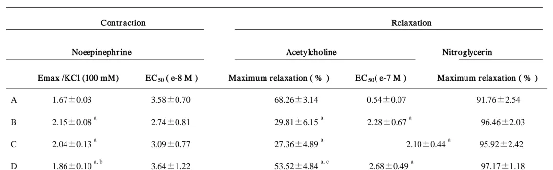
3.2. Measurement of contraction (A) and relaxation (B)
A: In the diabetic rat aortic rings, the contraction produced by norepinephrine was
augmented (Fig. 2A). With treatment of N-hexacosanol (8 mg/kg), the maximum
contraction was significantly attenuated toward that obtained using control rat aortic
rings (Fig. 2A). The maximum contraction (Emax) and EC50 values are shown in Table
B: The maximum relaxation (Emax) and EC50 values are shown in Table 2. In the
aortic rings precontracted by norepinephrine with intact endothelium obtained from all
groups, the relaxation was produced in a dose-dependent manner (Fig. 2B). The
relaxation was markedly reduced in the diabetic aorta (Fig. 2B). With treatment of
N-hexacosanol, the attenuated relaxation was recovered; data are shown in Fig. 2B.
Induction of diabetes significantly increased EC50 values for acetylcholine-induced
relaxation. Treatment with either dose of N-hexacosanol, however, did not ameliorate
these increased EC50 values. The nitroglycerin-induced relaxation was not changed in
any groups (Table 2).
3.3. Measurement of muscarinic M
3Fig. 3 shows the expression of muscarinic M
receptor, eNOS and iNOS mRNAs in
the aorta
3 receptor, eNOS and iNOS mRNAs in the
aorta. No significant difference in the eNOS mRNA levels was found between any
groups while the M3 receptor and iNOS mRNA levels were significantly higher in the
4. Discussion
In the diabetic state, the rat aorta exhibited enhanced vascular reactivity to
norepinephrine and impairment of endothelium-dependent-relaxation by acetylcholine.
While the eNOS mRNA level was not changed, the muscarinic M3 receptor and iNOS
mRNA levels were significantly increased in the diabetic aorta. Although
N-hexacosanol did not improve the general features or levels of serum glucose and
insulin in the diabetic rats, it significantly improved the enhanced contraction produced
by the α1
Previous reports demonstrated that aortic rings prepared from rats with
streptozotocin-induced diabetes showed increased contraction in response to
vasoconstrictor agents and reduced relaxation in response to endothelium-dependent
relaxant agents, but not to endothelium-independent relaxant agents (Oyama et al.,
1986; Pieper et al., 1998; Kinoshita et al., 2006). Previously, we demonstrated that
maximum relaxation in response to nitroglycerin was only around 50 % in Kinoshita’s
report, while maximum relaxation in response to nitroglycerin exceeded 90 % in this -agonist norepinephrine and the impairment of endothelium-dependent
study (Kinoshita et al., 2006). In Kinoshita’s reports, submaximal contraction was
induced by the thromboxane A2 analogue U46619 at a molar dose of 3x10⁻⁸, while in
this experiment, we chose the physiological agonist norepinephrine. Submaximal
contraction was induced by norepinephrine at a molar dose of 3x10⁻⁷. The extents of
contraction produced with agonists were distinct. The contraction produced by
U46619 was stronger than that by norepinephrine. The extent of relaxation by
nitroglycerin might be smaller than that by norepinephrine (Satoh et al., 1993). In this
study, we carefully observed the contractile responses induced by norepinephrine, and
decided that submaximal contraction was induced by norepinephrine at a molar dose of
3x10⁻⁷. This may be the reason we obtained maximum relaxation in response to
nitroglycerin exceeding 90 % in this study. In this study, although the degree of
relaxation induced by acetylcholine was significantly improved by the high dose of
N-hexacosanol, the EC50
Diabetes mellitus has been reported to induce hyperglycemia and injured vascular values for acetylcholine were not reversed by this dose.
These data suggest that the affinity of the endothelium for acetylcholine was not
endothelial cells and peripheral nerve cells (Oyama et al., 1986; Ozturk et al., 1996;
Shinozaki et al., 2003). Previous studies have shown that N-hexacosanol treatment has
no effect on serum glucose and insulin levels of diabetic rats (Satoh et al., 2005; Suzuki
et al., 2006; Kinoshita et al., 2006; saito et al., 2007). Therefore, the finding that this
drug does not influence the serum glucose levels indicates that the mechanisms of its
reversible effect on the diabetic rat aorta do not occur by reducing serum glucose.
To clarify the mechanism of the ameliorative effect of N-hexacosanol, we performed
quantification of muscarinic M3 receptor, eNOS and iNOS mRNAs by real-time PCR.
In the PCR studies, while the eNOS mRNA level was not changed, the muscarinic M3
receptor and iNOS mRNA levels exhibited significant increases in the induced diabetic
aorta. Previous studies suggested that arterial endothelium cells released acetylcholine
(Kawashima et al., 1990; Ikeda et al., 1994), and the endothelium dysfunction-induced
diabetic states might bring about a reduction in acetylcholine release. Therefore,
muscarinic M3 receptor mRNA might be upregulated in diabetes. And, because eNOS
is expressed in endothelial cells, endothelium dysfunctions reduce the eNOS expression.
constitutive NOS expression (Chakravarthy et al., 1998). Bojunga et al reported that
eNOS mRNA levels were decreased in the diabetic rat aorta (Bojunga et al., 2004). As
competitive reverse-transcriptase PCR is more sensitive than real-time PCR, the reasons
for these different results may be due to the small differences detected by real-time PCR.
The diabetic status of the animals in other experiments was different from that in ours.
Furthermore, the muscarinic M3 receptor activation on endothelial cells produces NO
via eNOS and PGI2 via phospholipase A2 and arachidonic acid (Jaiswal et al., 1991;
Kan et al., 1995; Maguire and Davenport, 2005; Triggle et al., 2003). The PG I2
pathway may be related to this event. The result obtained for the iNOS mRNA was in
agreement with that of a previous study (Bojunga et al., 2004). The results of the
present study demonstrate that eNOS and iNOS are regulated differentially in
experimental diabetes mellitus. Hyperglycemia and glucose-modified proteins may act
to reduce eNOS activity and at the same time increase iNOS gene expression with
enhanced NO release. It is also suggested that hyperglycemia markedly activates the
βII isoform of protein kinase C (PKC) in endothelial cells by promoting de novo
Activated PKC can indeed negatively influence transcription of eNOS while acting
positively on the iNOS gene (Bojunga et al., 2004). It is known that iNOS is induced
by inflammation, and that iNOS-derived NO exhibits a powerful protective activity
towards cellular stress conditions (Buttery et al., 1996). Moreover, other studies have
shown that iNOS-derived NO serves to modulate the expression of many different genes
that also affect protective responses during stress conditions (Ehrt et al., 2001;
Hemmrich et al., 2003). It is possible that iNOS does not contribute directly to the
induction of relaxation, but has other responses protecting the vascular endothelium.
Previous experimental evidence has suggested that tetrahydrobiopterin (BH4), the
natural and essential cofactor of NOS, plays a crucial role not only in increasing the rate
of NO generation by NOS, but also in controlling the formation of superoxide anions
(O2-) in endothelial cells (Baek et al., 1993). In fact, oral supplementation with BH4
restored endothelial function and relieved oxidative tissue damage through the
activation of eNOS in the aortas of insulin-resistant rats (Pieper et al., 1997; Shinozaki
et al., 2003). It is possible that the influence of eNOS-derived NO production on
Jover et al. reported that N-hexacosanol may act directly upon nerve terminals
inducing Ca2+ entry and hence promote neurohormone release (Jover et al., 2005). As
muscarinic M3 receptor subtypes, eNOS and iNOS are expressed in the aortic smooth
muscle (Ehlert, 2003). In order to reveal more details and the precise mechanisms of
this effect, further studies are needed. Taken together, our data demonstrate that
N-hexacosanol normalized the dysfunction of relaxation, and reversed diabetes-induced
upregulation of muscarinic M3 receptor and iNOS mRNAs.
Acknowledgments
This study was supported by a grant from the Ministry of Education, Science, and
Culture of Japan (#14704041) and by a research grant from the President of Tottori
Reference
Baek, K.J., Thiel, B.A., Lucas, S., Stuehr, D.J., 1993. Macrophage nitric oxide synthase
subunit: purification, characterization, and role of prosthetic groups and substrate in
regulating their association into a dimeric enzyme. J. Biol. Chem. 268, 21120-21129.
Bode-Boger, S.M., Boger, R.H., Kienke, S., Junker, W., Frolich, J.C., 1996. Elevated
L-arginine/dimethylarginine ratio contributes to enhanced systemic NO production by
dietary L-arginine in hyperholesterolemic rabbits. Biochem. Biophys. Res. Commun.
219, 598-603.
Brog. J., Kesslak, P.J., Cotman, C.W., 1990. Peripheral administration of a long-chain
fatty alcohol promotes septal cholinergic neurons survival after fimbria-fornix
transection. Brain Res. 518, 295-298.
Bojunga, J., Dresar-Mayert, B., Usadel, K., Kusterer, K., Zeuzem, S., 2004.
and attenuates tissue-cGMP activation in diabetic rats. Biochem. Biophys. Res.
Commun. 316, 771-780.
Boulanger, C.M., Morrison, K.J., Vanhoutte, P.M., 1994. Mediation by M3-muscarinic
receptors of both endothelium-dependent contraction and relaxation to acetylcholine in
the rat aorta f the spontaneously hypertensive rat. Br. J. Pharmacol. 112, 519-524.
Buttery, L.D., Springall, D.R., Chester, A.H., Evans, T.J., Standfield, E.N., Parums, D.V.,
Yacoub, M.H., Polak, J.M., 1996. Inducible nitric oxide synthase is present within
human atherosclerotic lesions and promotes the formation and activity of peroxynitrite.
Lab. Invest. 75, 77-85.
Chakravarthy, U., Hayes, R.G., Stitt, A.W., McAuley, E., Archer, D.B., 1998.
Constitutive nitric oxide synthase expression in retinal vascular endothelial cells is
Cooke, J.P., Tsao, P.S., 1997. Arginine: a new therapy for atherosclerosis? Circulation
95, 311-312.
De Vriese, A., Verbeuren, T.J., Van de Voorde, J., Lameire, N.H., Vanhoutte, P.M., 2000.
Endothelial dysfunction in diabetes. Br. J. Pharmacol. 130, 963-974.
The Diabetic Control and Complications Trial Research Group, 1993. The effect of
intensive treatment of diabetes on the development and progression of long-term
complications in insulin-dependent diabetes mellitus. N. Eng. J. Med.329, 977-986.
Ehlert, F.J., 2003. Pharmacological analysis of contractile role of M2 and M3
muscarinic receptors in smooth muscle. Receptors Channels 9, 261-277.
Ehrt, S., Schnappinger, D., Bekiranov, S., Drenkow, J., Shi, S., Gingeras, T.R.,
Gaasterland, T., Schoolnik, G., Nathan, C., 2001. Reprogramming of the macrophage
signaling roles of nitric oxide synthase-2 and phagocyte oxidase. J. Exp. Med. 194,
1123-1140.
Forstemann, U., Closs, E.I., Pollock, J.S., Nakane, M., Schwarz, P., Gath, I., Kleinert,
H., 1994. Nitric oxide synthase isozymes. Characterization, purification, molecular
cloning, and functions. Hypertension 23, 1121-1131.
Furchgott, R.F., Zawadzki, J.V., 1980. The obligatory role of endothelial cells in the
Relaxation of arterial smooth muscle by acetylcholine. Nature 288, 373-376.
Hemmrich, K., Suschek, C.V., Lerzynski, G., Kolb-Bachofen, V., 2003. iNOS activity
essential for endothelial stress gene expression protecting against oxidative damage. J.
Appl. Physiol. 95, 1937-1946.
Ikeda, C., Morita, I., Mori, A., Fujimoto, K., Suzuki, T., Kawashima, K., Murota, S.,
isolated from porcine cerebral microvessels. Brain Res. 655, 147-152.
Jaiswal, N., Jaiswal, R.K., Malik, K.U., 1991. Muscarinic receptor-mediated
prostacyclin and cGMP synthesis in cultured vascular cells. Mol. Pharmacol. 40,
101-106.
Jover, E., Gonzalez de Aguilar, J.L., Luu, B., Lutz-Bucher, B., 2005. Effect of a
cyclohexenonic long-chain fatty alcohol on calcium mobilization. Eur. J. Pharmacol.
516, 197-203.
Kan, H., Ruan, Y., Malk, K.U., 1995. localization and characterization of subtype(s) of
muscarinic receptor involved in prostacyclin synthesis in rabbit heart. J. Pharmacol. Exp.
Ther. 276, 934-941.
Kawashima, K., Watanabe, N., Oohata, H., Fujimoto, K., Suzuki, T., Ishizaki, Y., Morita,
endothelial cells. Neurosci. Lett. 119, 156-158.
Khurana, S., Chacon, I., Xie, G., Yamada, M., Wess, J., Raufman, J.P., Kennedy, R.H.,
2004. Vasodilatory effects of cholinergic agonists are greatly diminished in aorta from
M3R-/- mice. Eur. J. Pharmacol. 493, 127-132.
Kinoshita, Y., Saito, M., Satoh, I., Shomori, K., Suzuki, H., Yamada, M., Kono, T.,
Satoh, K., 2006. General administration of cyclohexenonic long-chain fatty alcohol
ameliorates hyperreactivity of STZ-induced diabetic rat aorta. Life Sci. 78, 1508-1514.
Maguire, J.J., Davenport, A.P., 2005. Regulation of vascular reactivity by established
and emerging GPCRs. Trends in Pharmacol. Sci. 26, 448-454.
Ozturk, Y., Altan, V.M., Yildizoglu-Ari, N., 1996. Effects of experimental diabetes and
Oyama, Y., Kawasaki, H., Hattori, Y., Kanno, M., 1986. Attenuation of
endothelium-dependent relaxation in aorta from diabetic rats. Eur. J. Pharmacol. 131,
75-78.
Pieper, G.M., Gross, G.J., 1988. Oxygen free radicals abolish endothelium-dependent
relaxation in diabetic rat aorta. Am. J. Physiol. 255, H825-H833.
Pieper, G.M., Langenstroer, P., Siebeneich, W., 1997. Diabetic-induced endothelial
dysfunction in rat aorta: role of hydroxyl radicals. Cardiovasc. Res. 34, 145-156.
Saito, M., Kinoshita, Y., Satoh, I., Shinbori, C., Suzuki, H., Yamada, M., Watanabe, T.,
Satoh, K., 2007. Ability of cyclohexenonic long-chain fatty alcohol to reverse
diabetes-induced cystopathy in the rat. Eur. Urol. 51, 479-488.
Satoh. I., Saito. M., Kinoshita, Y., Shomori, K., Suzuki, H., Yamada, M., Kono, T.,
trachea. Life Sci.77, 2030-2039.
Satoh, K., Mori, T., Yamada, H., Taira, N., 1993. Nicorandil as a nitrate, and
cromakalim as a potassium channel opener, dilate isolated porcine large coronary
arteries in an agonist-nonselective manner. Cardiovasc. Drugs Ther. 7, 691-699.
Senses, V., Ozyazgan, S., Ince, E., Tuncdemir, M., Kaya, F., Ozturk, M., Sultuybek, G.,
Akkan, A.G., 2001. Effect of 5-aminoimidazole-4-carbox-amide riboside (AICA-r) on
isolated thoracic aorta responses in streptozotocin-diabetic rats. J. Basic. Clin. Physiol.
Pharmacol. 12, 227-248.
Shinozaki, K., Kashiwagi, A., Masada, M., Okamura, T., 2003. Stress and Vascular
Responses: Oxidative stress and endothelial dtsfunction in the insulin-resistant state. J.
Pharmacol, Sci, 91, 187-191.
Yamada, M., Satoh, K., 2006. Preventive effects of cyclohexenonic long-chain fatty
alcohol on diabetic cystopathy in the rat. Can. J. Physiol. Pharmacol. 84, 195-201.
Triggle, C.R., Hollenberg, M., Anderson, T.J., Ding, H., Jiang, Y.-F., Ceroni, L., Wiehler,
W.B., Ng, E.S.M., Ellis, A., Andrews, K., McGuire, J.J., Pannirselvam, M., 2003. The
endothelium in health and disease- A target for therapeutic intervention. J. Smooth
Muscle Res. 39, 249-267.
Wilcox, J.N., Subramanian, R.R., Sundell, C.L., Preiser, J.C., 1997. Effects of multiple
isoforms of nitric oxide synthase in normal and atherosclerotic vessels. Arterioscler
Figure legends
Fig. 1. The chemical structure of N-hexacosanol used in this study.
Fig. 2. a) Contractile response of rat aortic rings to norepinephrine. The data of
contraction induced norepinephrine were normalized by the KCl (100 mM). NE:
norepinephrine. b) Endothelium-dependent relaxation of rat aortic rings to
acetylcholine. The data of relaxation were normalized by the nitroglycerine. Ach:
acetylcholine. A: control rats, B: diabetic rats, C: diabetic rats treated with
N-hexacosanol (2 mg/kg), and D: diabetic rats treated with N-hexacosanol (8 mg/kg).
Data are shown as mean ± S.E.M. of six to eight separated determinations in each group.
*) significantly different from the other groups. **) Significantly different from B
group. ***)Significantly different from B and C groups. (p < 0.05)
Fig. 3. a) Muscarinic M3 receptor mRNA normalized by β-actin. b) eNOS mRNA
normalized by β-actin. c) iNOS mRNA normalized by β-actin. A: control rats, B:
diabetic rats, C: diabetic rats treated with N-hexacosanol (2 mg/kg), and D: diabetic rats
treated with N-hexacosanol (8 mg/kg). Data are shown as mean ± S.E.M. of six to
Table 1. General features of the experimental rats
Body Weight (g)
8 weeks old 12 weeks old 16 weeks old Serum glucose (mg/dl) Serum insulin (µg/l)
A 242.9 ± 3.0 423.6 ± 5.9 498.8 ± 10.1 172.8 ± 13.9 2.39 ± 0.660
B 246.8 ± 6.4 259.5 ± 17.2 a 231.8 ± 23.5 a 405.0 ± 54.3 a 0.17 ± 0.009 a
C 250.4 ± 4.7 251.0 ± 17.0 a 229.0 ± 25.4 a 314.4 ± 46.9 a 0.17 ± 0.010 a
D 253.3 ± 5.1 257.0 ± 16.1 a 237.0 ± 17.1 a 364.8 ± 34.3 a 0.16 ± 0.004 a
A: control rats, B: diabetic rats treated with sham, C: diabetic rats treated with N-hexacosanol (2 mg/kg), and D: diabetic rats treated with N-hexacosanol (8 mg/kg). Data are shown as mean ± S.E.M. of six to eight separated determinations in each group. a) Significantly different from the A group.
Table 2. Data of functional studies in the rat aorta
Contr action Relaxation
Noeepinephr ine Acetylcholine Nitroglycer in Emax /KCl (100 mM) EC50 ( e-8 M ) Maximum relaxation ( % ) EC50
A 1.67±0.03 3.58±0.70 68.26±3.14 0.54±0.07 91.76±2.54
( e-7 M ) Maximum relaxation ( % )
B 2.15±0.08 a 2.74±0.81 29.81±6.15 a 2.28±0.67 a C 2.04±0.13 96.46±2.03 a 3.09±0.77 27.36±4.89 a 2.10±0.44 a D 1.86±0.10 95.92±2.42 a, b 3.64±1.22 53.52±4.84 a, c 2.68±0.49 a
A: control rat, B:diabetic rat, C: diabetic rat with treatment with N-hexacasanol (2mg/kg), and D: diabetic rat with treatment with N-hexacosanol (8mg/kg). The relaxation responses with submaximal contraction caused by 3 x 10
97.17±1.18
-7
M norepinephrine were expressed as percentages of nitroglycelin. Emax and EC50 values under contraction are for
norepinephrine. Maximum relaxation and EC50 values are for acetylcholine in aorta precontracted with norepinephrine. The relaxation responses to nitroglycerine as the
CH
2(CH
2)
14OH
CH
3O
H
3C
4-9 -8 -7 -6 -5 0. 0 0. 5 1. 0 1. 5 2. 0 2. 5 A B C D
*
*
Figure 2. Measurement of contraction and relaxation
-8 -7 -6 -5 -4 A B C D 0 20 40 60 80 100