Title
FURTHER EVIDENCE THAT GENOTYPE I AND
GENOTYPE II OF CRYPTOSPORIDIUM PARVUM ARE
DISTINCT( 本文(Fulltext) )
Author(s)
WU, Zhiliang; NAGANO, Isao; BOONMARS, Thidarut;
TAKAHASHI, Yuzo
Citation
[Tropical medicine and health] vol.[32] no.[1] p.[5]-[14]
Issue Date
2004-03-01
Rights
Japanese Society of Tropical Medicine (日本熱帯医学会)
Version
出版社版 (publisher version) postprint
URL
http://hdl.handle.net/20.500.12099/30117
FURTHER EVIDENCE THAT GENOTYPE I AND GENOTYPE II OF
CRYPTOSPORIDIUM PARVUM ARE DISTINCT
ZHILIANGWU, ISAONAGANO, THIDARUTBOONMARSand YUZOTAKAHASHI*
Accepted 3, February, 2004
Abstract: Three new genes of Cryptosporidium parvum were cloned, including a gene encoding methionine aminopeptidase, one encoding chaperonin containing T-complex protein 1 delta (TCP-1 delta) and one with un-known function. DNA sequence analysis indicated that these genes are quite conserved, but there were some base pair differences between genotype I and genotype II isolates. These differences were confirmed by PCR-restriction fragment length polymorphism (PCR-RFLP) analysis of the 3 genes from 41 isolates collected from different hosts and geographical origins. In brief, the band patterns generated by endonuclease Hind III or Hinf I restrictions of the gene of methionine aminopeptidase, Sac I restriction of the gene of chaperonin, or Ava II restriction of the un-known gene could differentiate the isolates of C. parvum into genotype I and genotype II. PCR primers based on these genes amplified only C. parvum genes. Even a single oocyst was detectable with these PCR primers. Thus the results provided further evidence that genotype I and genotype II are distinct, and our three new primers can be used to detect and characterize C. parvum isolates with high sensitivity.
Key Word: Cryptosporidium parvum, methionine aminopeptidase, chaperonin, genotype, PCR-RFLP
INTRODUCTION
An intestinal protozoan parasite, Cryptosporidium
par-vum is the major causative agent of cryptosporidiosis in
hu-mans and livestock. Outbreaks of human cryptosporidiosis are often caused by the contamination of water supplies with this parasite throughout the world. Because of the epi-demiological importance, it is desirable to develop a sensi-tive detection technique for this parasite and accurate ge-netic classification.
To this end, many efforts have been made to character-ize isolates of C. parvum in the past decade. The difference in virulence, infectivity, pathogenesis and drug sensitivity has indicated that C. parvum is not a uniform species or monophyletic (Morgan et al., 1999a; Okhuysen et al., 1999; Pereira et al., 2002). The present working hypothesis is that C. parvum is composed of two main genotypes, genotype I and genotype II. Genotype I (human genotype or anthroponotic genotype) is exclusively found in humans, while genotype II (bovine genotype or zoonotic genotype) was first found in cattle and has also been found in a wide range of mammals, including humans. Some other host-adapted-genotypes have been also found among C. parvum isolates, such as the pig, dog and mouse genotype (Morgan
et al., 1999b; Sulaiman et al., 2000; Xiao et al., 2000a)
With the enormous development of gene technology, gene diversities between genotype I and genotype II have
been reported. The evidence includes sequence and/or PCR-RFLP analysis of several gene loci, such as 18S rRNA (Morgan et al., 1997; Xiao et al., 1999), COWP (Spano et
al., 1997; Xiao et al., 2000b), HSP70 (Gobet and Toze,
2001; Sulaiman et al., 2000), TRAP (Spano et al., 1999; Sulaiman et al., 1998), and/Cpgp40/15 (Wu et al., 2003).
In the present study, we cloned 3 new genes of C.
par-vum adapting our previous method to design PCR primers
from random amplified polymorphic DNA (RAPD), and further confirmed the distinctness between genotype I and genotype II of C. parvum by the sequences and PCR-RFLP analysis of the isolates from different hosts with different geographical origins.
MATERIALS ANDMETHODS
Parasite isolates
Forty-one isolates of C. parvum were used. The details were published in our previous paper (Wu et al., 2003). The host and geographical origins are shown in Table 1.
All fecal samples were preserved in 2% K2Cr2O7and oocysts were isolated using a sucrose flotation method. The samples were refined, contaminated micro-organisms being reduced to a minimum level using an immuno-magnetic separation kit (Dynabeads anti-Cryptosporidium, Dynal A. S., Oslo, Norway).
Oocyst DNA was isolated by the method described Tropical Medicine and Health Vol. 32 No. 1, 2004, pp. 5-14
Copyright! 2004 by The Japanese Society of Tropical Medicine
Department of Parasitology, Gifu University School of Medicine, Tsukasa 40, Gifu 500-8705, Japan.
previously (Wu et al., 2000). In brief, oocysts were frozen and thawed repeatedly 5 times, treated at 100°C for 20 min, and then the samples were digested with proteinase K at a final concentration of 200µg/ml at 55°C for 3 hours. The reaction was stopped by heating at 95°C for 5 min. The treated samples were then directly used as a template for PCR.
Development of PCR primers
PCR primers were produced according to our previ-ously described methods (Nagano et al., 1996). In brief, RAPD was produced from C. parvum DNA (Code# CGJ2) by means of arbitrary primed PCR (AP-PCR) using 10 base
pair primers. The three target genes thus obtained were RAPD SB281, SB289 and SB012, which were produced by primers tgatgaccgc, gcgtgctcac and cggcccctgt, respectively. The DNA fragments were extracted and sequenced. Based on these sequences, 3 pairs of primers were developed as shown in Table 2.
Detection sensitivity and specificity of primers
The sensitivity of primers was tested by amplifying se-rially diluted template DNA. The purified oocyst DNA was diluted at 160, 40, 10, 2.5, 0.625, 0.156, 0.039 and 0.001 pg/µl. Tested primers included the primers SB281, SB289 and SB012, as well as 3 other primers to amplify the genes of COWP (GenBank accession No.: AF266273, Xiao et al., 2000b), HSP70 (GenBank accession No.: AF221535, Su-laiman et al., 2000), and TRAP1 (GenBank accession No.: AF017267, Spano et al., 1998), as shown in Table 2. PCR conditions were as follows: one cycle of initial denaturing at 94°C for 3 min; 35 cycles at 94°C for 30 sec, 51-56°C (see details in Table 2) for 30 sec, and 72°C for 1 min; and one cycle of final extension at 72°C for 10 min.
The specificity of the primers SB281, SB289 and SB012 was tested by detecting various kinds of template DNA, including C. parvum and control samples of C. muris, human, bovine, Entamoeba histolytica, Giardia lamblia,
Blastocystis hominis, Ascaris lumbricoides, Trichomonas vaginalis, Trichinella spiralis and Escherichia coli.
DNA sequencing and sequence analysis
The RAPD was produced by primer SB281, SB289 or SB012 from 16 isolates, which included 8 calf isolates (Code# CGJ2, CGJ5, CKJ1, CKJ3, CKJ7, CNJ1, CI2 and CI8) from Japan and Italy, one goat isolate (Code# GI1) from Italy and 7 human isolates (Code# HJ1, HJ2, HJ3, HI1, HI2, HN4 and HN6) from Japan, Italy and Nepal. DNA se-quence of the RAPD was determined using a Thermo Se-quenase cycle sequencing Kit (USB Corporation, Cleveland, USA) and an automatic sequencer (Model LIC-4200, Aloka Co., Ltd., Tokyo, Japan). The sequence data were analyzed using DNASIS Software (Hitachi Software Engineering, Tokyo, Japan). Homology searching on the nucleotide and protein database was carried out using the BLAST program at NCBI (Bethesda, MD, USA). Pairwise sequence align-ment and protein identities were performed using CLUS-TALW 1.8 software (Jeanmougin et al., 1998) and PHYLIP DNADIST software (distributed by Felsenstein, Department of Genetics, University of Washington, Seattle).
PCR-RFLP
PCR and RFLP were performed according to our pre-viously described methods (Wu et al., 1999). PCR
prod-Table 1. Isolates of C. parvum.
Code Host origin Geographical origin Genotype
CGJ1 Calf Gifu, Japan II
CGJ2 Calf Gifu, Japan II
CGJ3 Calf Gifu, Japan II
CGJ4 Calf Gifu, Japan II
CGJ5 Calf Gifu, Japan II
CGJ6 Calf Gifu, Japan II
CKJ1 Calf Kobe, Japan II
CKJ2 Calf Kobe, Japan II
CKJ3 Calf Kobe, Japan II
CKJ4 Calf Kobe, Japan II
CKJ5 Calf Kobe, Japan II
CKJ6 Calf Kobe, Japan II
CKJ7 Calf Kobe, Japan II
CKJ8 Calf Kobe, Japan II
CKJ9 Calf Kobe, Japan II
CKJ10 Calf Kobe, Japan II
CKJ11 Calf Kobe, Japan II
CNJ1 Calf Nagoya, Japan II
CI1 Calf Italy II
CI2 Calf Italy II
CI3 Calf Italy II
CI4 Calf Italy II
CI5 Calf Italy II
CI6 Calf Italy II
CI7 Calf Italy II
CI8 Calf Italy II
GI1 Goat Italy II
HJ1 Human Japan II
HJ2 Human Japan I
HJ3 Human Japan I
HI1 Human Italy I
HI2 Human Italy I
HI3 Human Italy I
HI4 Human Italy I
HI5 Human Italy I
HN1 Human Nepal I HN2 Human Nepal I HN3 Human Nepal I HN4 Human Nepal I HN5 Human Nepal I HN6 Human Nepal I 6
ucts were purified by ethanol precipitation, and then di-gested with an appropriate restriction endonuclease (Hind III, Hinf I, Sac I and Ava II) according to the manufacturer’s instructions. The digests were subjected to electrophoresis.
RESULTS
Sequence analysis
Three dense RAPD bands with an appropriate molecu-lar size of 610 (SB281), 600 (SB289) and 550 bp (SB012) were generated from the template DNA of the isolate CGJ2. The RAPD fragments were isolated from the gel and se-quenced (GenBank accession No: AY488139, AY488146, AY488153). Based on the 3 sequences, 3 pairs of primers were developed (Table 2). The RAPD from 16 isolates (in-cluding 8 calf isolates from Japan and Italy, one goat isolate from Italy and 7 human isolates from Japan, Italy and Ne-pal) were produced with the 3 developed primers and se-quenced (GenBank accession numbers are shown in Figs 1, 2 and 3).
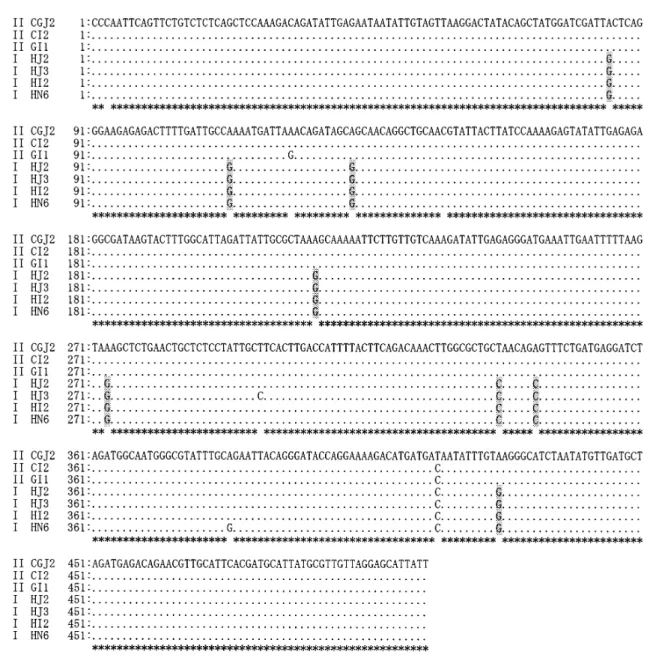
Homology analysis of the RAPD sequence showed that these genes were quite conserved among different iso-lates, as shown in Figs 1, 2 and 3. The identities of the se-quences of SB281 RAPD, SB289 RAPD, and SB012
RAPD were 98.0-100%, 97.6-100%, and 98.2-100% among the 16 isolates, respectively.
The animal isolates shared nearly the same sequence regardless of their origin. The identities of SB281 RAPD, SB289 RAPD, and SB012 RAPD among the 9 animal iso-lates were 99.3-100%, 99.6-100% and 99.0-100%, respec-tively. The same was true for human isolates. The identi-ties of RAPD SB281, RAPD SB289, and RAPD SB012 among the 7 human isolates were 98.4-99.6%, 98.0-100% and 99.3-100%, respectively.
Although DNA sequence was conserved, there were point mutations in human origin isolates (except the isolate HJ1) at identical nucleotide sites. The point mutation pat-tern was different from the animal origin isolates as shown in Figs 1, 2 and 3.
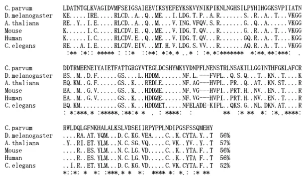
The DNA sequence of the RAPD fragments were de-duced into an amino acid sequence and subjected to Gen-Bank for blast searching. The blast results showed that SB281 was a protein gene encoding methionine aminopep-tidase. The deduced amino acid sequence shared 52-57% identities with the sequence of methionine aminopeptidase of other organisms, for example, Drosophila melanogaster,
Arabidopsis thaliana, human, mouse and Caenorhabditis elegans (Fig 4). The SB289 was a protein gene encoding
Table 2. Criteria of PCR Primers.
Primer Sequence Annealing
Tm
Product
size Target gene SB281 5’-gatagtgttccatttgagagg-3’ 5’-ttagatgcaacaaatacaggat-3’ 51.0 553 methionine aminopeptidase SB289 5’-cccaattcagttctgtctct-3’ 5’-aataatgctcctaacaacgc-3’ 51.0 505 chaperonin containing TCP-1 delta SB012 5’-ctccgttcgatgatgcagatg-3’ 5’-cggcccctgtagaaataagtca-3’ 51.3 434 unknown COWP 5’-ggttcctcctatgcctttct
5’-gtgggcattcctttgtgac 52.7 869 oocyst wall protein HSP70 5’-tcagttgccatcagtagagc
5’-tcttcttctcagcctcatca 52.5 841 heat shock protein TRAP1 5’-ggatgggtatcaggtaataagaa
5’-tagcccagttctgactctctg 52.0 1177
thrombospondin-related adhesive protein
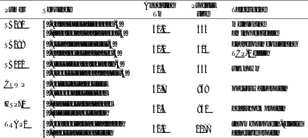
Table 3. Predicted RFLP pattern of RAPD SB281, SB289 and SB012 PCR products. Size of predicated bands
Isolate SB281 SB289 SB012 Genotype
Hae III Hinf I Sac I Ava II
CGJ2 59, 494 75, 478 505 152, 282 II CI2 59, 494 75, 478 505 152, 282 II IG1 59, 494 75, 478 505 152, 282 II HJ2 59, 204, 290 75, 138, 340 232, 273 434 I HJ3 59, 204, 290 75, 138, 340 232, 273 434 I HN6 59, 204, 290 75, 138, 340 232, 273 434 I HI2 59, 204, 290 75, 138, 340 232, 273 434 I 7
chaperonin containing TCP-1 delta subunit. The deduced amino acid sequence shared 54-61% identities with the se-quence of chaperonin containing TCP-1 delta subunit of other organisms, such as D. melanogaster, A. thaliana,
hu-man, mouse and C. elegans (Fig 5).
Differentiation of C. parvum isolates by the RFLP method
After PCR with the primer SB281, SB289 or SB012,
Figure 1. DNA sequence diversities among Cryptosporidium parvum isolates in SB281 (methionine aminopeptidase) gene. The genes from 16 isolates were sequenced and typed into genotype I and genotype II, as indicated in the left margin by I and II. Filled circles in the alignment indicate identical base pairs. The differences be-tween genotype I and genotype II are indicated by shading. GenBank accession numbers of CGJ2, CI2, GI1, HJ2, HJ3, HI2 and HN6 are AY488139, AY488140, AY488141, AY488142, AY488143, AY488144 and AY488145, respectively.
all 41 DNA samples produced the expected size bands of 553 bp, 505 bp and 434 bp, respectively. Thus 123 (3 times 41) RAPDs were analyzed by PCR-RFLP.
Two kinds of band patterns (Panels A and B in Fig 6) were produced by endonuclease Hind III and Hinf I restric-tion of the RAPD SB281. All of the calf isolates from both Japan and Italy, the isolate from the goat of Italy and one isolate from a Japanese patient (Code# HJ1) showed the same kind of RFLP pattern of genotype II. All of the
hu-man origin isolates, except one Japanese huhu-man isolate (Code# HJ1) produced the same kind of RFLP pattern of genotype I, which was different from that of genotype II.
The same results were obtained when the 41 isolates were analyzed by endonuclease Sac I restriction of RAPD SB289 (Panel C in Fig 6) and Ava II restriction of RAPD SB012 (Panel D in Fig 6).
Figure 2. DNA sequence diversities among Cryptosporidium parvum isolates in SB289 (chaperonin containing TCP-1 delta subunit) gene. The genes from 16 isolates were sequenced and typed into genotype I and genotype II, as indicated in the left margin by I and II. Filled circles in the alignment indicate identical base pairs. The differences between genotype I and genotype II are indicated by shading. GenBank accession numbers of CGJ2, CI2, GI1, HJ2, HJ3, HI2 and HN6 are AY488146, AY488147, AY488148, AY488149, AY488150, AY488151 and AY488152, respectively.
Detection sensitivity and specificity of primers
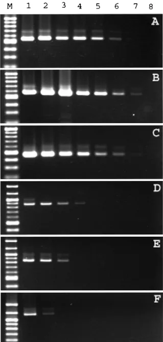
The detection sensitivity of primers was tested by means of PCR with serially diluted template DNA, as de-scribed in Materials and Methods. The density of bands tended to be faint when a lower concentration of template DNA was used. The primers SB281, SB289 and SB012 gave the highest sensitivity in detecting template DNA of C.
parvum. The minimum concentration of template DNA necessary for a positive reaction was 0.156 pg with the primer SB281, 0.039 pg with the primer SB289 and 0.156 pg with the primer SB012 (Panels A, B and C in Fig 7), while the control primers of COWP, HSP 70 and PRAP1 showed a minimum detection concentration of 10 pg, 10 pg and 40 pg, respectively (Panels D, E and F in Fig 7).
All 3 kinds of primers produced bands with the ex-pected size from C. parvum, while control samples pro-duced negative results, indicating the species specificity of these constructed primers.
DISCUSSION
Epidemiologically, molecular markers of pathogens are useful in determining the importance of responsible re-agents, for example, distinguishing between zoonotic and anthroponotic transmissions. Reportedly, both genotype I and genotype II of C. parvum are responsible for the spo-radic infection and outbreak of human cryptosporidiosis (McLauchlin et al., 1999; Ong et al., 1999; Sulaiman et al., 1998; Xiao et al., 2000a).
In the present study, 3 new target genes were proposed and assessed for genetic analysis. The sequence analysis of these genes from 41 isolates of C. parvum from different hosts and geographical origins indicated that there are iden-tical base pair differences between the isolates from human and animal origins. The PCR-RFLP analysis of the 3 genes confirmed that these 3 new genes are useful for the geno-typing of C. parvum, because all of the 3 genes showed 2 kinds of RFLP patterns which correspond to genotype I and
Figure 3. DNA sequence diversities among Cryptosporidium parvum isolates in SB012 gene. The genes from 16 iso-lates were sequenced and typed into genotype I and genotype II, as indicated in the left margin by I and II. Filled circles in the alignment indicate identical base pairs. The differences between genotype I and genotype II are indicated by shading. GenBank accession numbers of CGJ2, CI2, GI1, HJ2, HJ3, HI2 and HN6 are AY488153, AY488154, AY488155, AY488156, AY488157, AY488158, and AY488159, respectively.
genotype II. More important is the fact that the 3 primers amplified only C. parvum DNA with high sensitivity, mak-ing these candidate primers to detect and genotype C.
par-vum in water contamination at the required quality in terms
of sensitivity. Using our method, even low numbers of oo-cysts in sample water may be amplified with any one of the primers. RFLP analysis with a combination of any one of
the three genes can identify animal or human genotypes. Such examples are given by primer SB281/Hind III, SB281/ Hinf I, SB289/Sac I or SB012/Ava II.
We have reported that C. parvum specific primer SB012 is highly sensitive. The primer SB012 can detect the lowest amount of template DNA necessary for a positive re-action, which was 0.156 pg DNA or even one oocyst, or 50
Figure 4. Alignment of the deduced amino acid sequence of methionine aminopeptidase of C. parvum and the sequence of D. melanogaster, A. thaliana, human, mouse and C. elegans. Filled circles in the align-ment indicate identical base pair and hyphens indicate gaps. The identities of sequence are indicated at the ends of sequences.
Figure 5. Alignment of the deduced amino acid sequence of chaperonin containing TCP-1 delta subunit of C. parvum and the sequence of D. melanogaster, A. thaliana, human, mouse and C. elegans. Filled circles in the align-ment indicate identical base pairs and hyphens indicate gaps. The identities of sequence are indicated in the ends of sequences.
oocysts in raw water (Wu et al., 2000). The new primers (SB281 and SB289) had equivalent detection sensitivity to primer SB012. PCR detection indicates that these primers have a sensitivity 50 times higher than that of some other primers developed from COWP, HSP70 and TRAP1 and currently being used for genotyping Cryptosporidium.
Thus the analysis of the three RAPD genes provided the same genetic characterization to group the isolates to genotype I and genotype II, further evidence for the
distinc-tion between the two genotypes of C. parvum. Our new primers may also provide selectable methods for reliable elimination of C. parvum from tap water and for genotyp-ing of the contaminated oocysts for further epidemiological survey.
ACKNOWLEDGMENTS
We thank Dr. Uga (Department of Medical Technology,
Figure 6. PCR-RFLP analysis of SB281, SB289 and SB012 genes by the restriction of SB281 with Hind III (panel A) and Hinf I (panel B), SB289 with Sac I (panel C), and SB012 with Ava II (panel D). The isolates from Japanese calf (lanes 1-3), Italy calf (lane 4 and 5), Italy goat (lane 6) and one Japanese human (HJ1 in lane 7) give the same kind of band pattern of genotype. The isolates from the other Japanese human (HJ2 and HJ3) in lanes 8 and 9, Italy human (lane 10 and 11) and Nepal human (lanes 12 and 13) show the same kind of band pattern of genotype I. M is Base-Pair Ladder of mo-lecular weight marker.
Kobe University School of Medicine, Japan) and Dr. Pozio (Laboratorio de Parasitologia, Instituto Superiore de Sanita, Italy) who kindly provided Cryptosporidium isolates. This work was supported by a grant-in-aid (no. 13670243) from the Ministry of Education, Sports, Science and Technology, Japan.
REFERENCES
1)Gobet, P., and Toze, S. (2001): Sensitive genotyping of
Cryptosporidium parvum by PCR-RFLP analysis of the
70-kilodalton heat shock protein (HSP70) gene. FEMS Microbiol. Lett., 200, 37-41.
2)Jeanmougin, F., Thompson, J.D., Gouy, M., Higgins,
D.G. and Gibson, T.J. (1998): Multiple sequence align-ment with Clustal X. Trends Biochem. Sci., 23, 403-405.
3)McLauchlin, J., Pedraza-Diaz, S., Amar-Hoetzeneder, C.
and Nichols, G.L. (1999): Genetic characterization of
Cryptosporidium strains from 218 patients with diarrhea
diagnosed as having sporadic cryptosporidiosis. J. Clin. Microbiol., 37, 3153-3258.
4)Morgan, U.M., Constantine, C.C., Forbes, D.A. and
Thompson, R.C.A. (1997): Differentiation between hu-man and animal isolates of Cryptosporidium parvum us-ing rDNA sequencus-ing and direct PCR analysis. J. Parasi-tol., 83, 825-830.
5)Morgan, U., Xiao, L., Fayer, R., Lal, A.A. and Thompson,
R.C.A. (1999b): Variation in Cryptosporidium: towards a taxonomic revision of the genus. Inter. J. Parasitol., 29, 1733-1751.
6)Morgan, U., Xiao, L., Sulaiman, I., Weber, R., Lal, A.A.,
Thompson, R.C. and Deplazes, P. (1999a): Which geno-types/species of Cryptosporidium are humans susceptible to? J. Eukaryot. Microbiol., 46, 42S-43S.
7)Nagano, I., Wu, Z., Nakayama, M. and Takahashi, Y.
(1996): A simple method to design PCR primer to detect genomic DNA of parasites and its application to
Dirofi-laria immitis. Mol. Cell. Probes, 10, 423-425.
8)Okhuysen, P.C., Chappell, C.L., Crabb, J.H., Sterling,
C.R. and Dupont, H.L. (1999): Virulence of three distinct
Cryptosporidium parvum isolates for healthy adults. J.
Infect. Dis., 180, 1275-1281.
9)Ong, C.S., Eisler, D.L., Goh, S.H., Tomblin, J.,
Awad-El-Kariem, F.M., Beard, C.B., Xiao, L., Sulaiman, I., Lal, A., Fyfe, M., King, A., Bowie, W.R., Isaac-Renton, J.L. (1999): Molecular epidemiology of cryptosporidiosis outbreaks and transmission in British Columbia, Canada. Am. J. Trop. Med. Hyg., 61, 63-69.
10)Pereira, S.J., Ramirez, N.E., Xiao, L. and Ward, L.A.
(2002): Pathogenesis of human and bovine
Cryptosporid-ium parvum in gnotobiotic pigs. J. Infect. Dis., 186,
715-718.
11)Spano, F., Putignani, L., Guida, S. and Crisanti, A.
(1999): Cryptosporidium parvum: PCR-RFLP analysis of the TRAP-C1 (thrombosondin-related adhesive protein of
Cryptosporidium-1) gene discriminates between two
al-leles differentially associated with parasite isolates of ani-mal and human origin. Exp. Parasitol., 90, 195-198.
12)Spano, F., Putignani, L., McLauchlin, J., Casemore, D.P.
and Crisanti, A. (1997): PCR-RFLP analysis of the
Cryp-tosporidium oocyst wall protein (COWP) gene
discrimi-nates between C. wrairi and C. parvum, and between C.
parvum isolates of human and animal origin. FEMS
Mi-crobiol. Lett. 150, 209-217. Figure 7. The sensitivity of constructed primer pair SB281,
SB289 and SB012 by detecting template DNA and a comparison with other primers. M is 100 Base-Pair Ladder of molecular weight marker; lanes 1 to 8 are DNA amount of 160, 40, 10, 2.5, 0.625, 0.156, 0.039 and 0.001 pg, respectively. Panels A to F are primer SB281, SB289, SB012, COWP, HSP70 and TRAP1, respectively.
13)Spano, F., Putignani, L., Naitza, S., Puri, C., Wright, S.
and Crisanti, A. (1998): Molecular cloning and expres-sion analysis of a Cryptosporidium parvum gene encod-ing a new member of the thrombospondin family. J. Mol. Biochem. Parasitol., 92, 147-162.
14)Sulaiman, I.M., Morgan, U.M., Thompson, R.C.A., Lal,
A.A. and Xiao, L. (2000): Phylogenetic relationships of
Cryptosporidium parasites based on the 70-kilodalton
heat shock protein (HSP) gene. Appl. Environ. Micro-biol., 66, 2385-2391.
15)Sulaiman, I.M., Xiao, L., Yang, C., Escalante, L., Moore,
A., Beard, C.B., Arrowood, M.J. and Lal, A.A. (1998): Differentiating human from animal isolates of
Crypto-sporidium parvum. Emerg. Infect. Dis., 4, 681-685.
16)Wu, Z., Nagano, I., Boonmars, T. and Takahashi, Y.
(2003): Genotyping and intraspecies polymorphism of
Cryptosporidium parvum as revealed by restriction
frag-ment length polymorphism (RFLP) and single-strand conformational polymorphism (SSCP) analysis. Appl. Environ. Microbiol., 69, 4720-4726.
17)Wu, Z., Nagano, I., Matsuo, A., Pozio, E. and Takahashi,
Y. (1999): Polymerase chain reaction-restriction fragment length polymorphism (PCR-RFLP) for the identification of Trichinella isolates. Parasitology, 118, 211-218.
18)Wu, Z., Nagano, I., Matsuo, A., Uga, S., Kimata, I., Iseki,
M. and Takahashi, Y. (2000): Specific PCR primers for
Cryptosporidium parvum with extra high sensitivity.
Mol. Cell. Probes, 14, 33-39.
19)Xiao, L., Escalante, L., Yang, C., Sulaiman, I., Escalante,
A.A., Montali, R.J., Fayer, R., and Lal, A.A. (1999): Phy-logenetic analysis of Cryptosporidium parasites based on the small subunit ribosomal RNA gene locus. Appl. En-viron. Microbiol., 65, 1578-1583.
20)Xiao, L., Kimor, J., Morgan, U.M., Sulaiman, I.M.,
Thompson, R.C.A. and Lal, A.A. (2000b): Sequence dif-ferences in the diagnostic target region of the oocyst wall protein gene of Cryptosporidium parasites. Appl. Envi-ron. Microbiol., 66, 5499-5502.
21)Xiao L, Morgan, U.M., Fayer, R., Thompson, R.C.A. and
Lal, A.A. (2000a): Cryptosporidium systematics and im-plications for public health. Parasitol. Today, 16, 287-292.