1* Department of Community Health, Tokai University School of Medicine, Kanagawa 259–1193, Japan Department of Community Health, Tokai University School of Medicine, Isehara City, Kanagawa Prefec-ture 259–1193, Japan
E-mail: tewatana@is.icc.u-tokai.ac.jp
2 Department of Internal Medicine, Saiseikai Central Hospital, Tokyo 108–0073, Japan
CHANGE OF COMPONENTS OF THE METABOLIC SYNDROME
IN A WORKERS' HEALTH CHECKUP AFTER FIVE YEARS
―RELATION WITH ELEVATED LIVER ENZYMES, GENE
POLYMORPHISMS FOR ALDH 2, b3–AR AND LIFESTYLE
Noriko ISHIKAWA1, Chisato MURATA1,2, Hajime MIKURUBE1, Shun ITO1,
Reiichi HIGASHIYAMA1, Yoko KOMAKI1, Yoshihito ATSUMI2,
Kempei MATSUOKA2, and Tetsu WATANABE1*
We previously reported that the prevalence of elevated alanine aminotransferase (ALT) in-creases with accumulation of metabolic syndrome components, and a greater degree of involvement of aldehyde dehydrogenase 2 (ALDH2) than b3–adrenergic receptor gene ( b3–AR) polymor-phisms. The present study was designed to clarify the eŠect of aging, lifestyle and the two gene poly-morphisms on the relationship between 4 components of the metabolic syndrome (obesity, hyperten-sion, dyslipidemia and impaired glucose tolerance) and elevated ALT values in a subset of 73 out of 148 male workers who were 35 years of age in the baseline study and 40 years old in the present study. Study subjects completed questionnaires about drinking and smoking habits, and underwent urinalysis, physical examination and peripheral blood tests, blood chemistry, electrocardiogram and chest X-rays each year as required by Japanese law. Information from the questionnaires and physi-cal examinations, including liver function tests, were compared with previously reported ALDH2 andb3–AR genotypes for the 73 workers.
Of the 73 workers studied, 14 (19%) demonstrated decrease in metabolic syndrome compo-nents, 39 (53%) demonstrated no change, and 20 (27%) demonstrated an increase. Ten workers (14%) showed liver dysfunction at age 35 and 20 workers (27%) at age 40. Fourteen workers were newly diagnosed as having liver dysfunction at their 40-year checkup, thus being associated with the BMI and an active ALDH2 genotype. Accumulation of components of the metabolic syndrome were associated with the presence of liver dysfunction at 35 years.
In conclusion, these ˆndings indicate that ALDH2 genotyping as well as lifestyle habits may be important factors in causing metabolic syndrome with liver dysfunction.
Key words:metabolic syndrome, alanine aminotransferase (ALT), ALDH 2,b3–AR
I. Introduction
Recent studies have provided evidence that nonalcoholic fatty liver disease (NAFLD) including nonalcoholic steatohepatitis (NASH) is a common manifestation of the metabolic syndrome1~5), and the pathophysiology of both diseases seems to be
lar-gely attributable to insulin resistance. We reported that young male workers with elevated ALT might have NAFLD as an early manifestation of metabolic syndrome because they had two or more components of metabolic syndrome and the number of compo-nents increased with elevation of ALT. Moreover, persons with putative metabolic syndrome with elevated ALT demonstrated high body-mass-index (BMI), and serum triglycerides, as well as the active aldehyde dehydrogenase 2 (ALDH2) genotype6). An interesting ˆnding in the baseline study was that 4 subjects with normal weight but putatively the metabolic syndrome with elevated ALT had both ac-tive ALDH2 and Arg64b3–AR genotypes6).
As our baseline study was cross-sectional, the present investigation was conducted to assess
changes in metabolic syndrome components after an interval of 5 years in the same group of subjects6). In-terventions, including treatment for the metabolic syndrome and education, especially the necessity of increased physical activity and improvement of diet, have been provided for the subjects over the past 5 years. We hypothesized that lifestyle is involved in development of the metabolic syndrome more than the ALDH2 and b3–AR genotypes. To test this hypothesis, gene polymorphisms of both ALDH2 and b3–AR and accumulation of components of metabolic syndrome were compared for 73 workers from the baseline study linked with new information for physical ˆndings and lifestyle.
II. Subjects And Methods
Study Subjects
Of 148 car salesmen enrolled in the baseline study at 35 years6), test results of 73 subjects (at age of 40) were available for the study. Twenty eight subjects were missing because of job change and laboratory data of 45 subjects were unavailable as they transferred to branch operations or subsidiary companies. We excluded two subjects who have been treated for lung cancer or drug induced hepatitis. The subjects for the baseline study were enrolled during September 1998 to August 1999 and the present study was performed from September 2003 to August 2004. During this period subjects were provided with health education by registered nurses, if necessary, with a focus on the importance of lifestyle in order to prevent lifestyle-related problems such as metabolic disease. The subjects were treated as needed with medications for hypertension, dys-lipidemia, hyperglycemia, hyperuricemia and others by industrial physicians at the workplace, as well as by physicians at hospitals and/or clinics.
The industrial physician explained the purpose of the study to each worker. Each subject was asked to provide information on past medical history of illnesses and to ˆll out questionnaires to reveal drink-ing and smokdrink-ing habits, diet, health conditions, physical activity, sleeping time and current drug use. The subjects underwent a physical examination, conventional laboratory tests including urinalysis, peripheral blood examination (red and white blood cell counts, hemoglobin), clinical chemistry (glu-cose, total cholesterol, triglyceride, HDL-cholester-ol, aspartate aminotransferase (AST), alanine ami-notransferase (ALT), g–glutamyl transpeptidase (g–GTP)), electrocardiogram, and chest X-rays at their annual health checkup. The ethics committee of the Tokai University School of Medicine as well as the Health Care Committee of the company
ap-proved the study protocol, and informed consent was obtained from each worker before participating in the study.
Alcohol Consumption and Smoking Habits
The subjects ˆlled out questionnaires about al-cohol consumption and smoking habits. To avoid underreporting the amount of alcohol or tobacco use and to obtain accurate data, we ˆrst explained to the subjects that the questionnaires would be used only for research purposes.
To assess drinking habits, the questionnaire asked for the number of days each week that alcohol was consumed, and how much alcohol was con-sumed on those days.
Questions on smoking habits asked for the num-ber of cigarettes smoked per day and smoking history over the past 5 years.
Evaluation of Liver Dysfunction
The serum levels of three liver enzymes, i.e., AST, ALT, g–GTP, were used to determine liver dysfunction. A subject was considered to have liver dysfunction with AST>40 IU/l, ALT>40 IU/l, and/org–GTP>60 IU/l.
Evaluation of Components of the Metabolic Syndrome We evaluated the subjects for obesity, hyperten-sion, dyslipidemia and impaired glucose tolerance as the four key components of the metabolic syndrome. We categorized the subjects into the following three groups according to BMI criteria established by the Japan Society for the Study of Obesity ( JASSO): lean (BMI<18.5), normal (18.5BMI<25.0), and obese (25.0BMI). Hypertension (systolic blood pressure130 mmHg and/or diastolic blood pressure80 mmHg), dyslipidemia (total cholester-ol level220 mg/dl and/or triglyceride level150 mg/dl), and impairment of glucose tolerance (fast-ing plasma glucose level110 mg/dl) were also as-sessed.
Gene Polymorphisms for ALDH 2 and b–AR
The methodology for determination of both genes was reported previously6). The genotyping data from the baseline study were linked to the new information in this study. Subjects with the ALDH21/1 genotype were classiˆed as having ac-tive ALDH2, and subjects with the ALDH21/2 or ALDH22/2 genotype were classiˆed as having in-active ALDH2. The Arg64Arg and Trp64Arg poly-morphism of theb3–AR gene were classiˆed as the Arg64 genotype and the Trp64Trp genotype as the Trp64 genotype.
Statistical Analyses
Categorical variables were assessed by the chi-square or Fisher's exact tests. Quantitative values are expressed as means±standard deviations (SDs). The signiˆcance of diŠerences between groups was
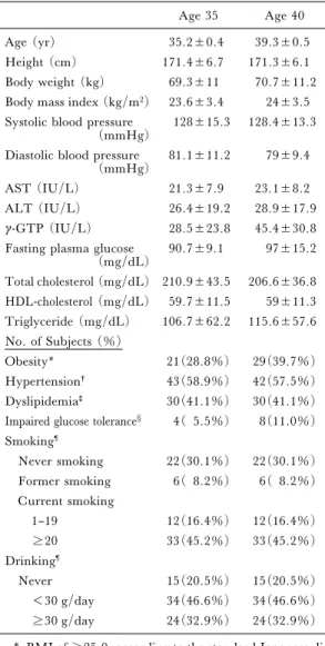
Table 1. Characteristics of the 73 male workers in the present follow-up study
Age 35 Age 40
Age (yr) 35.2±0.4 39.3±0.5
Height (cm) 171.4±6.7 171.3±6.1 Body weight (kg) 69.3±11 70.7±11.2 Body mass index (kg/m2) 23.6±3.4 24±3.5 Systolic blood pressure
(mmHg) 128±15.3 128.4±13.3 Diastolic blood pressure
(mmHg) 81.1±11.2 79±9.4
AST (IU/L) 21.3±7.9 23.1±8.2
ALT (IU/L) 26.4±19.2 28.9±17.9 g-GTP (IU/L) 28.5±23.8 45.4±30.8 Fasting plasma glucose
(mg/dL) 90.7±9.1 97±15.2 Total cholesterol (mg/dL) 210.9±43.5 206.6±36.8 HDL-cholesterol (mg/dL) 59.7±11.5 59±11.3 Triglyceride (mg/dL) 106.7±62.2 115.6±57.6 No. of Subjects (%) Obesity* 21(28.8%) 29(39.7%) Hypertension† 43(58.9%) 42(57.5%) Dyslipidemia‡ 30(41.1%) 30(41.1%) Impaired glucose tolerance§ 4( 5.5%) 8(11.0%) Smoking¶ Never smoking 22(30.1%) 22(30.1%) Former smoking 6( 8.2%) 6( 8.2%) Current smoking 1–19 12(16.4%) 12(16.4%) 20 33(45.2%) 33(45.2%) Drinking¶ Never 15(20.5%) 15(20.5%) <30 g/day 34(46.6%) 34(46.6%) 30 g/day 24(32.9%) 24(32.9%) * BMI of 25.0, according to the standard Japanese
di-agnosis of obesity.
†Systolic blood pressure 130 mm Hg and/or diastolic blood pressure 80 mm Hg.
‡Triglyceride level 150 mg/dl and/or HDL-choles-terol 40 mg/dL.
§Fasting plasma glucose level 110 mg/dl.
¶Smoking and drinking habits at 40 years were the same as those at 35 years.
analyzed by ANOVA, then a post hoc test (SheŠe's test). Logistic regression analysis was performed to predict factors that are associated with liver dysfunc-tion 5 years later or accumuladysfunc-tion of components of the metabolic syndrome. A level of p<0.05 was con-sidered signiˆcant. All analyses were performed with the computer program StatView 5.0 (SAS Institute Inc., Cary, NC) except for the logistic regression analysis, which was performed with SPSS 12.0 (SPSS Inc., Tokyo, Japan).
III. Results
Characteristics of the 73 Male Subjects
The physical and clinical chemistry data from the 73 male workers at age 35 and age 40 were com-pared (Table 1). None of the subjects had chronic liver disease caused by hepatitis B or C viral infec-tion, alcohol, drugs, or autoimmune hepatitis. In the baseline study, 28 of 148 (19.0%) subjects had liver dysfunction according to the criteria described in the Subjects and Methods. Of the 73 subjects studied in this study, 10 (14%) had liver dysfunction at age 35 and 20 (27%) at age 40.
Of the 148 subjects who were age 35 in the base-line study, 8 (5.4%) had elevated AST levels, 23 (15.5%) had elevated ALT levels, and 14 (9.5%) had elevatedg–GTP levels. In the 73 subjects stud-ied at both age 35 and age 40, AST levels were elevated in 2 (2.7%) at 35 years and 4 (5.4%) at 40 years, ALT levels were elevated in 9 (12.3%) at 35 years and 14 (19.2%) subjects at 40 years, and g–GTP levels were elevated in 6 (8.2%) at 35 years and 13 (17.8%) at 40 years, respectively. The proportion of subjects with each liver enzyme elevat-ed increaselevat-ed over the 5 year period. Patient charac-teristics at age 35 in the present study (n=73) were otherwise basically similar to those at age 35 in the baseline study (n=148).
Subjects with elevated liver enzymes underwent ultrasonography to conˆrm fatty inˆltration of the liver. Excluding other liver diseases by serological examination and the questionnaire regarding drink-ing habits (less than 30 g of alcohol per day), a majority of subjects were diagnosed as having nonal-coholic fatty liver disease (NAFLD).
The percentages of subjects who had obesity, hypertension, dyslipidemia, or impaired glucose tolerance were 28.8%, 58.9%, 41.1%, and 5.5% at age 35 and 39.7%, 57.5%, 41.1%, and 11.0% at 40, respectively (Table 1). There was only one subject with elevated liver enzymes without any of the four components of the metabolic syndrome at age 40. Comparison of Liver Function Among Four Groups Classi-ˆed According to the Number of Components of Metabolic
Syndrome at Ages 35 and 40
Seventeen (23.3%) of the 73 subjects had no components of metabolic syndrome, 21 (28.8%) had one component, 24 (32.9%) had two, 11 (15.1%) had more than three at age 35 (Table 2–a). Fifty-six subjects (76.7%) had one or more components of metabolic syndrome at age 35. At age
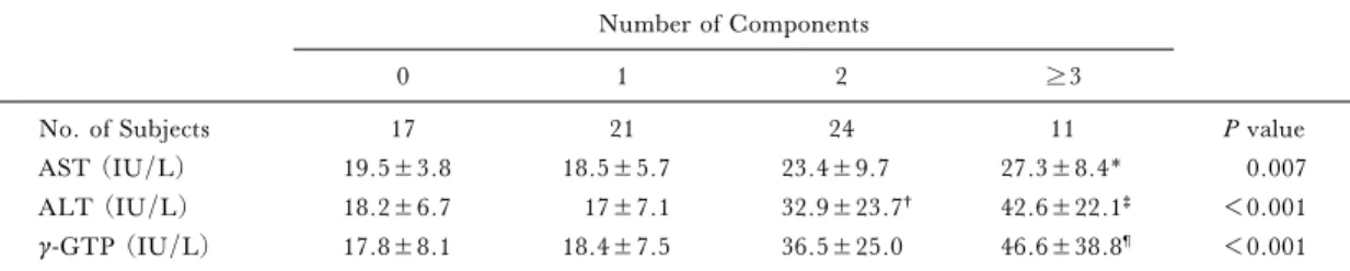
Table 2–a.Accumulation of components of the metabolic syndrome and the levels of AST, ALT, g-GTP at age 35. Number of Components
0 1 2 3
No. of Subjects 17 21 24 11 P value
AST (IU/L) 19.5±3.8 18.5±5.7 23.4±9.7 27.3±8.4* 0.007
ALT (IU/L) 18.2±6.7 17±7.1 32.9±23.7† 42.6±22.1‡ <0.001
g-GTP (IU/L) 17.8±8.1 18.4±7.5 36.5±25.0 46.6±38.8¶ <0.001
* P=0.021 v subjects having 1 component by ScheŠe's test. †P=0.002 v subjects having no component by ScheŠe's test.
‡P=0.001 v subjects having 1 component and P=0.005 v subjects having no component by ScheŠe's test. ¶P=0.010 v subjects having no component and P=0.009 v subjects having 1 component by ScheŠe's test.
Table 2–b.Accumulation of components and levels of AST, ALT, g-GTP at age 40 Number of Components
0 1 2 3
No. of Subjects 15 26 17 15 P value
AST (IU/L) 20.1±9.9 21.5±6.3 32.2±5.9 29.9±8.6* 0.002
ALT (IU/L) 18.1±9.7 23.6±10 31.7±19.5 45.6±21.1† <0.001
g-GTP(IU/L) 29.6±13.7 36±16.1 51.3±37.6‡ 70.6±38‡ <0.001
* P=0.008, 0.01, and 0.008 v subjects with no component, subjects with 1 component, and subjects having 2 compo-nents, respectively by ScheŠe's test.
†P=<0.0001 v subjects with non component and subjects with 1 component by ScheŠe's test. ‡P=0.001 and 0.003 v subjects with no component and subjects with 1 component by ScheŠe's test
Table 3. Prevalence of ALDH2 and b3-AR geno-types in subjects with or without liver dysfunction
Subjects With Liver Dysfunction (n=20) Subjects Without Liver Dysfunction (n=53) ALDH2 genotype ALDH21/1 15(75.0%) 26(49.1%) ALDH21/2 4(20.0%) 24(45.3%) ALDH22/2 1( 5.0%) 3( 5.7%) ALDH2 activity Active(ALDH21/1) 15(75.0%) 26(49.1%) x2=3.97 Inactive(ALDH21/2 +ALDH22/2) 5(25.0%) 27(50.9%) P=0.046 b3-AR genotype Trp64Trp 15(75.0%) 43(81.1%) Trp64Arg 4(20.0%) 9(17.0%) Arg64Arg 1( 5.0%) 1( 1.9%) b3-AR allele Trp64 15(75.0%) 43(81.1%) x2=0.33 Arg64 5(25.0%) 10(18.9%) P=0.563
40, 26 (35.6%) had one component, 17 (23.3%) had two components, and 15 (20.5%) had more than three (Table 2–b). The values of AST and ALT were similar in both age groups, butg–GTP levels increased after 5 years even in workers with no components. The AST, ALT andg–GTP levels in-creased with the number of components of metabolic syndrome in both age groups (at age 35; AST,P= 0.007; ALT,P=0.0001; g–GTP, P=0.0005; at age 40, AST,P=0.023; ALT, P<0.0001; g–GTP, P= 0.0003).
Association of Liver Dysfunction with Frequency of the ALDH2 and b3–AR Genotypes
Table 3 shows the association of liver dysfunc-tion with the ALDH2 andb3–AR genotypes. Active ALDH2 was seen more frequently among those with liver dysfunction than among those who did not (P= 0.046, chi-square test), although this relationship was not signiˆcant in the baseline study (P= 0.0745). No relationship between theb3–AR gene polymorphism and liver function was evident among the subjects in either the baseline or present studies. Association of Liver Dysfunction with Frequency of ALDH2 and b3–AR Genotypes Among Subjects with Normal BMI In the normal BMI group, persons with elevat-ed ALT (>40) were more likely to have active
ALDH2 and the Arg64 genotype ofb3–AR (18.5 BMI<25.0) in the baseline study. In the lean and obese BMI groups, there was no correlation between liver dysfunction and prevalence of the b3–AR or
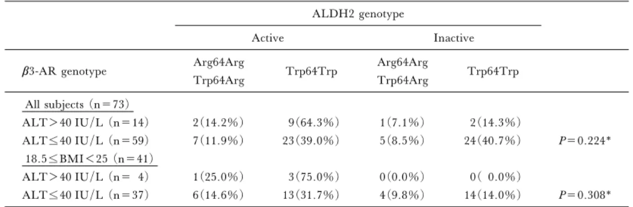
Table 4. Relationship between elevated ALT and the ALDH2 and b3-AR genotypes ALDH2 genotype
Active Inactive
b3-AR genotype Arg64Arg Trp64Trp Arg64Arg Trp64Trp
Trp64Arg Trp64Arg All subjects (n=73) ALT>40 IU/L (n=14) 2(14.2%) 9(64.3%) 1(7.1%) 2(14.3%) ALT40 IU/L (n=59) 7(11.9%) 23(39.0%) 5(8.5%) 24(40.7%) P=0.224* 18.5BMI<25 (n=41) ALT>40 IU/L (n= 4) 1(25.0%) 3(75.0%) 0(0.0%) 0( 0.0%) ALT40 IU/L (n=37) 6(14.6%) 13(31.7%) 4(9.8%) 14(14.0%) P=0.308* * Frequency analysis was performed with an extended Fisher's exact test.
Table 5. Factors associated with development of liver dysfunction* by logistic regression analysis Independent
variables Oddsratio 95% conˆdentinterval P value
BMI 11.88 2.56–55.16 0.002
ALDH2 (active) 5.44 1.10–26.90 0.038 * Fourteen workers were newly diagnosed as having
liver dysfunction in the present study. The checkup data of these 14 workers were compared with those for 49 workers who did not have liver dysfunction in the present study.
ALDH2 genotypes. In the present study, all 4 sub-jects with elevated ALT without obesity had the ac-tive ALDH2 genotype (Table 4).
Association of ALT Levels with the ALDH2 and b3–AR Genotypes in Subjects of the Normal BMI Group (18.5 BMI<25.0)
We analyzed the relationship between ALT lev-els and the ALDH2 orb3–AR genotype in the nor-mal BMI group (18.5BMI<25.0) by two-way ANOVA and post hoc test in both studies. The ALT levels of subjects with the Arg/Arg or Trp/Arg geno-type ofb3–AR were signiˆcantly higher than those of subjects with the Trp/Trp genotype ofb3–AR in the baseline study, but not higher in the present study (P =0.0390 in the baseline study vs.P=0.3585 in the present, ScheŠe's test).
The ALT level of subjects with active ALDH2 was also higher than that of subjects with inactive ALDH2 (P=0.0286 in the baseline study vs. P= 0.0592 in the present, ScheŠe's test).
The AST level was signiˆcantly higher among those with the Arg/Arg or Trp/Arg genotype of b3–AR compared to those with the Trp/Trp geno-type in the baseline study but not in the present study.
Association of Alcohol Drinking, Smoking Habits, and Ap-pearance of Liver Dysfunction with Number of Metabolic Syndrome Components
As in the baseline study, there were no sig-niˆcant diŠerences in the amount of daily alcohol in-take and smoking habit between those who did and those who did not have liver dysfunction. Of the 73 workers studied, 14 (19%) demonstrated decrease in metabolic syndrome components, 39 (53%) demonstrated no change, and 20 (27%) demonstrat-ed an increase. There were neither signiˆcant diŠer-ences in alcohol drinking nor in smoking habits among the three groups.
Fourteen workers were newly diagnosed as hav-ing liver dysfunction at their 40-year checkup. To select the factors signiˆcantly associated with de-veloping liver dysfunction in these 14, a logistic regression analysis was performed. Drinking and
smoking habits, ALDH2 and b3–AR genotypes
(categorical variables), and BMI (continuous bles) at 40 years were chosen as independent varia-bles. By forward stepwise variable selection, the BMI at 40 years and the active ALDH2 genotype were in-dependently associated with development of liver dysfunction at 40 years (Table 5).
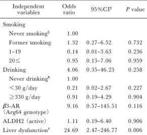
In the present study 15 out of 73 workers had more than 3 components of the metabolic syndrome. To clarify the factors associate with accumulation of components of the metabolic syndrome, logistic regression analysis was performed with variables in-cluding smoking and drinking habits at 40 years, ALDH2 and b3–AR genotypes, and presence of liver dysfunction at 35 years (categorical variables). Among these factors liver dysfunction at 35 years was signiˆcantly associated with accumulation of metabolic syndrome components (Table 6).
Table 6. Factors associated with accumulation of the metabolic syndrome components*
Independent
variables Oddsratio 95%CI‡ P value Smoking Never smoking§ 1.00 Former smoking 1.32 0.27–6.52 0.732 1–19 0.14 0.01–3.63 0.236 20 0.95 0.13–7.06 0.959 Drinking 4.06 0.35–46.23 0.258 Never drinking¶ 1.00 <30 g/day 0.21 0.02–2.67 0.227 330 g/day 0.91 0.19–4.29 0.904 b3-AR (Arg64 genotype) 9.16 0.57–145.51 0.116 ALDH2 (active) 1.11 0.19–6.40 0.906 Liver dysfunction† 24.69 2.47–246.77 0.006
* Subjects having 3 or more components of the metabol-ic syndrome in the present study. Forced entry: All variables entered in a single step.
†Liver dysfunction at the baseline. ‡Conˆdence interval.
§Never smoking used as the reference. ¶Never drinking used as the reference.
IV. Discussion
A close association of liver dysfunction and the metabolic syndrome, related to a polymorphism in the ALDH2 gene was demonstrated in both the present and baseline studies6), although the numbers of subjects did not provide su‹cient statistical evi-dence for unequivocal conˆrmation. At the begin-ning of the present study, we hypothesized that ALDH2 andb3–AR genotypes are involved in the development of metabolic syndrome in younger per-sons, especially normal weight perper-sons, and that ac-cumulation of unhealthy habits may aŠect its occur-rence in the middle and older aged persons because it has been demonstrated that the prevalence of meta-bolic syndrome increases with age7,8). In order to clarify this hypothesis we enrolled 148 workers in this study when they were 35 years old, as a blood test was performed at this age for the ˆrst time after join-ing the company, and 73 of 148 workers could be fol-lowed up after 5 years. Among them, a total of 20 workers had liver dysfunction, and 4 of them showed putative metabolic syndrome with elevated ALT with normal weight. Contrary to our baseline study6), all 4 workers had active ALDH2 but not the Arg64 genotype ofb3–AR gene. The results indicate that theb3–AR gene polymorphism is not involved in development of the metabolic syndrome with
elevated ALT in middle-aged persons.
Workers with elevated liver enzymes who had no viral markers or other causes such as alcohol, drug, or autoimmune mechanisms, exhibited in-creased numbers of metabolic syndrome components (abdominal obesity, dyslipidemia, hypertension, and glucose intolerance) in both the present and baseline study6). As mentioned previously, the depo-sition of fat in the abdominal fat cells may cause ‰ux of free fatty acid to the liver, resulting in the develop-ment of fatty liver1~5). Therefore, one might expect to observe elevated liver enzymes at the beginning of the metabolic syndrome, though conclusive evidence was not obtained in the present study. We are still trying to conˆrm this theory, because early detection of metabolic syndrome in young adults using liver enzymes would be very useful. In fact, insulin resist-ance evaluated by HOMA-IR was present in youn-ger adults in a cross-sectional study in Spain8). They suggested that these young persons with insulin re-sistance may progress to metabolic syndrome in mid-dle or older age. ALT might replace HOMA-IR to predict the occurrence of metabolic syndrome.
``Syndrome X'' by Reaven9), ``Deadly Quar-tet'' by Kaplan10), ``Insulin Resistance Syndrome'' by DeFronzo11), ``Visceral Obesity Syndrome'' by Matsuzawa12), ``Metabolic Syndrome X'' by Matsuzawa13), ``Multiple Metabolic Syndrome'' by Liese14), ``Multiple Risk Factor Syndrome''15)have a common pathophysiology, that is insulin resistance, and are now collectively called metabolic syndrome. A WHO working group established a deˆnition and diagnostic criteria for the disease16). The Third Na-tional Health and Nutrition Examination Survey (NHANES III, Adult Treatment Panel III report) reported new criteria deˆning metabolic syndrome to consist of more than 3 of the following 5 risk factors: (1) waist circumference≧102 cm in men and≧88 cm in women, (2) TG≧150 mg/dl, (3) HDL–C≦ 40 mg/dl, (4) arterial blood pressure≧130/85 mmHg, and (5) fasting blood sugar≧110 mg/dl7). Very recently, Matsuzawa announced the following new diagnostic criteria for metabolic syndrome for Japanese on behalf of eight Japanese academic societies17). A waist circumference of ≧85 cm in men and ≧90 cm in women is necessary for the diagno-sis, and the two of the following three conditions should be present17). The Adult Treatment Panel III report as well as the new criteria for Japanese empha-sized the presence of abdominal obesity among other risks. As periodic health checks regulated by law (the Community Health Law and the Labor Safety and Health Law) in Japan do not require the measure-ment of waist circumference, we here used BMI in-stead of waist circumference. If waist circumference
were available, a relation between the increase in the number of the metabolic syndrome components and/or elevated ALT and gene polymorphism might be observed. A relationship between the ALDH2 gene polymorphism and the occurrence of metabolic syndrome with elevated liver enzymes was seen among normal BMI persons in the baseline study as well as in the present study.
The b3–AR gene polymorphism is thought to be related to abdominal obesity and insulin resist-ance18~22). Several reports have described a relation-ship between fatty liver and insulin resistance2). Therefore, b3–AR gene polymorphism may be in-volved in the development of fatty liver in the early phase. Though few reports have described relation-ships between obesity and theb3–AR gene polymor-phism, di‹culty in losing weight in people with the Arg64 genotype has been descrived23,24). We selected the ALDH2 gene as another candidate gene for fatty liver because the ALDH2 genotype is related to the alcohol drinking habit25~27). As observed in the base-line study6)the present study showed a high preva-lence of active ALDH2 among workers with elevated ALT. Our results are similar to those of a study that revealed that habitual drinkers with active ALDH2 showed liver dysfunction more often than subjects with inactive ALDH228). ALDH2 may participate in the metabolism of not only alcohol but also other chemicals with aldehyde radicals, resulting in altera-tion of lipid metabolism as menaltera-tioned previously6).
EŠects of smoking and/or drinking alcohol on prevalence of the metabolic syndrome, especially on the dynamic condition of insulin resistance, have been reported29~34). We could not conˆrm any in-‰uence of either alcohol consumption or smoking on the development of metabolic syndrome in the present study. The reason may be that the number of subjects was too small. Another is that we used rela-tively young persons as subjects, and it takes time to accumulate components of the metabolic syndrome. The present study did show that liver dysfunction at 35 years was associated with accumulation of compo-nents of the metabolic syndrome at 40, indicating that liver dysfunction is an early symptom. Other lifestyle related factors such as diet and physical ac-tivity might be involved. The present study lacks in-formation concerning these lifestyle related factors. Increased physical activity should be evaluated for the treatment of metabolic syndrome with liver dys-function and ALDH2 gene polymorphism, though health education is very di‹cult35).
Most reports on metabolic syndrome have been cross-sectional studies36), and there have been no prospective studies to date. A longitudinal study is necessary to clarify the involvement of genetic factors
in development of the metabolic syndrome, and tem-poral relationship with fatty liver. Therefore, we will continue to follow-up workers and accumulate health data in order to develop prevention guidelines for metabolic syndrome based on the impact of gene polymorphism.
Acknowledgment
We thank Professor Isao Okazaki for stimulat-ing our research and providstimulat-ing useful suggestions for this manuscript.
References
1) Marceau P, Biron S, Hould FS, et al. Liver patholo-gy and the metabolic syndrome X in severe obesity. J Clin Endocrinol Metab 1999; 84: 1513–1517. 2) Marchesini G, Brizi M, Morselli-Labate AM, et al.
Association of nonalcoholic fatty liver disease with insu-lin resistance. Am J Med 1999; 107: 450–455. 3) Chitturi S, Abeygunasekera S, Farrell GC, et al.
NASH and insulin resistance: Insulin hypersecretion and speciˆc association with the insulin resistance syn-drome. Hepatology 2002; 35: 373–379.
4) Pagano G, Pacini G, Musso G, et al. Nonalcoholic steatohepatitis, insulin resistance, and metabolic syn-drome: further evidence for an etiologic association. Hepatology 2002; 35: 367–372.
5) Ioannou GN, Weiss NS, Boyko EJ, et al. Contribu-tion of metabolic factors to alanine aminotransferase ac-tivity in persons with other causes of liver disease. Gas-troenterology 2005; 128: 627–635.
6) Murata C, Watanabe T, Furuya H, et al. Aldehyde dehydrogenase 2 and b3–adrenergic receptor gene poly-morphisms: Their association with elevated liver en-zymes and metabolic syndrome. Metabolism 2003; 52: 1096–1101.
7) Ford ES, Giles WH, Dietz WH. Prevalence of the metabolic syndrome among US adults. Findings from the Third National Health and Nutrition Examination Survey. JAMA 2002; 287: 356–359.
8) Ascaso JF, Romero P, Real JT, et al. Abdominal obesity, insulin resistance, and metabolic syndrome in a southern European population. Europ J Int Med 2003; 14: 101–106.
9) Reaven GM. Role of insulin resistance in human dis-ease. Diabetes 1988; 37: 1595–1607.
10) Kaplan NM. The deadly quartet. Arch Intern Med 1989; 149: 1514–20.
11) DeFronzo RA, Ferrannini E. Insulin resistance: a multifaceted syndrome responsible for NIDDM, obesi-ty, hypertension, dyslipidemia and atherosclerotic heart disease. Diabetes Care 1991; 14: 173–94.
12) Fujioka S, Matsuzawa Y, Tokunaga K, Tarui S. Contribution of intraabdominal fat accumulation to the impairment of glucose and lipid metabolism. Metabolism 1987; 36: 54–59.
13) Matsuzawa Y, Funahashi T, Nakamura T. Molecu-lar mechanism of metabolic syndrome X: Contribution
of adipocytokines adipocyte-derived bioactive sub-stances. Ann NY Acad Sci 1999; 892: 146–154. 14) Liese AD, Mayer-Davis EJ, Tyroler HA, et al.
De-velopment of the multiple metabolic syndrome in the ARIC cohort: joint contribution of insulin, BMI, and WHR. Ann Epidemiol 1997; 7: 407–416.
15) Multiple Risk Factor Intervention Trial Research Group. Multiple Risk Factor Intervention Trial. Risk factor changes and mortality results. JAMA 1982; 248: 1465–1477.
16) Alberti KG, Zimmet PZ. Deˆnition, diagnosis and classiˆcation of diabetes mellitus and its complications. Part 1: Diagnosis and Classiˆcation of Diabetes Melli-tus, provisional report of a WHO consultation. Diabet Med 1998; 15: 539–553.
17) The expert committee for the formation of criteria on metabolic syndrome. The deˆnition and criteria for metabolic syndrome. J Jap Soc Intern Med 2005; 94: 188–203.
18) Sakane N, Yoshida T, Yoshioka K, et al. Trp64Arg mutation of beta3–adrenergic receptor and non-insulin dependent diabetes mellitus. Intern Med 1998; 37:345. 19) Walston J, Silver K, Bogardus C, et al. Time of onset of non-insulin-dependent diabetes mellitus and genetic variation in the beta3–adrenergic-receptor gene. N Engl J Med 1995; 333: 343–347.
20) Kadowaki H, Yasuda K, Iwamoto K, et al. A muta-tion in the beta3–adrenergic receptor gene is associated with obesity and hyperinsulinemia in Japanese subjects. Biochem Biophys Res Commun 1995; 215: 555–560. 21) Widen E, Lehto M, Kanninen T, et al. Association
of a polymorphism in the beta3–adrenergic-receptor gene with features of the insulin resistance syndrome in Finns. N Engl J Med 1995; 333: 348–351.
22) Shima Y, Tsukada T, Nakanishi K, et al. Association of the Trp64Arg mutation of the beta3–adrenergic receptor with fatty liver and mild glucose intolerance in Japanese subjects. Clin Chim Acta 1998; 274: 167–176. 23) Shiwaku K, Nogi A, Annurad E, et al. Di‹culty in losing weight by behavioral intervention for women with Trp64Arg polymorphism of the beta3–adrenergic receptor gene. Int J Obes Relat Metab Disord 2003; 27: 1028–1036.
24) Kim OY, Cho EY, Park HY, et al. Additive eŠect of the mutations in the beta3–adrenoceptor gene and
UCP3 gene promoter on body fat distribution and glycemic control after weight reduction in overweight subjects with CAD or metabolic syndrome. Int J Obes Relat Metab Disord. 2004; 28: 434–441.
25) Thomasson HR, Edenberg HJ, Crabb DW, et al. Al-cohol and aldehyde dehydrogenase genotypes and alco-holism in Chinese men. Am J Hum Genet 1991; 48: 677–681.
26) Harada S, Zhang S. New strategy for detection of ALDH2 mutant. Alcohol Alcohol 1993; Suppl 1A: 11–13.
27) Takeshita T, Morimoto K, Mao XQ, et al. Pheno-typic diŠerences in low Km aldehyde dehydrogenase in Japanese workers. Lancet 1993; 341: 837–838. 28) Takeshita T, Yang X, Morimoto K: The ALDH2
genotype, alcohol intake, and liver-function biomarkers among Japanese male workers. Hum Genet 2000; 106: 589–593.
29) Facchini FS, Hollenbeck CB, Jeppesen J, et al. Insu-lin resistance and cigarette smoking. Lancet 1992; 339: 1128–30.
30) Eliasson B, Mero N, Taskinen M-R, et al. The insu-lin resistance syndrome and postprandial lipid intoler-ance in smokers. Atherosclerosis 1997; 129: 79–88. 31) Geslain-Biquez C, Voi S, Tichet J, et al. The
meta-bolic syndrome in smokers. The D.E.S.I.R. study. Dia-betes Metab 2003; 29: 226–34.
32) Facchini FS, Chen YD, Reaven GM. Light-to moderate alcohol intake is associated with enhanced in-sulin sensitivity. Diabete Care 1994; 17: 115–119. 33) Goude D, Fagerberg B, Hulthe J. Alcohol
consump-tion, the metabolic syndrome and insulin resistance in 58-year-old clinically healthy men (AIR study). Clin Sci 2002; 102: 345–352.
34) Rosell M, de Faire U, Hellenius M-L. Low preven-tion of the metabolic syndrome in wine drinkers -is it the alcohol beverage or the life style? Eur J Clin Nutr 2003; 57: 227–234.
35) Ito S, Furuya H, et al. Comparative socio-cultural analysis of smoking behavior and di‹culty of quitting smoking in Japan and Thailand. Jpn J Pub Hlth, 2004 Nov; 51(11): 975–985.
36) Reaven GM. Syndrome X: 6 years later. J Int Med 1994; 236 (Suppl 736): 13–22. 571–633.