INTRODUCTION
Prostate cancer continues to be the most common cancer and the third - leading cause of cancer - related deaths among men in the United States. Projections for 2017 indicated 161,360 new cases of prostate cancer, along with 26,730 deaths from the disease (1, 2). Prostate cancer incidence is much lower in Asian countries including Japan than that of Western countries. However, prostate cancer incidence in Asian countries has been on a steady increase during the past few decades (3, 4). Extraprostatic extension of prostate cancer is defined as pT3a in the tumor, lymph node, and metastasis staging system for prostate cancer, and it is well -established as being associated with a poor prognosis. This feature must be identified accurately for optimal patient management after radical prostatectomy (5 - 7). The prostate is surrounded by pe-riprostatic adipose tissue. Therefore, an admixture of tumor cells with periprostatic adipose tissue (Figure 1) is the most common finding of extraprostatic extension (8, 9).
Adipose tissue is composed mainly of adipocytes, although addi-tional cell types are present, including pericytes, monocytes, macrophages, lymphocytes, fibroblasts, vascular endothelial cells, and pluripotent stem cells. Previously, adipose tissue was thought to play limited physiological roles mainly as energy storage and protection from cold temperatures. Currently, adipose tissue is rec-ognized as an active endocrine organ, secreting growth factors, chemokines, or pro - inflammatory molecules termed ‘adipokines’ that can regulate metabolism and the immune system. Physiologi-cally, adipokines regulate appetite, lipid metabolism, glucose ho-meostasis, insulin sensitivity, angiogenesis, blood pressure, and
inflammatory processes (10). Obesity is defined by increased adi-pose mass arising from an energy imbalance. Adipocyte hypertro-phy during obesity causes adipose dysfunction and inflammation by increasing the secretion of pro - inflammatory adipokines from adipocytes (11). Alterations of adipocyte biology associated with adipocyte hypertrophy affect systemic organs. Epidemiologically, obesity is related to the risk of many types of cancers, including esophageal, gastric, colorectal, biliary, pancreatic breast, en-dometrial, ovarian, and kidney cancers (12 - 14). In addition, obe-sity is associated with the progression of many cancers, including prostate, breast, endometrial, kidney, pancreatic, esophageal, and thyroid cancers (15 - 19). Furthermore, direct crosstalk between normal adipocytes and cancer cells can exist at the front of cancer invasion to adipose tissue (20 - 22). Thus, growing evidence sug-gests crucial roles for adipose tissue in the development of several cancers.
In this article, we review the contribution of adipose tissue to prostate cancer development, including bone metastasis.
1. OBESITY AND PROSTATE CANCER
It is estimated that overweight and obesity could account for 14% of all cancer deaths in men and 20% of all cancer deaths in women in the United States (23). Epidemiologic studies have found that the relationship between obesity and the incidence of prostate cancer is unclear. On the other hand, obesity is associated with prostate cancer mortality (12 - 14). In addition, obesity and hyper-tension were each associated with an increased risk of biochemical recurrence of prostate cancer after radical prostatectomy, inde-pendent of age at diagnosis and tumor pathological features (24). Interestingly, several studies suggest that obesity reduces the risk of localized, low - grade, and nonaggressive prostate cancer, al-though it increases the risk of advanced, high - grade, and aggres-sive prostate cancer (25, 26). Obesity has also been associated with
REVIEW
Adipose tissue : Critical contributor to the development of
prostate cancer
Hisanori Uehara, Tomoko Kobayashi, Minoru Matsumoto, Shunsuke Watanabe, Akiko Yoneda, and Yoshimi Bando
Division of Pathology, Tokushima University Hospital, Tokushima, Japan
Abstract : The prostate is surrounded by periprostatic adipose tissue. Although adipose tissue was thought to play limited physiological roles, it has recently been recognized as an active endocrine organ, secreting growth factors and adipokines. Epidemiologically, obesity is associated with prostate cancer progression. A major mechanism to explain the link between obesity and cancer includes the insulin and insulin -like growth factor (IGF) -1 axis, sex steroids, and adipokines. When prostate cancer cells invade periprostatic adipose tissue, adipose tissue contributes to create the tumor microenvironment, mainly via adipokine secretion. Furthermore, direct crosstalk between adipocytes and cancer cells can exist. We showed that fatty acid -binding protein 4 (FABP4) re-leased from adipocytes was taken up into prostate cancer cells and may act as a carrier of an energy source for the invasion. Bone is an adipocyte-rich organ and is the common metastatic site of prostate cancer. In the mi-croenvironment of bone metastases, tumor cells, osteoblasts, osteoclasts, adipocytes, and other stromal cells are interacting with one another and organizing a complex system. Thus, growing evidence implicates adipose tissue as a critical contributor to the development of prostate cancer. A deeper understanding of the mechanisms leads to more effective therapeutic strategies for prostate cancer. J. Med. Invest. 65 : 9-17, February, 2018
Keywords : adipose tissue, prostate cancer, adipokine, bone metastasis
Received for publication October 11, 2017 ; accepted October 31, 2017. Address correspondence and reprint requests to Hisanori Uehara, M.D., Ph.D., Division of Pathology, Tokushima University Hospital, 2 - 50 - 1 Kuramoto - cho, Tokushima - shi, Tokushima 770 - 8503, Japan, and Fax : +81 - 88 - 633 - 7067.
the presence of prostatic intraepithelial neoplasia in benign speci-mens and with future prostate cancer risk after an initial benign finding. Therefore, obesity should be taken into account in the clinical follow - up plan after a benign biopsy (27).
A major mechanism to explain the link between obesity and cancer includes insulin and the IGF - 1 axis, sex steroids, and adipose tissue - derived cytokines. They are linked through insulin resistance (28). Other possible mechanisms include fatty acid -induced inflammation, oxidative stress, endoplasmic reticulum stress, and hypoxia.
Insulin-IGF-1 axis
Circulating insulin levels correlate positively with increasing body mass index, and many obese persons are insulin - resistant. The evidence is mounting that insulin resistance is a risk factor for cancer development. It is suggested that hyperinsulinemia may contribute to cancer development through the growth - promoting effect of elevated insulin levels (29, 30). Obesity and prolonged hyperinsulinemia are associated with increases in the levels of free or bioactive IGF - 1 due to reduced production of IGF - binding pro-teins, which normally bind IGF - 1 and inhibit its action. Through the interaction with the IGF receptor, IGF activates downstream signaling pathways that affect the growth of cancer cells by the promotion of mitogenic pathways, induction of neovascularization, and inhibition of apoptosis. Insulin itself has anabolic, antiapop-totic, and mitotic effects (12, 31, 32).
Epidemiological data suggest that high levels of circulating IGF - 1 are associated with an increased risk of prostate cancer develop-ment (33). IGF - 1 signaling is elevated in prostate cancer com-pared to prostate epithelium and is associated with tumor progres-sion (34, 35). In addition, overexpresprogres-sion of the IGF - 1 receptor has been shown in prostate cancer (36).
Sex hormones
Androgens have been known to play an important role in normal prostate development and function, as well as for the growth and progression of prostate cancer. However, it has been suggested that there is no association between the plasma concentration of androgens and prostate cancer risk (37). Obesity is associated with a decrease in testosterone levels (38) and the synthesis of sex hormone - binding globulin (SHBG), which binds to sex hormones
including testosterone and dihydrotestosterone and regulates their effects (39). A significant association was shown between low serum testosterone levels and tumor stage and extraprostatic tumor spread in prostate cancer patients (40). Furthermore, the levels of testosterone were significantly lower in patients with pros-tate cancer than in those with benign prostatic hyperplasia (41). These results suggest that the low testosterone levels in obese men may be related to prostate cancer development. However, the exact mechanisms are still unknown.
Adipose tissue-derived cytokines
Adipose tissues in obesity are infiltrated by a large number of inflammatory cells (e.g. macrophages and leukocytes), and this recruitment is linked to systemic inflammation and insulin resis-tance (42, 43). The inflammation induces the generation of reactive oxygen species that act as tumor promoters at a low concentrations (44). Adipocytes, other stromal cells, and infiltrating inflammatory cells in adipose tissue secrete several adipokines and other cytoki-nes, which have been implicated to play a pivotal role in the development of obesity - related cancer (45). Adipokines are de-fined as hormone - like polypeptides that are actively secreted by white adipose tissue, and they include cytokines (e.g. interleukin - 6 (IL - 6) and tumor necrosis factor (TNF) -α), angiogenic factors (e.g. vascular endothelial growth factor (VEGF) and apelin), and other factors (e.g. leptin and adiponectin) (46). Several adipokines have been recognized to have multiple effects on prostate cancer cells.
2. MAJOR ADIPOKINES (LEPTIN, IL-6, AND
ADI-PONECTIN) AND PROSTATE CANCER
Leptin
Leptin, a polypeptide hormone that is mainly produced by adipo-cytes, acts as a major regulator for appetite and energy homeostasis via its action on specific receptors expressed in the hypothala-mus (47). The levels of leptin in plasma are correlated with the percentage of body fat (48, 49). This observation suggests that most obese persons are insensitive to endogenous leptin production. Epidemiologically, the association between serum leptin levels and prostate cancer risk is controversial. Stattin et al. reported that Figure 1. Extraprostatic extension of prostate cancer. (a) Prostate cancer invasion into periprostatic adipose tissue. Scale = 400μm. PC, prostate cancer cells ; A, adipose tissue. (b) Prostate cancer cells grow in proximity to adipocytes. Scale = 100μm. PC, prostate cancer cells ; A, adipose tissue.
moderately elevated plasma leptin concentrations are associated with later development of prostate cancer (50). Another study showed that elevated plasma leptin concentrations are associated with an increased risk of high - volume prostate cancer (tumors" 0.5 cc in volume or with histologic evidence of extraprostatic exten-sion but without metastases) (51). On the other hand, several studies suggest that there is no association between serum leptin levels and prostate cancer risk (52, 53).
In human prostate tissue, leptin receptors have been detected in normal epithelium, prostatic high - grade intraepithelial neoplasia, and carcinoma by immunohistochemistry (50). DU145 and PC - 3 human prostate cancer cells have also been found to express leptin receptors, and leptin treatment had mitogenic and anti - apoptotic effects on these cells with phosphatidyl - inositol 3 - kinase (PI3K) and mitogen - activated protein kinase (MAPK) pathway activation (54, 55). Moreover, long - term exposure to leptin enhanced prolif-eration, invasion, and migration of prostate cancer cells with in-creased cyclin D1 expression and dein-creased p21 expression, sug-gesting the participation of leptin in cell cycle progression (56). Single nucleotide polymorphism (SNP) in exon 4 of the leptin receptor gene was significantly associated with prostate cancer -specific mortality (57).
IL-6
Serum IL - 6 has demonstrated the strongest relationship with obesity and insulin resistance (58). In adipose tissue, the main sources of IL - 6 are infiltrating macrophages and stromal cells. However, adipocytes also produce IL - 6 (59). Serum IL - 6 levels were significantly higher in patients with metastatic or hormone -refractory prostate cancer than in normal controls or patients with localized disease (60, 61), and they were associated with shorter survival time in patients with hormone - refractory prostate cancer (62). Exogenous IL - 6 stimulated growth in hormone - sensitive LNCaP prostate cancer cells, but not hormone - insensitive DU145 and PC - 3 cells (63). In a tumor xenograft model, when LNCaP cells continuously treated with IL - 6 were inoculated into nude mice, tumor volumes were larger than those of their controls cultured without IL - 6 treatment (64). Increased IL - 6 receptor expression was observed in human prostate cancer tissue, compared with nor-mal prostate tissue, and high levels of IL - 6 receptor expression in prostate cancer were associated with higher rates of cell prolifera-tion on immunohistochemistry (65). Thus, IL - 6 appears to act primarily as a paracrine growth factor for hormone - sensitive prostate cancer. However, its main action may shift to an autocrine mode as hormone resistance develops in prostate cancer.
Adiponectin
Adiponectin is a protein displaying structural similarities to collagen and TNF -α, and it is mainly secreted by adipocytes (66) and regulates glucose and lipid metabolism, vascular remodeling, and bone homeostasis. In addition, adiponectin has defensive ef-fects against inflammation and insulin resistance (67 - 70). In hu-mans, plasma concentrations of adiponectin are significantly lower in obesity and insulin - resistant states (71, 72). Adiponectin can bind to three receptors, AdipoR1, AdipoR2, and T - cadherin (73). Through the interaction with AdipoR1 and AdipoR2, adi-ponectin shows antidiabetic effects (74).
Epidemiological studies have reported that plasma adiponectin levels were significantly lower in patients with prostate cancer than in benign prostatic hyperplasia (BPH) patients or healthy con-trols (75, 76). Tan et al. examined the immunohistochemical ex-pressions of adiponectin in BPH cases and prostate cancer cases with low Gleason score (!7) Gleason score 7 or high Gleason score ("7). The results suggested that decreased adiponectin expres-sion was associated with prostate cancer progresexpres-sion. Further-more, silencing of adiponectin induced proliferation and invasion
in 22RV1 human prostate cancer cells via the epithelial - to - mese-nchymal transition process (77). AdipoR2 expression levels in prostate cancer cells were positively associated with cell prolifera-tion, with expression of fatty acid synthase, and with angiogenesis (78). These results suggests a positive relationship between AdipoR2 and prostate cancer development.
3. TUMOR MICROENVIRONMENT AND ADIPOSE
TISSUE
Periprostatic adipose tissue was present on 48% of all prostatic surfaces. The distribution of adipose tissue in the anterior, poste-rior, right, and left prostatic surfaces was 44%, 36%, 59%, and 57%, respectively (79). When prostate cancer cells invade into pe-riprostatic adipose tissue, adipose tissue contributes to create the tumor microenvironment. It has been recognized that the tumor microenvironment is crucial for tumor progression and metastasis. In such cases, adipokines play a central role. Adipokines act in paracrine, autocrine, and endocrine manners (80). However, paracrine and autocrine signaling may be a key action in the tumor microenvironment. For example, IL - 6 levels in periprostatic adi-pose tissue harvested from prostate cancer patients undergoing radical prostatectomy were approximately 375 times higher than in patient- matched serum. In addition, a higher Gleason score was correlated with high IL - 6 levels (81). Fain et al. reported that the levels of VEGF and IL - 6 released from visceral adipose tissue were significantly higher than those from subcutaneous adipose tissue (82). Thus, in cases of extraprostatic extension, prostate cancer cells are directly exposed to huge amounts of adipokines released from periprostatic adipose tissue. Furthermore, increased pe-riprostatic adipose tissue due to obesity etc. may modify the tumor microenvironment and accelerate tumor progression. Actually, in-creasing periprostatic adipose tissue thickness, measured by transrectal ultrasonography as the distance between the prostate and the pubic bone, was associated with prostate cancer and high -grade prostate cancer (83).
In the tumor microenvironment, matrix metalloproteinases (MMPs) play an important role in cancer progression, as well as adipokines. Periprostatic adipose tissue from prostate cancer pa-tients released higher amounts of pro - MMP - 9 than that from BPH patients (84). Supernatants from whole periprostatic adipose tissue showed increased activities of both MMP2 and MMP9, and they promoted proliferation and migration of PC - 3 cells, compared with those from the stromal vascular fraction of adipose tissue (85). These findings suggest that MMPs from periprostatic adipose tissue modulate the tumor microenvironment and promote pros-tate cancer cell survival and migration.
When cancer cells invade into adipose tissue, direct contact between cancer cells and adipocytes may occur. Dirat et al. re-ported that adipocytes adjacent to cancer cells at the invasive front demonstrated a decrease in more differentiated adipose markers and overexpressions of several inflammatory cytokines in breast cancer. In addition, the levels of IL - 6 in tumor surrounding adipo-cytes were higher in cases with tumors of larger size and/or with lymph node involvement. These adipocytes were termed “cancer -associated adipocytes” (20). Thus the cross - interactions between breast cancer cells and adipocytes modify each other’s characteris-tics/phenotypes, leading cancer cells to become more aggressive. Nieman et al. indicated another mechanism of cancer progression by direct contact between cancer cells and adipocytes. Co -culture of adipocytes and ovarian cancer cells promoted in vitro and in vivo tumor growth. In this culture system, the direct transfer of lipids from adipocytes to ovarian cancer cells, lipolysis in adipocytes, andβ-oxidation in cancer cells were induced, suggest-ing that adipocytes act as an energy source for the cancer cells.
From the results of protein array and immunohistochemical analy-ses in omental metastaanaly-ses compared to primary ovarian tumors, FABP4 was found to play an important role in ovarian cancer me-tastasis (86). FABP4, also known as aP2, is a member of the cyto-plasmic fatty acid binding protein multigene family that is associ-ated with insulin resistance, type 2 diabetes mellitus, and cardio-vascular disease (87 - 89). FABP4 is released from adipocytes and is abundantly present in human serum. Serum FABP4 levels are linked to obesity (87). We showed the involvement of FABP4 in human prostate cancer cell progression. In an in vitro study, FABP4 treatment promoted prostate cancer cell invasion, and the promot-ing effects were reduced by a FABP4 inhibitor, which inhibits FABP4 binding to fatty acids. Uptake of FABP4 into prostate cancer cells following FABP4 treatment was observed on immunohisto-chemistry. A FABP4 inhibitor also reduced subcutaneous growth and lung metastasis of prostate cancer cells in vivo. These results suggest that FABP4 may act as a carrier of an energy source for the prostate cancer cells. In addition, FABP4 treatment activated the phosphoinositide 3 - kinase (PI3K)/Akt pathway, with or without a FABP4 inhibitor, suggesting that FABP4 exerts its effect on pros-tate cancer cells through several pathways (90). Thus, FABP4 might be a key molecule to understand the mechanisms underly-ing the adipose tissue - prostate cancer progression link. Possible mechanisms involving FABP4 in prostate cancer progression are shown in Figure 2.
4. BONE MARROW ADIPOCYTES AND CANCER
METASTASIS
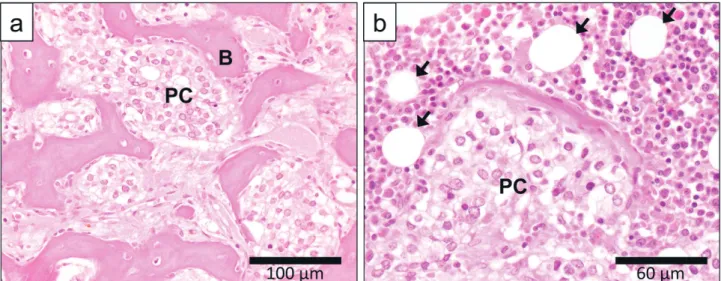
Bone is the common metastatic site of prostate cancer, and bone metastasis is seen in 90% of patients with metastatic prostate cancer. In fact, 86% of these patients had only bone metastases (91). Bone metastasis of prostate cancer is a multistep process including detachment of cancer cells from the primary tumor, travel of the cells through the blood vessels or lymphatics, attachment to bone tissue, and development of metastatic tumor in the bone. Figure 3
shows the histology of prostate cancer bone metastases. Prostate cancer metastases cause an osteoblastic, osteolytic, or mixed bone response (92 - 94). In osteolytic metastases, cancer cells produce osteoclastic factors such as parathyroid hormone - related protein (PTHrP) and transforming growth factorβ (TGFβ), which stimu-late osteoblasts and stromal cells to express receptor activator of NF kappa B (54) ligand (RANKL). The binding of RANKL to its receptor, RANK, on osteoclasts drives bone resorption and release of growth factors from the matrix, which supports cancer cell growth (95, 96). On the other hand, osteoblastic metastases are formed by cancer cell growth with new bone formation. Some tumor - associated factors, including endothelin - 1 and Wnts, have been proposed to stimulate osteoblast activity directly, and other factors including urokinase - type plasminogen activator activate proteases such as PSA, which enhance the osteosclerotic process either via activation of the quiescent forms of TGFβ or via degrada-tion of PTHrP (95, 97, 98).
Although bone contains few adipocytes at birth, aging causes an increase in the number of marrow adipocytes (99). In addition, obesity is associated with an increase of bone marrow adipose tissue (100). Therefore, it is possible that marrow adipose tissue may affect the growth and survival of metastatic cancer cells. Actually, when breast cancer cells were co - cultured with cancel-lous human bone tissue fragments, cancer cell migration toward tissue - conditioned medium was enhanced in association with in-creasing levels of leptin and IL - 1β. Immunohistochemistry of frag-ments showed breast cancer cell colonization within the marrow adipose tissue compartment (101).
IGF - 1 is the most abundant growth factor that is deposited in the bone matrix (102). Although IGF - 1 is a key factor in the endocrine regulation of body composition, IGF - 1 also plays an important role in the maintenance of bone mineral density. IGF - 1 is released from the bone matrix in response to bone resorption. IGF - 1 in-duces the differentiation of osteoblasts and stimulates expression and secretion of RANKL in osteoblasts. RANKL promotes osteo-clastogenesis. As a result, new bone is formed at bone resorption sites (103 - 105). Furthermore, IGF - 1 stimulates LNCaP cell growth (106). C4 - 2 human prostate cancer cells, a subline of LNCaP hav-ing a proclivity to form osteoblastic bone metastases, produced higher amounts of IGF - 1 than LNCaP. IGF - I mRNA expression in C4 - 2 cells was substantially increased in the presence of exoge-nous IGF - I. These results suggest the contribution of the IGF - I axis to the osteoblastic metastases of prostate cancer (107).
Whereas IL - 6 is one of the major adipokines, it also plays a profound role in bone metabolism. Acting via stromal/osteoblastic cells, IL - 6 stimulates osteoclastogenesis. Moreover, IL - 6 can pro-mote differentiation of osteoblasts toward a more mature pheno-type (108). Therefore, in the bone microenvironment, metastatic cancer cells may be exposed to large amounts of IL - 6 from both stromal/osteoblastic cells and adipocytes. Actually, it has been suggested that IL - 6 is involved with the mechanisms of bone me-tastasis. In a mouse model of bone metastasis, administration of a high - fat diet increased melanoma cell growth in the bone marrow with induction of osteopontin and IL - 6. Immunofluorescence stain-ing of IL - 6 showed that higher numbers of bone marrow adipo-cytes expressing IL - 6 were observed in the vicinity of tumor cells (109). Culture medium from PC - 3 human prostate cancer cells in-duced IL - 6 gene expression in osteoblast- like MC3T3 - E1 cells and promoted osteoclastogenesis in vitro. IL - 6 was highly ex-pressed in PC - 3 cells growing in the bones of SCID mice and human bone metastases (110). On the other hand, culture medium from human osteoblast- like HOBIT cells induced proliferation and PSA expression in human prostate cancer cells, LNCaP, C4 - 2B, and VCaP. These effects were inhibited by treatment with anti - IL - 6 antibody, suggesting that IL - 6 secreted from osteoblasts promotes prostate cancer growth (111).
Figure 2. Possible mechanisms involving fatty acid - binding protein 4 (FABP4) in prostate cancer progression. FABP4 binding to free fatty acids (FFAs) is taken up into prostate cancer cells, and FFAs may be used as an energy source for prostate cancer development. In addition, FABP4 can activate the phosphatidyl - inositol 3 - kinase (PI3K)/Akt path-way without being taken up into prostate cancer cells. Obesity increases the serum levels of FABP4.
As thus far described, in the microenvironment of bone metasta-ses, tumor cells, osteoblasts, osteoclasts, adipocytes, and other stromal cells interacting with each other and organize a complex system (Figure 4). Because aging is associated with both cancer risk and amount of bone marrow adipose tissue, bone marrow adipose tissue may be one of the key factors in bone metastasis. However, its roles are poorly understood.
5. CONCLUSIONS
The data presented in this review suggest that adipose tissue plays an important role in the development of prostate cancer, including tumor growth, invasion, and metastasis. Prostate cancer cells with extracapsular invasion form a new microenvironment in periprostatic adipose tissue. In the process, cancer cells are thought to have direct or indirect interactions with adipocytes. Moreover, obesity may modify these interactions and promote tumor progression. Bone is a favorite metastatic site of prostate cancer and contains increasing adipose tissue with age. Bone marrow adipose tissue interacts with tumor cells, osteoblasts, and other stromal cells, and it participates in the organization of the tumor microenvironment. Whereas adipokines seem to be key molecules in the relationship between cancer cells and adipose tissue, several other mechanisms are suggested. A deeper under-standing of the roles of adipose tissue in prostate cancer progres-sion will lead to more effective therapeutic strategies for prostate cancer.
CONFLICT OF INTEREST
None of the authors has any conflicts of interest to declare.
ACKNOWLEDGEMENTS
This work was supported by JSPS KAKENHI Grant Number 15K 08403.
REFERENCES
1. Miller KD, Siegel RL, Lin CC, Mariotto AB, Kramer JL, Rowland JH, Stein KD, Alteri R, Jemal A : Cancer treatment and survivorship statistics, 2016. CA Cancer J Clin 66 : 271 - 289, 2016
2. Siegel RL, Miller KD, Jemal A : Cancer Statistics, 2017. CA Cancer J Clin 67 : 7 - 30, 2017
3. Center MM, Jemal A, Lortet- Tieulent J, Ward E, Ferlay J, Figure 3. Bone metastases of prostate cancer. (a) Prostate cancer cells grow with bone formation. Scale = 100μm. (b) At the periphery of bone metastases, prostate cancer cells are facing normal bone marrow cells including several adipocytes. Scale = 60μm. PC, prostate cancer cells ; B, bone ; arrows, bone marrow adipocytes.
Figure 4. Interactions of prostate cancer cells with osteoblasts, osteo-clasts, and adipocytes in the microenvironment of bone metastases. In osteolytic metastases, cancer cells produce osteoclastic factors such as parathyroid hormone - related protein (PTHrP) that stimulate os-teoblasts to express receptor activator of NF kappa B (54) ligand (RANKL). The binding of RANKL to RANK on osteoclasts drives bone resorption and release of growth factors from the matrix, which supports cancer cell growth. In osteoblastic metastases, some tumor - associated factors including endothelin - 1 (ET - 1) and Wnts have been proposed to stimulate osteoblast activity directly, and other factors, including urokinase - type plasminogen activator (uPA), activate proteases such as PSA that enhance the osteosclerotic process, either via activation of the quiescent forms of transforming growth factorβ (TGFβ) or via degrada-tion of PTHrP. Adipose tissue is presumably involved in these proc-esses via adipokine secretion.
Brawley O, Bray F : International variation in prostate cancer incidence and mortality rates. Eur Urol 61 : 1079 - 1092, 2012 4. Kitagawa Y, Namiki M : Prostate - specific antigen - based popu-lation screening for prostate cancer : current status in Japan and future perspective in Asia. Asian J Androl 17 : 475 - 480, 2015
5. Epstein JI, Pound CR, Partin AW, Walsh PC : Disease progres-sion following radical prostatectomy in men with Gleason score 7 tumor. J Urol 160 : 97 - 100 ; discussion 101, 1998 6. Kausik SJ, Blute ML, Sebo TJ, Leibovich BC, Bergstralh EJ,
Slezak J, Zincke H : Prognostic significance of positive surgical margins in patients with extraprostatic carcinoma after radical prostatectomy. Cancer 95 : 1215 - 1219, 2002
7. Swindle P, Eastham JA, Ohori M, Kattan MW, Wheeler T, Maru N, Slawin K, Scardino PT : Do margins matter? The prognostic significance of positive surgical margins in radical prostatectomy specimens. J Urol 174 : 903 - 907, 2005 8. Nopajaroonsri C : Intraprostatic fat. Hum Pathol 29 : 887, 1998 9. Sung MT, Eble JN, Cheng L : Invasion of fat justifies
assign-ment of stage pT3a in prostatic adenocarcinoma. Pathology 38 : 309 - 311, 2006
10. Sethi JK, Vidal - Puig AJ : Thematic review series : adipocyte biology. Adipose tissue function and plasticity orchestrate nutritional adaptation. J Lipid Res 48 : 1253 - 1262, 2007 11. Ouchi N, Parker JL, Lugus JJ, Walsh K : Adipokines in
inflam-mation and metabolic disease. Nat Rev Immunol 11 : 85 - 97, 2011
12. De Pergola G, Silvestris F : Obesity as a major risk factor for cancer. J Obes 2013 : 291546, 2013
13. Kyrgiou M, Kalliala I, Markozannes G, Gunter MJ, Paraskevaidis E, Gabra H, Martin - Hirsch P, Tsilidis KK : Adiposity and cancer at major anatomical sites : umbrella review of the litera-ture. Bmj 356 : j477, 2017
14. Wolin KY, Carson K, Colditz GA : Obesity and cancer. Oncologist 15 : 556 - 565, 2010
15. Buschemeyer WC, 3rd, Freedland SJ : Obesity and prostate cancer : epidemiology and clinical implications. Eur Urol 52 : 331 - 343, 2007
16. Majed B, Moreau T, Senouci K, Salmon RJ, Fourquet A, Asselain B : Is obesity an independent prognosis factor in woman breast cancer? Breast Cancer Res Treat 111 : 329 - 342, 2008
17. Zhu Y, Wang HK, Zhang HL, Yao XD, Zhang SL, Dai B, Shen YJ, Liu XH, Zhou LP, Ye DW : Visceral obesity and risk of high grade disease in clinical t1a renal cell carcinoma. J Urol 189 : 447 - 453, 2013
18. Ansary - Moghaddam A, Huxley R, Barzi F, Lawes C, Ohkubo T, Fang X, Jee SH, Woodward M : The effect of modifiable risk factors on pancreatic cancer mortality in populations of the Asia- Pacific region. Cancer Epidemiol Biomarkers Prev 15 : 2435 - 2440, 2006
19. Gilbert CA, Slingerland JM : Cytokines, obesity, and cancer : new insights on mechanisms linking obesity to cancer risk and progression. Annu Rev Med 64 : 45 - 57, 2013
20. Dirat B, Bochet L, Dabek M, Daviaud D, Dauvillier S, Majed B, Wang YY, Meulle A, Salles B, Le Gonidec S, Garrido I, Escourrou G, Valet P, Muller C : Cancer -associated adipo-cytes exhibit an activated phenotype and contribute to breast cancer invasion. Cancer Res 71 : 2455 - 2465, 2011
21. Wang YY, Lehuede C, Laurent V, Dirat B, Dauvillier S, Bochet L, Le Gonidec S, Escourrou G, Valet P, Muller C : Adipose tissue and breast epithelial cells : a dangerous dynamic duo in breast cancer. Cancer Lett 324 : 142 - 151, 2012
22. Andarawewa KL, Motrescu ER, Chenard MP, Gansmuller A, Stoll I, Tomasetto C, Rio MC : Stromelysin - 3 is a potent negative regulator of adipogenesis participating to cancer cell
-adipocyte interaction/crosstalk at the tumor invasive front. Cancer Res 65 : 10862 - 10871, 2005
23. Calle EE, Rodriguez C, Walker - Thurmond K, Thun MJ : Overweight, obesity, and mortality from cancer in a prospectively studied cohort of U.S. adults. N Engl J Med 348 : 1625 -1638, 2003
24. Asmar R, Beebe - Dimmer JL, Korgavkar K, Keele GR, Cooney KA : Hypertension, obesity and prostate cancer biochemical recurrence after radical prostatectomy. Prostate Cancer Prostatic Dis 16 : 62 - 66, 2013
25. Discacciati A, Orsini N, Wolk A : Body mass index and inci-dence of localized and advanced prostate cancer - - a dose - res-ponse meta- analysis of prospective studies. Ann Oncol 23 : 1665 - 1671, 2012
26. Vidal AC, Howard LE, Moreira DM, Castro - Santamaria R, Andriole GL, Jr., Freedland SJ : Obesity increases the risk for high - grade prostate cancer : results from the REDUCE study. Cancer Epidemiol Biomarkers Prev 23 : 2936 - 2942, 2014 27. Rundle A, Jankowski M, Kryvenko ON, Tang D, Rybicki BA :
Obesity and future prostate cancer risk among men after an initial benign biopsy of the prostate. Cancer Epidemiol Biomarkers Prev 22 : 898 - 904, 2013
28. Renehan AG, Frystyk J, Flyvbjerg A : Obesity and cancer risk : the role of the insulin - IGF axis. Trends Endocrinol Metab 17 : 328 - 336, 2006
29. Giovannucci E : Insulin and colon cancer. Cancer Causes Control 6 : 164 - 179, 1995
30. McKeown - Eyssen G : Epidemiology of colorectal cancer revisited : are serum triglycerides and/or plasma glucose associated with risk? Cancer Epidemiol Biomarkers Prev 3 : 687 - 695, 1994
31. Gallagher EJ, LeRoith D : The proliferating role of insulin and insulin - like growth factors in cancer. Trends Endocrinol Metab 21 : 610 - 618, 2010
32. Samani AA, Yakar S, LeRoith D, Brodt P : The role of the IGF system in cancer growth and metastasis : overview and recent insights. Endocr Rev 28 : 20 - 47, 2007
33. Roddam AW, Allen NE, Appleby P, Key TJ, Ferrucci L, Carter HB, Metter EJ, Chen C, Weiss NS, Fitzpatrick A, Hsing AW, Lacey JV, Jr., Helzlsouer K, Rinaldi S, Riboli E, Kaaks R, Janssen JA, Wildhagen MF, Schroder FH, Platz EA, Pollak M, Giovannucci E, Schaefer C, Quesenberry CP, Jr., Vogelman JH, Severi G, English DR, Giles GG, Stattin P, Hallmans G, Johansson M, Chan JM, Gann P, Oliver SE, Holly JM, Donovan J, Meyer F, Bairati I, Galan P : Insulin - like growth factors, their binding proteins, and prostate cancer risk : analysis of individual patient data from 12 prospective studies. Ann Intern Med 149 : 461 - 471, w483 - 468, 2008
34. DiGiovanni J, Kiguchi K, Frijhoff A, Wilker E, Bol DK, Beltran L, Moats S, Ramirez A, Jorcano J, Conti C : Deregulated expres-sion of insulin - like growth factor 1 in prostate epithelium leads to neoplasia in transgenic mice. Proc Natl Acad Sci U S A 97 : 3455 - 3460, 2000
35. Krueckl SL, Sikes RA, Edlund NM, Bell RH, Hurtado - Coll A, Fazli L, Gleave ME, Cox ME : Increased insulin - like growth factor I receptor expression and signaling are components of androgen - independent progression in a lineage - derived prostate cancer progression model. Cancer Res 64 : 8620 - 8629, 2004
36. Hellawell GO, Turner GD, Davies DR, Poulsom R, Brewster SF, Macaulay VM : Expression of the type 1 insulin - like growth factor receptor is up - regulated in primary prostate cancer and commonly persists in metastatic disease. Cancer Res 62 : 2942 - 2950, 2002
37. Roddam AW, Allen NE, Appleby P, Key TJ : Endogenous sex hormones and prostate cancer : a collaborative analysis of 18
prospective studies. J Natl Cancer Inst 100 : 170 - 183, 2008 38. Williams G : Aromatase up - regulation, insulin and raised
in-tracellular oestrogens in men, induce adiposity, metabolic syndrome and prostate disease, via aberrant ER - alpha and GPER signalling. Mol Cell Endocrinol 351 : 269 - 278, 2012 39. Zumoff B, Strain GW, Miller LK, Rosner W, Senie R, Seres DS,
Rosenfeld RS : Plasma free and non sex hormone binding -globulin - bound testosterone are decreased in obese men in proportion to their degree of obesity. J Clin Endocrinol Metab 71 : 929 - 931, 1990
40. Schnoeller T, Jentzmik F, Rinnab L, Cronauer MV, Damjanoski I, Zengerling F, Ghazal AA, Schrader M, Schrader AJ : Cir-culating free testosterone is an independent predictor of ad-vanced disease in patients with clinically localized prostate cancer. World J Urol 31 : 253 - 259, 2013
41. Mearini L, Zucchi A, Nunzi E, Villirillo T, Bini V, Porena M : Low serum testosterone levels are predictive of prostate can-cer. World J Urol 31 : 247 - 252, 2013
42. Xu H, Barnes GT, Yang Q, Tan G, Yang D, Chou CJ, Sole J, Nichols A, Ross JS, Tartaglia LA, Chen H : Chronic inflammation in fat plays a crucial role in the development of obesity -related insulin resistance. J Clin Invest 112 : 1821 - 1830, 2003 43. Weisberg SP, McCann D, Desai M, Rosenbaum M, Leibel
RL, Ferrante AW, Jr. : Obesity is associated with macrophage accumulation in adipose tissue. J Clin Invest 112 : 1796 - 1808, 2003
44. Ziech D, Franco R, Pappa A, Panayiotidis MI : Reactive oxygen species (ROS) - - induced genetic and epigenetic alterations in human carcinogenesis. Mutat Res 711 : 167 - 173, 2011 45. Roberts DL, Dive C, Renehan AG : Biological mechanisms
linking obesity and cancer risk : new perspectives. Annu Rev Med 61 : 301 - 316, 2010
46. Kershaw EE, Flier JS : Adipose tissue as an endocrine organ. J Clin Endocrinol Metab 89 : 2548 - 2556, 2004
47. Li MD : Leptin and beyond : an odyssey to the central control of body weight. Yale J Biol Med 84 : 1 - 7, 2011
48. Rosenbaum M, Nicolson M, Hirsch J, Heymsfield SB, Gallagher D, Chu F, Leibel RL : Effects of gender, body composition, and menopause on plasma concentrations of leptin. J Clin Endocrinol Metab 81 : 3424 - 3427, 1996
49. Considine RV, Sinha MK, Heiman ML, Kriauciunas A, Stephens TW, Nyce MR, Ohannesian JP, Marco CC, McKee LJ, Bauer TL, Caro JF : Serum immunoreactive - leptin concentrations in normal weight and obese humans. N Engl J Med 334 : 292 -295, 1996
50. Stattin P, Soderberg S, Hallmans G, Bylund A, Kaaks R, Stenman UH, Bergh A, Olsson T : Leptin is associated with increased prostate cancer risk : a nested case - referent study. J Clin Endocrinol Metab 86 : 1341 - 1345, 2001
51. Chang S, Hursting SD, Contois JH, Strom SS, Yamamura Y, Babaian RJ, Troncoso P, Scardino PS, Wheeler TM, Amos CI, Spitz MR : Leptin and prostate cancer. Prostate 46 : 62 - 67, 2001
52. Hsing AW, Chua S, Jr., Gao YT, Gentzschein E, Chang L, Deng J, Stanczyk FZ : Prostate cancer risk and serum levels of insulin and leptin : a population - based study. J Natl Cancer Inst 93 : 783 - 789, 2001
53. Li H, Stampfer MJ, Mucci L, Rifai N, Qiu W, Kurth T, Ma J : A 25 - year prospective study of plasma adiponectin and leptin concentrations and prostate cancer risk and survival. Clin Chem 56 : 34 - 43, 2010
54. Somasundar P, Frankenberry KA, Skinner H, Vedula G, McFadden DW, Riggs D, Jackson B, Vangilder R, Hileman SM, Vona- Davis LC : Prostate cancer cell proliferation is influ-enced by leptin. J Surg Res 118 : 71 - 82, 2004
55. Somasundar P, Yu AK, Vona- Davis L, McFadden DW :
Differ-ential effects of leptin on cancer in vitro. J Surg Res 113 : 50 - 55, 2003
56. Noda T, Kikugawa T, Tanji N, Miura N, Asai S, Higashiyama S, Yokoyama M : Longterm exposure to leptin enhances the growth of prostate cancer cells. Int J Oncol 46 : 1535 - 1542, 2015
57. Lin DW, FitzGerald LM, Fu R, Kwon EM, Zheng SL, Kolb S, Wiklund F, Stattin P, Isaacs WB, Xu J, Ostrander EA, Feng Z, Gronberg H, Stanford JL : Genetic variants in the LEPR, CRY1, RNASEL, IL4, and ARVCF genes are prognostic markers of prostate cancer - specific mortality. Cancer Epidemiol Biomarkers Prev 20 : 1928 - 1936, 2011
58. Kern PA, Ranganathan S, Li C, Wood L, Ranganathan G : Adipose tissue tumor necrosis factor and interleukin - 6 ex-pression in human obesity and insulin resistance. Am J Physiol Endocrinol Metab 280 : E745 - 751, 2001
59. Fried SK, Bunkin DA, Greenberg AS : Omental and subcutane-ous adipose tissues of obese subjects release interleukin - 6 : depot difference and regulation by glucocorticoid. J Clin Endocrinol Metab 83 : 847 - 850, 1998
60. Adler HL, McCurdy MA, Kattan MW, Timme TL, Scardino PT, Thompson TC : Elevated levels of circulating interleukin - 6 and transforming growth factor - beta1 in patients with metastatic prostatic carcinoma. J Urol 161 : 182 - 187, 1999
61. Drachenberg DE, Elgamal AA, Rowbotham R, Peterson M, Murphy GP : Circulating levels of interleukin - 6 in patients with hormone refractory prostate cancer. Prostate 41 : 127 -133, 1999
62. George DJ, Halabi S, Shepard TF, Sanford B, Vogelzang NJ, Small EJ, Kantoff PW : The prognostic significance of plasma interleukin 6 levels in patients with metastatic hormone -refractory prostate cancer : results from cancer and leukemia group B 9480. Clin Cancer Res 11 : 1815 - 1820, 2005 63. Okamoto M, Lee C, Oyasu R : Interleukin - 6 as a paracrine and
autocrine growth factor in human prostatic carcinoma cells in vitro. Cancer Res 57 : 141 - 146, 1997
64. Steiner H, Godoy - Tundidor S, Rogatsch H, Berger AP, Fuchs D, Comuzzi B, Bartsch G, Hobisch A, Culig Z : Accelerated in vivo growth of prostate tumors that up - regulate interleukin - 6 is associated with reduced retinoblastoma protein expression and activation of the mitogen - activated protein kinase pathway. Am J Pathol 162 : 655 - 663, 2003
65. Giri D, Ozen M, Ittmann M : Interleukin - 6 is an autocrine growth factor in human prostate cancer. Am J Pathol 159 : 2159 - 2165, 2001
66. Wang Y, Xu A, Knight C, Xu LY, Cooper GJ : Hydroxylation and glycosylation of the four conserved lysine residues in the colla-genous domain of adiponectin. Potential role in the modulation of its insulin sensitizing activity. J Biol Chem 277 : 19521 -19529, 2002
67. Hada Y, Yamauchi T, Waki H, Tsuchida A, Hara K, Yago H, Miyazaki O, Ebinuma H, Kadowaki T : Selective purification and characterization of adiponectin multimer species from human plasma. Biochem Biophys Res Commun 356 : 487 -493, 2007
68. Berg AH, Combs TP, Du X, Brownlee M, Scherer PE : The adipocyte - secreted protein Acrp30 enhances hepatic insulin action. Nat Med 7 : 947 - 953, 2001
69. Ouchi N, Kihara S, Arita Y, Okamoto Y, Maeda K, Kuriyama H, Hotta K, Nishida M, Takahashi M, Muraguchi M, Ohmoto Y, Nakamura T, Yamashita S, Funahashi T, Matsuzawa Y : Adiponectin, an adipocyte - derived plasma protein, inhibits endothelial NF - kappaB signaling through a cAMP - depen-dent pathway. Circulation 102 : 1296 - 1301, 2000
70. Tilg H, Moschen AR : Adipocytokines : mediators linking adi-pose tissue, inflammation and immunity. Nat Rev Immunol 6 :
772 - 783, 2006
71. Arita Y, Kihara S, Ouchi N, Takahashi M, Maeda K, Miyagawa J, Hotta K, Shimomura I, Nakamura T, Miyaoka K, Kuriyama H, Nishida M, Yamashita S, Okubo K, Matsubara K, Muraguchi M, Ohmoto Y, Funahashi T, Matsuzawa Y : Paradoxical de-crease of an adipose - specific protein, adiponectin, in obesity. Biochem Biophys Res Commun 257 : 79 - 83, 1999
72. Weyer C, Funahashi T, Tanaka S, Hotta K, Matsuzawa Y, Pratley RE, Tataranni PA : Hypoadiponectinemia in obesity and type 2 diabetes : close association with insulin resistance and hyperinsulinemia. J Clin Endocrinol Metab 86 : 1930 - 1935, 2001
73. Dalamaga M, Diakopoulos KN, Mantzoros CS : The role of adi-ponectin in cancer : a review of current evidence. Endocr Rev 33 : 547 - 594, 2012
74. Kadowaki T, Yamauchi T, Kubota N, Hara K, Ueki K, Tobe K : Adiponectin and adiponectin receptors in insulin resistance, diabetes, and the metabolic syndrome. J Clin Invest 116 : 1784 -1792, 2006
75. Michalakis K, Venihaki M, Mantzoros C, Vazaiou A, Ilias I, Gryparis A, Margioris AN : In prostate cancer, low adiponectin levels are not associated with insulin resistance. Eur J Clin Invest 45 : 572 - 578, 2015
76. Goktas S, Yilmaz MI, Caglar K, Sonmez A, Kilic S, Bedir S : Prostate cancer and adiponectin. Urology 65 : 1168 - 1172, 2005
77. Tan W, Wang L, Ma Q, Qi M, Lu N, Zhang L, Han B : Adiponectin as a potential tumor suppressor inhibiting epithe-lial - to - mesenchymal transition but frequently silenced in prostate cancer by promoter methylation. Prostate 75 : 1197 -1205, 2015
78. Rider JR, Fiorentino M, Kelly R, Gerke T, Jordahl K, Sinnott JA, Giovannucci EL, Loda M, Mucci LA, Finn S : Tumor ex-pression of adiponectin receptor 2 and lethal prostate cancer. Carcinogenesis 36 : 639 - 647, 2015
79. Hong H, Koch MO, Foster RS, Bihrle R, Gardner TA, Fyffe J, Ulbright TM, Eble JN, Cheng L : Anatomic distribution of pe-riprostatic adipose tissue : a mapping study of 100 radical prostatectomy specimens. Cancer 97 : 1639 - 1643, 2003 80. Booth A, Magnuson A, Fouts J, Foster M : Adipose tissue,
obesity and adipokines : role in cancer promotion. Horm Mol Biol Clin Investig 21 : 57 - 74, 2015
81. Finley DS, Calvert VS, Inokuchi J, Lau A, Narula N, Petricoin EF, Zaldivar F, Santos R, Tyson DR, Ornstein DK : Periprostatic adipose tissue as a modulator of prostate cancer aggressive-ness. J Urol 182 : 1621 - 1627, 2009
82. Fain JN, Madan AK, Hiler ML, Cheema P, Bahouth SW : Comparison of the release of adipokines by adipose tissue, adipose tissue matrix, and adipocytes from visceral and subcutaneous abdominal adipose tissues of obese humans. Endocrinology 145 : 2273 - 2282, 2004
83. Bhindi B, Trottier G, Elharram M, Fernandes KA, Lockwood G, Toi A, Hersey KM, Finelli A, Evans A, van der Kwast TH, Fleshner NE : Measurement of peri - prostatic fat thickness using transrectal ultrasonography (TRUS) : a new risk factor for prostate cancer. BJU Int 110 : 980 - 986, 2012
84. Sacca PA, Creydt VP, Choi H, Mazza ON, Fletcher SJ, Vallone VB, Scorticati C, Chasseing NA, Calvo JC : Human periprostatic adipose tissue : its influence on prostate cancer cells. Cell Physiol Biochem 30 : 113 - 122, 2012
85. Ribeiro R, Monteiro C, Cunha V, Oliveira MJ, Freitas M, Fraga A, Principe P, Lobato C, Lobo F, Morais A, Silva V, Sanches - Magalhaes J, Oliveira J, Pina F, Mota- Pinto A, Lopes C, Medeiros R : Human periprostatic adipose tissue promotes prostate cancer aggressiveness in vitro. J Exp Clin Cancer Res 31 : 32, 2012
86. Nieman KM, Kenny HA, Penicka CV, Ladanyi A, Buell -Gutbrod R, Zillhardt MR, Romero IL, Carey MS, Mills GB, Hotamisligil GS, Yamada SD, Peter ME, Gwin K, Lengyel E : Adipocytes promote ovarian cancer metastasis and provide energy for rapid tumor growth. Nat Med 17 : 1498 - 1503, 2011 87. Xu A, Wang Y, Xu JY, Stejskal D, Tam S, Zhang J, Wat NM, Wong WK, Lam KS : Adipocyte fatty acid - binding protein is a plasma biomarker closely associated with obesity and meta-bolic syndrome. Clin Chem 52 : 405 - 413, 2006
88. Hotamisligil GS, Johnson RS, Distel RJ, Ellis R, Papaioannou VE, Spiegelman BM : Uncoupling of obesity from insulin resis-tance through a targeted mutation in aP2, the adipocyte fatty acid binding protein. Science 274 : 1377 - 1379, 1996 89. Boord JB, Fazio S, Linton MF : Cytoplasmic fatty acid - binding
proteins : emerging roles in metabolism and atherosclerosis. Curr Opin Lipidol 13 : 141 - 147, 2002
90. Uehara H, Takahashi T, Oha M, Ogawa H, Izumi K : Exogenous fatty acid binding protein 4 promotes human prostate cancer cell progression. Int J Cancer 135 : 2558 - 2568, 2014 91. Hess KR, Varadhachary GR, Taylor SH, Wei W, Raber MN,
Lenzi R, Abbruzzese JL : Metastatic patterns in adenocarci-noma. Cancer 106 : 1624 - 1633, 2006
92. Charhon SA, Chapuy MC, Delvin EE, Valentin - Opran A, Edouard CM, Meunier PJ : Histomorphometric analysis of sclerotic bone metastases from prostatic carcinoma special reference to osteomalacia. Cancer 51 : 918 - 924, 1983 93. Cheville JC, Tindall D, Boelter C, Jenkins R, Lohse CM,
Pankratz VS, Sebo TJ, Davis B, Blute ML : Metastatic prostate carcinoma to bone : clinical and pathologic features associ-ated with cancer - specific survival. Cancer 95 : 1028 - 1036, 2002
94. Roudier MP, True LD, Higano CS, Vesselle H, Ellis W, Lange P, Vessella RL : Phenotypic heterogeneity of end - stage prostate carcinoma metastatic to bone. Hum Pathol 34 : 646 - 653, 2003 95. Guise TA : The vicious cycle of bone metastases. J Musculoskelet
Neuronal Interact 2 : 570 - 572, 2002
96. Gartrell B, ASaad F : Managing bone metastases and reducing skeletal related events in prostate cancer. Nat Rev Clin Oncol 11 : 335 - 345, 2014
97. Guise TA, Mundy GR : Cancer and bone. Endocr Rev 19 : 18 -54, 1998
98. Hall CL, Bafico A, Dai J, Aaronson SA, Keller ET : Prostate cancer cells promote osteoblastic bone metastases through Wnts. Cancer Res 65 : 7554 - 7560, 2005
99. Moerman EJ, Teng K, Lipschitz DA, Lecka- Czernik B : Aging activates adipogenic and suppresses osteogenic programs in mesenchymal marrow stroma/stem cells : the role of PPAR -gamma2 transcription factor and TGF - beta/BMP signaling pathways. Aging Cell 3 : 379 - 389, 2004
100. Ambrosi TH, Scialdone A, Graja A, Gohlke S, Jank AM, Bocian C, Woelk L, Fan H, Logan DW, Schurmann A, Saraiva LR, Schulz TJ : Adipocyte Accumulation in the Bone Marrow during Obesity and Aging Impairs Stem Cell - Based Hema-topoietic and Bone Regeneration. Cell Stem Cell 20 : 771 - 784.e 776, 2017
101. Templeton ZS, Lie WR, Wang W, Rosenberg - Hasson Y, Alluri RV, Tamaresis JS, Bachmann MH, Lee K, Maloney WJ, Contag CH, King BL : Breast Cancer Cell Colonization of the Human Bone Marrow Adipose Tissue Niche. Neoplasia 17 : 849 - 861, 2015
102. Seck T, Scheppach B, Scharla S, Diel I, Blum WF, Bismar H, Schmid G, Krempien B, Ziegler R, Pfeilschifter J : Concentra-tion of insulin - like growth factor (IGF) - I and - II in iliac crest bone matrix from pre - and postmenopausal women : rela-tionship to age, menopause, bone turnover, bone volume, and circulating IGFs. J Clin Endocrinol Metab 83 : 2331 - 2337,
1998
103. Agnusdei D, Gentilella R : GH and IGF - I as therapeutic agents for osteoporosis. J Endocrinol Invest 28 : 32 - 36, 2005 104. Belfiore A, Frasca F, Pandini G, Sciacca L, Vigneri R : Insulin
receptor isoforms and insulin receptor/insulin - like growth factor receptor hybrids in physiology and disease. Endocr Rev 30 : 586 - 623, 2009
105. Crane JL, Cao X : Function of matrix IGF - 1 in coupling bone resorption and formation. J Mol Med (Berl) 92 : 107 - 115, 2014 106. Lee HL, Pienta KJ, Kim WJ, Cooper CR : The effect of bone -associated growth factors and cytokines on the growth of prostate cancer cells derived from soft tissue versus bone me-tastases in vitro. Int J Oncol 22 : 921 - 926, 2003
107. Rubin J, Chung LW, Fan X, Zhu L, Murphy TC, Nanes MS, Rosen CJ : Prostate carcinoma cells that have resided in bone have an upregulated IGF - I axis. Prostate 58 : 41 - 49, 2004 108. Bellido T, Borba VZ, Roberson P, Manolagas SC : Activation of
the Janus kinase/STAT (signal transducer and activator of transcription) signal transduction pathway by interleukin - 6 -type cytokines promotes osteoblast differentiation. Endocrinology 138 : 3666 - 3676, 1997
109. Chen GL, Luo Y, Eriksson D, Meng X, Qian C, Bauerle T, Chen XX, Schett G, Bozec A : High fat diet increases melanoma cell growth in the bone marrow by inducing osteopontin and inter-leukin 6. Oncotarget 7 : 26653 - 26669, 2016
110. Morrissey C, Lai JS, Brown LG, Wang YC, Roudier MP, Coleman IM, Gulati R, Vakar - Lopez F, True LD, Corey E, Nelson PS, VessellaRL : The expression of osteoclastogenesis -associated factors and osteoblast response to osteolytic pros-tate cancer cells. Prospros-tate 70 : 412 - 424, 2010
111. Lu Y, Zhang J, Dai J, Dehne LA, Mizokami A, Yao Z, Keller ET : Osteoblasts induce prostate cancer proliferation and PSA ex-pression through interleukin - 6 - mediated activation of the an-drogen receptor. Clin Exp Metastasis 21 : 399 - 408, 2004