INTRODUCTION
It has been reported that G-CSF administration
im-proves cardiac function by reducing the area of the infarct, leading to a decreased mortality in a rat myo-cardial infarction model. In 2001, inhibition of postin-farction cardiac remodeling, improvement in cardiac function, and a reduction in the mortality rate by the administration of granulocyte colony-stimulating factor (G-CSF) and stem cell factor (SCF) were re-ported (1).
ORIGINAL
The effect of G-CSF in a myocardial ischemia reperfusion
model rat
Kiyoshi Ieishi, Masahiro Nomura
*, Tomohito Kawano
**, Sayuri Fujimoto
**,
Hiroyuki Ikefuji
**, Yoshiko Noda, Akiyoshi Nishikado, and Susumu Ito
**Division of Cardiology, Kagawa Prefectural Shirotori Hospital ; Kagawa, Japan*
Faculty of Integrated Art and Sciences, Department of Human and Social Sciences, and**
Department of Digestive and Cardio-vascular Medicine, Institute of Health Biosciences, The University of Tokushima Graduate School, Tokushima, Japan
Abstract : Purpose : It has been reported that G-CSF administration improves cardiac function by reducing the area of the infarct in a myocardial infarction model rat. In the present study, myocardial infarction model rats, produced by ligation of the left anterior coronary artery, were prepared. The G-CSF effect for treating cardiac muscle cell disor-ders by ischemia reperfusion was studied. Methods : Myocardial infarction model rats were produced by ligation of the left anterior descending coronary artery in 12-week-old Wis-tar rats. G-CSF was administered subcutaneously daily at a dose of 100μg/kg/day for 5
days to rats with a complete ligation (MI-G group, n=6) and rats in which the ligated coro-nary artery was reperfused 30 minutes after the ligation (R-G group, n=6). Physiological saline was subcutaneously administered to rats with a complete ligation and reperfusion (MI-C and R-C groups, respectively, n=6 each), as controls. After 4 weeks, the infarct area ratio (%%), cardiac function on echocardiography (left ventricular ejection fraction), and a myocardial histopathological diagnosis were carried out and the results compared among the groups. Results : No significant differences were found in the proportion of the re-sidual heart muscle in the infarct lesion, myocardial wall thickness, or left ventricular ejection fraction between the MI-G and MI-C groups. In contrast, the infarct area, myocar-dial wall thickness, and left ventricular ejection fraction were significantly improved in the R-G group compared to the R-C group (p<0.05). Conclusions : Any inhibitory effect of G-CSF on the infarct lesion was found in the myocardial infarction reperfusion model rat, but only a small effect was found in rats with a complete ligation-induced myocardial infarction. The findings in the present study, therefore, suggest that G-CSF is effective for treating cardiac muscle cell disorders by ischemia reperfusion. J. Med. Invest. 54 : 177-183, February, 2007
Keywords : G-CSF, myocardial infarction, reperfusion, coronary artery
Received for publication November 30, 2006 ; accepted January 10, 2007.
Address correspondence and reprint requests to Dr. Masahiro Nomura, M.D., PhD., Faculty of Integrated Art and Sciences, De-partment of Human and Social Sciences, The University of Tokushima, Minami Jyosanjima-cho, Tokushima 760-8502, Japan and Fax : +81-88-656-6173.
The Journal of Medical Investigation Vol. 54 2007
In the present study, myocardial infarction model rats were prepared by ligation of the left anterior descending coronary artery, and G-CSF was admin-istered to rats with a complete ligation and with reperfusion of the ligation-induced infarction. In the present study, the G-CSF effect for treating cardiac muscle cell disorders by ischemia reperfusion was studied.
METHODS
1) Myocardial infarction and the reperfusion rat model
After the induction of general anesthesia by a in-traperitoneal injection of 35 mg/kg pentobarbital (Nembutal!, Dainippon-Sumitomo Pharmaceutical Co., Ltd., Osaka, Japan) in Wistar rats (12-weeks-old, 250 g body weight), a midline skin incision was made in the anterior cervical region, and the ster-nohyoid muscle was divided in the middle to expose the trachea. Half circumference of an inter-tracheal cartilage region was incised, and an artificial respi-ration Y-piece (2.0Ф) was inserted and fixed by su-turing. A respirator (SN-480-7, Shinano manufac-turing Co., Ltd. Tokyo, Japan) was set to 1 ml/100 g body weight and a respiratory rate of 80 times/ min. The thorax was opened via the left 4-5th inter-costal region, using a rib spreader (retractor, MY-9454S, Matsuyoshi & Co., Ltd., Tokyo, Japan). Elec-trocardiography (ECG) electrodes were attached to the bilateral front legs and rear ankles with ECG paste (Keratin cream, Fukuda Denshi, Tokyo, Japan), and a limb-led ECG was recorded (LABO-SYSTEM ZS-501, Fukuda ME, Tokyo, Japan). The retractor was detached during the ECG recording.
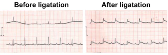
Fig. 1 shows an example of a limb-lead ECG be-fore and after ligation of the left anterior descending coronary artery on thoracotomy under artificial
res-piration. No ST elevation was noted on the lead I and II ECG before the ligation. Myocardial ischemia-associated ST elevation was noted on the lead II ECG after the ligation. The success of the ligation of the anterior descending branch of the coronary artery was confirmed, as evidenced by ST elevation on the ECG in all rats.
The left anterior descending coronary artery was visually confirmed, and ligated using a suturing needle with thread (vascular round needle, No. 008 mm, black braid silk No. 6-0, Natsume Seisakusho, Tokyo, Japan). After ligation, ST elevation was con-firmed on ECG, and 12 rats without removal of the ligation (complete ligation group ; MI group) and 12 rats in which the ligation was removed for reper-fusion 30 minutes after ligation (reperreper-fusion group ; R group) were prepared. For reperfusion of the coro-nary artery, a 0.6-mm rubber thread was placed under the ligation thread so that the ligation thread could be easily cut. The opened thorax was closed by suturing with 2.0 silk thread (alfresa, Tokyo, Ja-pan), and the rats were maintained at the animal experiment facility of The University of Tokushima. The present study was approved by The Univer-sity of Tokushima Animal Experiment Committee, and performed in accordance with The University of Tokushima Guidelines for Animal Experiments (enforced on June 30, 1989, revised on April 15, 2005) in conformity with the Law concerning Ani-mal Protection and Management (Law No. 105 in 1973, partially revised on December 22, 1999) and the Standards concerning Animal Maintenance and Storage for Animal Experiments (Notice from the Prime Minister’s Office No. 6, March 1980, partially revised on May 28, 2002).
2) Grouping of animals for the administration of G-CSF and physiological saline
The MI and R groups were divided into two groups that received an intraperitoneal injection of 0.5 ml of physiological saline daily for 5 days (MI-C and R-C groups, n=6 each) and groups that received an intraperitoneal injection of recombinant G-CSF (nar-tograstim, Neu-up!, Kyowa Hakko Industry Ltd., Tokyo, Japan) at 100 μg/kg/day (single dose : 25
μg of G-CSF dissolved in 0.5 ml of physiological
saline) daily for 5 days (MI-G and R-G groups, n=6 each). G-CSF was administrated 60 min after the preparation of the myocardial infarction.
3) Echocardiography
For the ultrasonic instrument and probe, an Aplio
Fig. 1 Example of a limb-lead ECG before and after ligation of the left anterior descending coronary artery on thoracotomy under artificial respiration.
K. Ieishi, et al. G-CSF effect on reperfusion ischemia
80 (Toshiba Medical Systems SSA-770A) and a 10 MHz linear transducer were used. Echocardiogra-phy was performed by the M and B modes 4 weeks after the administration of G-SCF or saline in the MI-G, MI-C, R-G, and R-C groups, and the left tricular end-diastolic dimension (LVDd), left ven-tricular diameter at end systole (LVDs), interven-tricular septum thickness (LVST) and left ventricu-lar posterior wall thickness (LVPWT) were calcu-lated. These data were stored on a hard disc. For echocardiography, the minimum dose of pentobar-bital (0.2 ml) was intraperitoneally injected for an-esthesia. When the depth of anesthesia was insuf-ficient, an additional 0.1 ml was administered. LVDd, the proportion of the left ventricular wall thickness ([infarct wall thickness]/[non-infarct wall thick-ness]), and the left ventricular ejection fraction (EF) were compared among the MI-G, MI-C, R-G, and R-C groups after 4 weeks. Echocardiography was measured under these conditions ; room tempera-ture, 20 degrees Celsius and a heart rate between 300 to 400/min.
4) Histological examination
After echocardiography, the animals were sacri-ficed by an intraperitoneal injection of 2 ml of pen-tobarbital (overdose anesthesia). The heart was ex-cised, a portion was fixed with 10% formalin, and paraffin-embedded sections were prepared. These sections were stained with hematoxylin-eosin (HE) and Azan for a histological examination. To meas-ure the infarct area, one slice of the central part of the ventricle, was manually measured using ImageJ (Image Processing and Analysis in Java, Wayne Ras-band, National Institutes of Health, USA). The in-farct area ratio (%) on Azan staining were compared among the MI-G, MI-C, R-G, and R-C groups. 5) Statistical analysis
The measured values are presented as the means
±standard deviation (SD) and were analyzed
us-ing the unpaired t-test. P<0.05 was regarded as sig-nificant.
RESULTS
1) Echocardiography findings
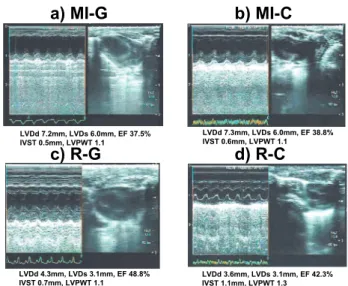
Fig. 2 shows examples of echocardiography find-ings for the MI-G (panel a), MI-C (panel b), R-G (panel c), and R-C (panel d) groups. In the MI-G group (panel a), the anterior wall was thinned on the
B mode, and akinetic motion was noted on the M mode. In the MI-C group (panel b), thinning of the anterior wall on the B mode was milder than that in the MI-G group, and the anterior wall motion re-mained hypokinetic on the M mode. In the R-G group (panel c), the thickness of the anterior wall was maintained on the B mode, and no abnormal wall motion was noted. In the R-C group (panel d), the anterior wall thickness was normal on the B mode, and the anterior wall motion was normal on the M mode.
Table 1 shows a comparison of the echocardiogra-phy findings among the MI-G, MI-C, R-G, and R-C groups. LVDd increased in the MI-G (7.1±1.4 mm)
and MI-C (7.1±1.1 mm) groups, showing no
sig-nificant difference between the groups. In contrast, LVDd in the R-G group (3.6±0.4 mm) was
signifi-cantly smaller than that in the R-C group (4.2 ±
0.8 mm).
LVDs showed a tendency similar to that for LVDd. LVDs increased in the MI-G (6.0 ± 1.2 mm) and
MI-C (6.1±1.4 mm) groups, showing no significant
difference between the groups. In contrast, LVDs in the R-G group (1.4±0.4 mm) was significantly
smaller than that in the R-C group (2.1±0.6 mm).
The proportion of left ventricular wall thickness (infarct wall thickness/non-infarct wall thickness) was 0.5 ±0.1 and 0.4±0.2 in the MI-G and MI-C
groups, respectively, showing thinning of the inter-ventricular septum, and no significant difference was
Fig. 2 Examples of echocardiography findings for the MI-G (panel a), MI-C (panel b), R-G (panel c), and R-C (panel d) groups.
LVDd, left ventricular end-diastolic dimension ; LVDs, left ven-tricular diameter at end systole ; IVST, intervenven-tricular septum thickness ; LVPWT, left ventricular posterior wall thickness ; EF, left ventricular ejection fraction.
noted between the groups. In contrast, the propor-tion for the R-G group (0.9±0.2) was significantly
higher than that for the R-C group (0.7±0.4).
EF was markedly decreased in the MI-G (38.6±
10.2%) and MI-C (35.4±8.8%) groups, showing no
significant difference between the groups. In con-trast, sufficient EF was maintained in the R-G (64.4
±12.4%) and R-C (52.8±10.8%) groups, and the EF
in the R-G group was significantly higher than that in the R-C group.
2) Histological findings
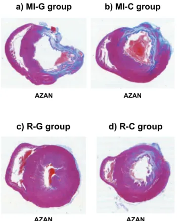
Fig. 3 shows a comparison of Azan-stained
ven-tricular cross sections among the MI-G (panel a), MI-C (panel b), R-G (panel c), and R-C (panel d) groups. In the MI-G and MI-C groups, blue-stained collagen fibers, considered to be the fibrosed region of myocardial infarction-associated necrosis, were noted. In contrast, the ventricular wall thickness was relatively maintained in the R-G and R-C groups, but collagen fibers were noted in spaces between car-diomyocytes.
Fig. 4 shows the histological findings on HE and Azan staining for the MI-G (panel a), MI-C (panel b), R-G (panel c), and R-C (panel d) groups. In the MI-C group, decreased cytoplasmic muscle fibers and the formation of many fissures between the heart muscle fibers were noted on HE staining. On Azan staining, numerous blue-stained collagen fi-bers were noted. In the MI-G group, the decrease in cytoplasmic muscle fibers was less than that in the MI-C group, but numerous fissures were formed between the heart muscle fibers. Azan staining showed numerous blue-stained collagen fibers, as in the MI-C group. In R-G group, the cytoplasmic muscle fibers stained red with eosin were homoge-nously maintained on HE staining, and no fissure formation along cardiomyocytes was noted. No
blue-Fig. 3 Comparison of Azan-stained ventricular cross sections among the MI-G (panel a), MI-C (panel b), R-G (panel c), and R-C (panel d) groups.
Fig. 4 Histological findings on HE and Azan staining for the MI-G (panel a), MI-C (panel b), R-G (panel c), and R-C (panel d) groups.
Table 1 Comparison of the echocardiography findings among the MI-G, MI-C, R-G, and R-C groups
group MI-G MI-C R-G R-C
LVDd (mm) 7.1±1.4 7.1±1.1 3.6±0.4* 4.2±0.8
LVDs (mm) 6.0±1.2 6.1±1.4 1.4±0.4* 2.1±0.6
IVST/LVPWT 0.5±0.1 0.4±0.2 0.9±0.2* 0.7±0.4
EF(%) 38.6±10.2 35.4±8.8 64.4±12.4* 52.8±10.8
LVDd, left ventricular end-diastolic dimension ; LVDs, left ven-tricular diameter at end systole ; IVST, intervenven-tricular septum thickness ; LVPWT, left ventricular posterior wall thickness ; EF, left ventricular ejection fraction.
mean±SD,*p<0.05 vs R-C group
K. Ieishi, et al. G-CSF effect on reperfusion ischemia
stained collagen fibers were noted on Azan staining. In the R-C group, the cytoplasmic muscle fibers were stained red with eosin, numerous fissures were formed along the heart muscle fibers, and the in-terstitium was widely infiltrated by inflammatory cells dominated by monocytes on HE staining. Blue-stained collagen fibers were noted on Azan stain-ing, but to a lesser degree than that noted in the MI-G and MI-C groups.
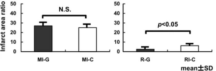
Fig. 5 shows the infarct area ratio (%). The infarct area ratio (%) was high in the MI-G (26.9±6.1%)
and MI-C (25.2±6.3%) groups, showing no
signifi-cant difference between the groups. In contrast, the ratio was significantly lower in the R-G group (2.6
±1.0%) than in the R-C group (6.2±1.1%).
DISCUSSION
G-CSF administration did not change the infarct area ratio, left ventricular wall thickness, or left ven-tricular ejection fraction in rats with a complete liga-tion of the coronary artery, whereas G-CSF signifi-cantly reduced the infarct area ratio and left ven-tricular thickness and improved the left ventricu-lar ejection fraction in rats with reperfusion of the ligation-induced infarction.
The areas of the myocardial infarction by ligation release and complete ligation are different in this animal model. Moreover, myocardial infarction is caused by different mechanisms, because the MI-C group and R-MI-C group are coagulative necrosis and ischemia reperfusion models, respectively. The find-ings in the present study, therefore, suggest that G-CSF is effective for treating cardiac muscle cell disorders by ischemia reperfusion.
In recent years with the marked advances in re-generative medicine, the early clinical application of myocardial regenerative treatment would be ex-pected to be promising for increasing myocardial contractility. Regenerative medicine for myocardial
infarctions is roughly divided into cellular treatments aimed at vascular regeneration (2-4) and myocardial regeneration in which cells are transplanted in the infarct region of the heart muscle (5, 6). As a clini-cal application of vascular regeneration, G-CSF ad-ministration has been reported to significantly in-crease collateral flow in patients with coronary ar-terial disease, showing a vascularization effect (7).
Various approaches for myocardial regeneration have been investigated, and studies of regenerated cardiomyocytes, pluripotent stem cells, and skele-tal myoblasts using embryonic and bone marrow so-matic stem cells as tools are currently underway. Myocardial sheets have been developed by tissue engineering groups (8-11). No regeneration of car-diomyocytes or arterialization in the border zone was found in this experiment and this does not prove that G-CSF does not have a regenerative effect.
The possibility of treatment with cytokines, such as G-CSF, during the acute phase of a myocardial infarction to induce the migration of stem cells to the infarct region and regeneration of the injured myocardial region has been reported (12). Orlic, et
al. (1) reported that the concomitant administra-tion of G-CSF and stem cell factor mobilized bone marrow cells to the infarct tissue at a high rate in a mouse acute myocardial infarction model, and that these cells regenerated cardiomyocytes and vascu-lar endothelial cells, reducing the myocardial infarct size by 40% and the mortality rate by 68%.
Minatoguchi, et al. (13) reported that the admini-stration of G-CSF alone at a dose close to the typi-cal clinitypi-cal dose after the preparation of a myocar-dial infarction had a therapeutic effect, in which G-CSF significantly increased cardiomyocytes, vascu-lar endothelium, vascuvascu-lar smooth muscle, and my-ofibroblasts, which may have reduced the infarct area, inhibited cardiac remodeling, and improved cardiac function, but very few bone marrow-derived cardiomyocytes were found (13, 14), suggesting that G-CSF not only promotes myocardial regeneration by the migration of bone marrow cardiomyocytes but also directly protects existing cardiomyocytes.
Harada, et al. (15) also reported that G-CSF pro-motes survival of cardiac myocytes and prevents left ventricular remodeling after myocardial infarc-tion through the funcinfarc-tional communicainfarc-tion between cardiomyocytes and noncardiomyocytes. Therefore, in this study, the mechanism of G-CSF effect for treating cardiac muscle cell disorders by ischemia reperfusion is not coronary arteries patency, but the direct action on cardiomyocytes after myocardial
in-Fig. 5 Infarct area ratio (%).
farction.
G-CSF is clinically used as a therapeutic drug for the treatment of leukemia and leucopenia after can-cer chemotherapy. G-CSF has various actions, and the major actions are to promote the proliferation and differentiation of bone marrow precursor cells and to immobilize bone marrow stem cells. G-CSF administration promotes the induction of stem cells in the bone marrow and their release into the cir-culation in rats.
Since G-CSF can be readily administere, such as by subcutaneous and intravenous injection, it is less invasive for patients. In addition, fewer ethical prob-lems are involved because it mobilizes the patient’s own cells. Thus, G-CSF may become a new thera-peutic strategy for treating myocardial infarctions in the near future (16). However, it has been reported that the stent re-stenosis rate increased after G-SCF administration, although it improved cardiac func-tion after coronary arterial stenting for myocardial infarction (17).
In the present study, G-CSF was administered in-traperitoneally, and a sufficient effect may be pro-duced by intravenous administration, if the blood vessel responsible for the infarction is patent, al-though it cannot be directly injected into the heart muscle. Because of these possible actions, no sig-nificant differences were noted in the infarct area ratio, left ventricular wall thickness, or left ventricu-lar ejection fraction between the MI-G and MI-C groups with a complete ligation of the coronary ar-tery, whereas a significant reduction in the infarct area and improvement of the left ventricular wall thickness and left ventricular ejection fraction were noted in R-G and R-C groups with reperfusion. Based on these findings, G-CSF is effective for treating car-diac muscle cell disorders by ischemia reperfusion.
REFERENCES
1. Orlic D, Kajstura J, Chimenti S, Limana F, Jakoniuk I, Quaini F, Nadal-Ginard B, Bodine DM, Leri A, Anversa P : Mobilized bone mar-row cells repair the infarcted heart, improving function and survival. Proc Natl Acad Sci USA 98 : 10344-49, 2001
2. Takeshita S, Zheng LP, Brogi E, Kearney M, Pu LQ, Bunting S, Ferrara N, Symes JF, Isner JM : Therapeutic angiogenesis. A single intraar-terial bolus of vascular endothelial growth factor augments revascularization in a rabbit ischemic
hind limb model. J Clin Invest 93 : 662-70, 1994 3. Morishita R, Nakamura S, Hayashi S, Taniyama Y, Moriguchi A, Nagano T, Taiji M, Noguchi H, Takeshita S, Matsumoto K, Nakamura T, Higaki J, Ogihara T : Therapeutic angiogene-sis induced by human recombinant hepatocyte growth factor in rabbit hind limb ischemia model as cytokine supplement therapy. Hyper-tension 33 : 1379-84, 1999
4. Losordo DW, Vale PR, Isner JM : Gene therapy for myocardial angiogenesis. Am Heart J 138 (2 Pt 2) : S132-41, 1999
5. Menasche P, Hagege AA, Scorsin M, Pouzet B, Desnos M, Duboc D, Schwartz K, Vilquin JT, Marolleau JP : Myoblast transplantation for heart failure. Lancet 357 : 279-80, 2001
6. Kamihata H, Matsubara H, Nishiue T, Fujiyama S, Tsutsumi Y, Ozono R, Masaki H, Mori Y, Iba O, Tateishi E, Kosaki A, Shintani S, Murohara T, Imaizumi T, Iwasaka T : Implantation of bone marrow mononuclear cells into ischemic myo-cardium enhances collateral perfusion and re-gional function via side supply of angioblasts, angiogenic ligands, and cytokines. Circulation 104 : 1046-52, 2001
7. Seiler C, Pohl T, Wustmann K, Hutter D, Nicolet PA, Windecker S, Eberli FR, Meier B : Promotion of collateral growth by granulocyte-macrophage colony-stimulating factor in pa-tients with coronary artery disease : a random-ized, double-blind, placebo-controlled study. Cir-culation 104 : 2012-7, 2001
8. Badylak SF, Kochupura PV, Cohen IS, Doronin SV, Saltman AE, Gilbert TW, Kelly DJ, Ignotz RA, Gaudette GR : The use of extracellular ma-trix as an inductive scaffold for the partial re-placement of functional myocardium. Cell Trans-plant 15(Suppl 1) : S29-40, 2006
9. Miyahara Y, Nagaya N, Kataoka M, Yanagawa B, Tanaka K, Hao H, Ishino K, Ishida H, Shimizu T, Kangawa K, Sano S, Okano T, Kitamura S, Mori H : Monolayered mesenchy-mal stem cells repair scarred myocardium after myocardial infarction. Nat Med 12 : 459-65, 2006 10. Miyagawa S, Sawa Y, Sakakida S, Taketani S, Kondoh H, Memon IA, Imanishi Y, Shimizu T, Okano T, Matsuda H : Tissue cardiomyoplasty using bioengineered contractile cardiomyocyte sheets to repair damaged myocardium : their integration with recipient myocardium. Trans-plantation 80 : 1586-95, 2005
11. Menasche P, Hagege AA, Scorsin M, Pouzet
K. Ieishi, et al. G-CSF effect on reperfusion ischemia
B, Desnos M, Duboc D, Schwartz K, Vilquin JT, Marolleau JP : Myoblast transplantation for heart failure. Lancet 357 : 279-80, 2001
12. Lim SY, Kim YS, Ahn Y, Jeong MH, Rok LS, Kim JH, Kim KH, Park HW, Kim W, Cho JG, Park JC, Kang PM, Schwartz RS, Kang JC : The effects of granulocyte-colony stimulating factor in bare stent and sirolimus-eluting stent in pigs following myocardial infarction. Int J Cardiol 2006 ; [Epub ahead of print]
13. Minatoguchi S, Takemura G, Chen XH, Wang N, Arai M, Misao Y, Lu C, Uno Y, Koda M, Goto K, Takahashi T, Kosai K : Myocardial in-farction itself induces cardiomyocyte regenera-tion from bone marrow cells, and post-ischemic G-CSF treatment improves cardiac dysfunc-tion via acceleradysfunc-tion of the process. Circuladysfunc-tion 106(suppl II) : II-132, 2002
14. Minatoguchi S, Takemura G, Chen XH, Wang N, Uno Y, Koda M, Arai M, Misao Y, Lu C, Koji S, Goto K, Komada A, Takahashi T, Kosai K, Fujiwara T, Fujiwara H : Acceleration of the healing process and myocardial regeneration may be important as a mechanism of improve-ment of cardiac function and remodeling by postinfarction granulocyte colony-stimulating
factor treatment. Circulation 109 : 2572-80, 2004 15. Harada M, Qin Y, Takano H, Minamino T, Zou Y, Toko H, Ohtsuka M, Matsuura K, Sano M, Nishi J, Iwanaga K, Akazawa H, Kunieda T, Zhu W, Hasegawa H, Kunisada K, Nagai T, Nakaya H, Yamauchi-Takihara K, Komuro I : G-CSF prevents cardiac remodeling after myo-cardial infarction by activating the Jak-Stat path-way in cardiomyocytes. Nat Med 11 : 305-311, 2005
16. Takano H, Ohtsuka M, Akazawa H, Toko H, Harada M, Hasegawa H, Nagai T, Komuro I : Pleiotropic effects of cytokines on acute myo-cardial infarction : G-CSF as a novel therapy for acute myocardial infarction. Curr Pharm Des 9 : 1121-7, 2003
17. Kang HJ, Kim HS, Zhang SY, Park KW, Cho HJ, Koo BK, Kim YJ, Soo Lee D, Sohn DW, Han KS, Oh BH, Lee MM, Park YB : Effects of intracoronary infusion of peripheral blood stem-cells mobilised with granulocyte-colony stimulating factor on left ventricular systolic function and restenosis after coronary stent-ing in myocardial infarction : the MAGIC cell randomised clinical trial. Lancet 363 : 751-6, 2004