Original article (Secondary publication)
Reliability and validity of the patient
disability-oriented diagnostic nomenclature system for
prosthetic dentistry
§
Yoshizo Matsuka DDS, PhD
a,b,*
, Yoshiyuki Hagiwara DDS, PhD
c,
Katsushi Tamaki DDS, PhD
d, Hisahiro Takeuchi DDS, PhD
b,
Masanori Fujisawa DDS, PhD
a,e, Takahiro Ono DDS, PhD
f,
Yoshihiro Tsukiyama DDS, PhD
g, Kan Nagao DDS, PhD
h,
Kazuhiro Tsuga DDS, PhD
i, Hideki Aita DDS, PhD
a,j,
Hisatomo Kondo DDS, PhD
k, Kenji Fueki DDS, PhD
l,
Hiroaki Tsukasaki DDS, PhD
m, Keisuke Nishigawa DDS, PhD
b,
Shogo Ozawa DDS, PhD
a,n, Rika Kuwatsuru DDS, PhD
a,g,
Hajime Minakuchi DDS, PhD
a,o, Toshimitsu Iinuma DDS, PhD
a,p,
Takashi Matsuura DDS, PhD
a,q, Kanji Ishibashi DDS, PhD
r,
Shigehisa Fujii DDS, PhD
s, Toshihiro Hirai DDS, PhD
t,
Keiichi Sasaki DDS, PhD
u, Hirofumi Yatani DDS, PhD
v,
Yoshimasa Igarashi DDS, PhD
w, Yuji Sato DDS, PhD
x,
Tetsuo Ichikawa DDS, PhD
h, Tetsuo Yamamori DDS, PhD
y,
Takuo Kuboki DDS, PhD
o, Kazuyoshi Baba DDS, PhD
m,
Kiyoshi Koyano DDS, PhD
g, Hironobu Sato DDS, PhD
a,q,
Hideo Matsumura DDS, PhD
caClinical Guideline Committee, Japan Prosthodontic Society, Japan
bDepartment of Stomatognathic Function and Occlusal Reconstruction, [5_TD$DIFF]Graduate School of Biomedical Sciences, Tokushima University[1_TD$DIFF], Japan
c
Department of Fixed Prosthodontics, Nihon University School of Dentistry, Japan
dDepartment of Prosthodontic Dentistry for Function of TMJ and Occlusion, Kanagawa Dental University, Japan eDivision of Fixed Prosthodontics, Meikai University School of Dentistry, Japan
fDivision of Comprehensive Prosthodontics, Niigata University Graduate School of Medical and Dental Sciences, Japan gSection of Implant and Rehabilitative Dentistry, Faculty of Dental Science, Kyushu University, Japan
hDepartment of Oral & Maxillofacial Prosthodontics,[5_TD$DIFF]Graduate School of Biomedical Sciences, Tokushima University[1_TD$DIFF], Japan
iDepartment of Advanced Prosthodontics, Institute of Biomedical & Health Sciences, Hiroshima University, Japan
§
A Japanese-language version of this report has been published previously[4], and this is an English translation.
* Corresponding author at: Department of Stomatognathic Function and Occlusal Reconstruction,[5_TD$DIFF]Graduate School of Biomedical Sciences, Tokushima University[1_TD$DIFF], 3-18-15 Kuramoto-cho, Tokushima 770-8504, Japan. Tel.: +81 88 633 7350; fax: +81 88 633 7391.
E-mail address:matsuka@tokushima-u.ac.jp(Y. Matsuka).
Abbreviations: JPS, Japan Prosthodontic Society; DNS, diagnostic nomenclature system; CGC, Clinical Guideline Committee.
Available online at
www.sciencedirect.com
Journal of Prosthodontic Research
journal homepage:www.elsevier.com/locate/jporhttp://dx.doi.org/10.1016/j.jpor.2016.06.005
1883-1958/# 2016 The Authors. Published by Elsevier Ltd on behalf of Japan Prosthodontic Society. This is an open access article under the CC BY-NC-ND license (http://creativecommons.org/licenses/by-nc-nd/4.0/).
jDepartment of Occlusion and Removable Prosthodontics, Health Sciences University of Hokkaido School of Dentistry, Japan
kDepartment of Prosthodontics and Oral Implantology, School of Dentistry, Iwate Medical University, Japan lRemovable Partial Denture Prosthodontics, Tokyo Medical and Dental University, Japan
mDepartment of Prosthodontics, Showa University School of Dentistry, Japan
nDepartment of Removable Prosthodontics, School of Dentistry, Aichi Gakuin University, Japan o
Department of Oral Rehabilitation and Regenerative Medicine, Okayama University Graduate School of Medicine, Dentistry and Pharmaceutical Sciences, Japan
pDepartment of Complete Denture Prosthodontics, Nihon University School of Dentistry, Japan qSection of Fixed Prosthodontics, Department of Oral Rehabilitation, Fukuoka Dental College, Japan rSchool of Dentistry, Iwate Medical University, Japan
sDepartment of Partial and Complete Denture, The Nippon Dental University School of Life Dentistry, Japan t
Health Sciences University of Hokkaido, Japan
uDivision of Advanced Prosthetic Dentistry, Tohoku University Graduate School of Dentistry, Japan vDepartment of Fixed Prosthodontics, Osaka University Graduate School of Dentistry, Japan wOsaka Dental University, Japan
xDepartment of Geriatric Dentistry, Showa University School of Dentistry, Japan y
Department of Prosthetic Dentistry, Ohu University School of Dentistry, Japan
1.
Introduction
Current disease names used for prosthodontic treatment generally reflect oral condition, and are not based on logical diagnoses. The terminology includes dental caries, missing teeth, ill-fitting or fracture of artificial denture, irregularity of residual ridge (such as abnormality of basal seat mucosa), decubital ulcer or fibrous proliferation of palatal mucosa. In some cases, missing teeth do not cause any disability in daily life, and terms that simply describe the irregular condition of
oral structures do not provide insight into the underlying pathogenesis.
On January 18, 2008, a conference on disease nomencla-ture for prosthodontic treatment was held to discuss new diagnostic strategies based on a patient-oriented system[1] and to specify treatment tailored to patient disability[2]. At this conference, our group proposed a new diagnostic nomenclature system (DNS) for prosthodontic dentistry, based on pathogenesis and etiology (Fig. 1), that would enhance prosthodontic treatment in Japan. This system specifies patient disability and indicates the factor causing
a r t i c l e
i n f o
Article history:
Received 9 January 2016 Received in revised form 24 May 2016
Accepted 23 June 2016 Available online 8 August 2016
Keywords:
Diagnostic nomenclature system Prosthodontic treatment Reliability
Validity
a b s t r a c t
Purpose: The Japan Prosthodontic Society (JPS) has proposed a new diagnostic nomenclature system (DNS), based on pathogenesis and etiology, to facilitate and improve prosthodontic treatment. This system specifies patient disability and the causative factor (i.e. ‘‘B (disability) caused by A (causative factor)’’). The purpose of this study was to examine the reliability and validity of this DNS.
Study selection: The JPS Clinical Guideline Committee assessed mock patient charts and formulated disease names using the new DNS. Fifty validators, comprising prosthodontic specialists and dental residents, made diagnoses using the same patient charts. Reliability was evaluated as the consistency of the disease names among the validators, and validity was evaluated using the concordance rate of the disease names with the reference disease names.
Results: Krippendorff’s a was 0.378 among all validators, 0.370 among prosthodontic spe-cialists, and 0.401 among dental hospital residents. Krippendorff’s a for 10 validators (3 specialists and 7 residents) with higher concordance rates was 0.524. Two validators (1 specialist and 1 resident) with the highest concordance rates had a Krippendorff’s a of 0.648. Common disease names had higher concordance rates, while uncommon disease names showed lower concordance rates. These rates did not show correlation with clinical experience of the validator or time taken to devise the disease name.
Conclusions: High reliability was not found among all validators; however, validators with higher concordance rates showed better reliability. Furthermore, common disease names had higher concordance rates. These findings indicate that the new DNS for prosthodontic dentistry exhibits clinically acceptable reliability and validity.
#2016 The Authors. Published by Elsevier Ltd on behalf of Japan Prosthodontic Society. This is an open access article under the CC BY-NC-ND license (http:// creativecommons.org/licenses/by-nc-nd/4.0/).
[(Fig._1)TD$FIG]
Basic disease name structural format: B (disability) caused by A (causative factor) Examples of A (causative factor)
(1) Abnormal condition of tooth:
Dental caries, fracture of tooth crown, fracture of tooth root, tooth crack, attrition, abrasion, dentin hyperesthesia, discolored tooth, stained tooth, enamel hypoplasia, etc. (2) Dental pulp and periodontal disease:
Pulpitis, marginal periodontitis, apical periodontitis, hypermobility of tooth, traumatic occlusion, etc.
(3) Defect:
Missing tooth (with tooth number), defect of jaw, defect of tongue, etc. (4) Malocclusion:
Maxillary protrusion, mandibular protrusion, bimaxillary protrusion, occlusal interference, open bite, cross bite, crowding, inclined tooth, rotation, extrusion, etc. (5) Temporomandibular joint disease:
Temporomandibular disorders, analogical disease of temporomandibular disorders (fibromyalgia, rheumatism, hyperplasia of coronoid process, elongated styloid process, burning mouth syndrome) , etc.
(6) Problem with prosthesis:
Problem with crown restoration, fracture of crown restoration, problem with fixed prosthesis, fracture of fixed prosthesis, problem with partial denture, fracture of partial denture, problem with complete denture, fracture of complete denture, etc.
(7) Other factors Examples of B (disability) (1) Masticatory dysfunction (2) Swallowing disability (3) Pronunciation disability (4) Esthetic disturbance
(5) Abnormal sensation: abnormal sensation, paresthesia or dysesthesia with prosthesis (6) Pain
(7) Ahead sick (patient might be sick in the future): health condition that might cause illness without appropriate treatment.
(8) Other disability
Procedure for diagnosis
Medical interview, consultation, examination
Evaluate chief complaint of the patient through medical interview and consultation. Then carry out appropriate examinations of the patient. Recognize etiopathology using data from the interview or examinations.
Diagnosis:
Diagnose disease with the following procedure:
(1) List all disease names for the patient. If patient has symptoms in a localized area, describe the regions and tooth number.
Examples: 1 Esthetic disturbance caused by fracture of facing crown; Masticatory dysfunction caused by temporomandibular disorders.
the disability (i.e. ‘‘B (disability) caused by A (causative factor)’’).
Before clinical application of this DNS, its reliability and validity should be studied in the clinical setting. We prepared mock patient charts, and the Clinical Guideline Committee (CGC) of the Japan Prosthodontic Society (JPS) examined these patient charts to formulate diagnoses and devise disease names (used as reference). The same patient charts were also studied by validators that consisted of specialists in prostho-dontics of the JPS and dental residents from university hospitals. Reliability of the DNS was evaluated by the consistency of the disease names among the validators, and validity was evaluated using the concordance rates of the disease names with the reference disease names devised by the CGC.
2.
Materials and methods
This research was approved by the Epidemiological Research Ethics Committee of Okayama University Graduate School of Medicine, Dentistry and Pharmaceutical Sciences. Mock patient charts were prepared from clinical information for real patients who visited Crown Bridge Prosthodontics, Okayama University Hospital from January 1 to June 30, 2010. A poster was displayed at Crown Bridge Prosthodontics asking for cooperation for this research project on prosthodontic disease names (Fig. 2). This poster also contained information about the purpose and methods of this study. Patient information, including age, gender, family history, past history, clinical findings and radiography, was used for the research with anonymity.
[(_)TD$FIG]
(2) If multiple disease names on the list contain the same B (disability), unite these disease names. Single disease names should not contain more than three As (causative factors) or Bs (disabilities).
Examples: 567 Pain and masticatory dysfunction caused by marginal periodontitis and temporomandibular disorders; Masticatory dysfunction caused by fracture of 765 partial denture; 5 Esthetic disturbance, masticatory dysfunction and abnormal sensation caused by fracture of prosthesis.
(3) For patients with an unclear causative factor, use disease name “B (disability) probably caused by A (causative factor)”. For patients requiring diagnosis by physicians, use the same expression.
Examples: 7 Pain and masticatory dysfunction probably caused by apical periodontitis; Sleep disorder probably caused by psychiatric factor.
[(Fig._2)TD$FIG]
We request your cooperation for a study on the reliability and validity of the diagnostic nomenclature system for prosthodontic dentistry (for patients who visited our clinic and had prosthodontic treatment from January 1 to June 30, 2010).
Research institute: Oral Rehabilitation and Regenerative Medicine, Okayama University Graduate School of Medicine, Dentistry and Pharmaceutical Sciences
Primary investigator: Professor Takuo Kuboki, Department of Oral Rehabilitation and Regenerative Medicine, Okayama University Graduate School of Medicine, Dentistry and Pharmaceutical Sciences
Corroborative investigator: Associate Professor Yoshizo Matsuka, Department of Oral Rehabilitation and Regenerative Medicine, Okayama University Graduate School of Medicine, Dentistry and Pharmaceutical Sciences
1. Objective of this study
We are developing a new disease naming system for prosthodontic treatment such as denture and crown-bridge restoration. The disease name expresses “B (disability) caused by A (causative factor)”. It is a logical disease naming system that should help facilitate appropriate treatment. In the current study, a group of dentists will make diagnoses based on patient charts according to this diagnostic nomenclature system. We will investigate how these diagnosed disease names will be concordant, and also test the validity of this naming system.
2. Methods
Participants: Patients who visited Crown Bridge Prosthodontics, Okayama University Hospital.
Research period: From January 1 to June 30, 2010.
3. Procedures
We will make mock patient charts using clinical information for patients who visited the Department of Prosthodontics in this hospital from January 1 to June 30, 2010. Dentists who belong to Okayama University, Meikai University, Nihon University, Kanagawa Dental University, Osaka University, Hiroshima University, Tokushima University and Kyushu University will make their diagnoses based on these patient charts
using the new diagnostic nomenclature system. These dentists will be selected randomly, and we will investigate whether the diagnoses by these dentists are concordant or not.
4. Materials
Your clinical information such as age, gender, family history, medical history, examination findings and radiography images will be used to make mock patient charts. Personal information that might distinguish you will be carefully deleted, and only anonymous data will be used for the investigation.
5. Protection of information
All information in this study will be carefully guarded in the Department of Oral Rehabilitation and Regenerative Medicine, Okayama University Graduate School of Medicine, Dentistry and Pharmaceutical Sciences. The computer that stores the electronic information will be protected with a password, and other information will be kept in a locked safe box. The results of this investigation will be published at an academic meeting, and all personal information that might distinguish any individual participant will be hidden. If you have any questions, pleases contact us at the following address. If you do not want to be involved in this investigation, please let us know at the same address by January 31, 2010. We will not include any patient who does not to this investigation. Whether you participate or not, you will not have any advantage or disadvantage regarding the clinical service at this hospital. If you do not object, we will take it as your sign of consent to participate in this investigation.
Contact address:
Crown Bridge Prosthodontics, Okayama University Hospital Takuo Kuboki, Yoshizo Matsuka
TEL: 086-235-6682; FAX: 086-235-6684
Fig. 2 – Poster asking for patient cooperation in this study on the reliability and validity of the diagnostic nomenclature system for prosthetic dentistry.
[(Fig._3)TD$FIG]
Patient 1: 81-year-old male Chief complaints:
Bad looking with missing mandibular incisor Difficulty chewing with left mandibular molar
Occasional difficulty chewing caused by loose right mandibular molar History of present illness:
The patient had prosthodontic treatment with placement of removable mandibular denture 3 months ago at his local dental clinic. He did not feel good with the denture, and felt even when he occluded with it. He therefore stopped using the denture. One month ago, he visited his home physician for consultation on his dental problem, and the doctor referred him to the university hospital.
Past history:
Reflux esophagitis: Examined 30 years ago. Visiting physician when he felt bad. Cataract: Examined 8 years ago. He had cataract operation.
Blood pressure: 163/88 mmHg
Medications: Omeprazole, Naftopidil, Bifidobacterium, Mepenzolate bromide, Diclofenac sodium, Rebamipide
Allergy: nothing particular Bleeding tendency: nothing particular
Local anesthesia at dental clinic: Yes (nothing remarkable) Experience of tooth extraction: Yes (nothing remarkable) Present illness:
Temporary crown was attached on the left mandibular central incisor
1 temporary prosthesis.
Diagnosis:
Esthetic disturbance caused by mandibular frontal tooth missing Masticatory dysfunction caused by mandibular molar missing Masticatory dysfunction caused by marginal periodontitis
[(_)TD$FIG]
Patient 2: 47-year-old female Chief complaints:
Abnormal sensation on face Headache
History of present illness:
The patient had a car accident 30 years ago and lost her incisors at that time. Subsequently, she started to have a strange sensation on her face and the temporomandibular joint area (typically on the right side). She was aware of tooth clenching during both daytime and nighttime. Because she had terrible headaches in the morning, she took Loxoprofen as soon as she woke up. She had tried a mouthpiece, but it did not make her symptoms better. Because she had severe pain on the face and temporomandibular joint, she occasionally took more than 100 analgesic tablets per month. When she consulted a local doctor about her symptons, the doctor recommended her to undergo examination at the university hospital and referred her to our clinic.
Past history:
Cushing syndrome: One year ago, she had left adrenalectomy surgery. Blood pressure: 150/84 mmHg
Pulse rate: 64 Weight: 57 kg
Medication: Hydrocortisone, Loxoprofen Allergy: none
Infectious disease: none Bleeding tendency: none
Local anesthesia at dental clinic: Yes (nothing remarkable) Experience of tooth extraction: Yes (nothing remarkable) Present illness:
She became worried about the space between her central incisors. Because she tended to breathe air through that space, she clenched her teeth to stop the breathing habit during daytime. She had severe headache localized to the right temple, with a throbbing pain that worsened while taking a bath. She felt nausea and had photosensitivity. Drinking 5–6 cups of coffee a day and smoking a pack of cigarettes a day.
Tenderness was positive on both sides of the TMJ, masseter and temple. TMJ pain was positive during jaw opening and closing. Joint noise was negative. Range of maximum jaw opening without pain: 47 mm; maximum voluntary jaw opening: 52 mm; maximum passive jaw opening: 53 mm; no lateral deviation was found during jaw opening. Clear pressed mark was found on tongue and cheek mucosa.
No tenderness was found at the elbow, knee or ankle joint. Diagnosis:
Pain caused by temporomandibular disorders through bruxism Migraine caused by excess intake of analgesic, caffeine, nicotine
Patient 3: 72-year-old female Chief complaint:
Pain at mandibular incisor
History of present illness:
One year ago, a resin-facing crown was inserted on the mandibular incisor at a local dental clinic, and she also had prosthodontic treatment with a mandibular partial denture for a missing bilateral molar teeth. However, the partial denture did not fit well. She felt pain and stopped using the denture. One month ago, she had terrible biting pain at the mandibular incisor. The pain reached the center of her head. One day before, she also felt spontaneous pain at the left maxillary tooth, and she visited the university hospital.
[(_)TD$FIG]
Past history:
Herpes: Examined 20 years ago
Thyroid gland cancer and left lobar excision, 17 years ago Lymphadenitis: Examined 15 years ago
Thyroid gland cancer and total extirpation (with bilateral recurrent nerve paralysis), 9 years ago
Tracheotomy, 6 years ago
Cataract operation of left eye, 5 years ago Hypercalcemia, 4 years ago
Sialoadenitis of submandibular gland, 2 years ago Parkinson’s disease, 1 year ago
Medication: Calcium lactate, Levothyroxine, Alfacalcidol, L-Carbocisteine, Famotidine, Repamide, Amlodipine, Sensodyne, Clotiazepam, Antibiotic-resistant lactic acid, Flavin, Bromhexine, Serrapeptase, Salicylamide, Acetaminophen, Dextromethorphan, Ambroxol hydrochloride, Limaprost Alfadex, Etdolac, Solifenacin succinate, Azithromycin, Levodopa, Demperidone
Allergy: none Infectious disease: none Bleeding tendency: none
Local anesthesia at dental clinic: Yes (nothing remarkable) Experience of tooth extraction: Yes (nothing remarkable)
Present illness:
She felt occlusal interference at the lower incisor with pain. She had a tooth clenching habit. She felt pain caused by food contacting at residual ridge of the mandibular molar
region. Dental caries was found at the maxillary left first premolar. Radiograph showed radiolucency at the apical area of the mandibular incisor. She had dry mouth.
[(_)TD$FIG]
Patient 4: 71-year-old female Chief complaint:
Difficulty in speech and swallowing
History of present illness:
One year ago, she had cerebral infarction. After that, she had slight paralysis of the right arm and leg. Recently, she had prosthodontic treatment, but she had trouble with speech and swallowing. She visited the university hospital.
Past history: Cerebral infarction Osteoporosis Blood pressure: 158/93 mmHg Pulse rate: 82 Weight: 47 kg
Medication: Warfarin, Alendronate, Nifedipine Allergy: Pollinosis with primrose
Infectious disease: none Bleeding tendency: none
Local anesthesia at dental clinic: Yes (nothing remarkable)
Experience of tooth extraction: Yes (nothing remarkable before suffering cerebral infarction)
Present illness:
She had difficulty in swallowing and speech even when not wearing the denture. When she wore the denture, these difficulties worsened. She also had a strange sensation in her mouth with the denture. Repetitive saliva swallowing test and modified water swallow test revealed slight swallowing disorder, but deglutition dysfunction was not found. Food swallowing test revealed clear swallowing sound. Her pronunciation was not clear, but possible to catch. It became a little unclear when she wore the denture.
Diagnosis:
Swallowing disability caused by cerebral infarction Pronunciation disability caused by cerebral infarction
Swallowing disability caused by ill-fitting denture Pronunciation disability caused by ill-fitting denture
Diagnosis:
Patient privacy was maintained, and the patient could refuse participation in the study at any time. During the survey period, no patient objected to participation in this study.
2.1. Preparation of mock patient charts
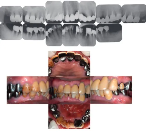
Five patients with chief complaint of difficulty in mastication/ swallowing/speaking, esthetic disturbance, abnormal sensa-tion and pain were selected from the patients who visited Crown Bridge Prosthodontics, Okayama University Hospital, from January 1 to June 30, 2010. The mock patient charts were prepared from patient information, and included chief complaint, present/past history, social history and family history ([6_TD$DIFF]Fig.[2_TD$DIFF] 3[3_TD$DIFF]). These patient charts were reviewed and assessed by the CGC of the JPS. The committee devised reference disease names, i.e. ‘‘B (disability) caused by A (causative factor)’’, for each patient diagnosed.
2.2. Sampling of the validators
Thirty prosthodontic specialists from the JPS were selected from Okayama University, Nihon University and Kanagawa
Dental University. The random sampling function of a spreadsheet application (Excel, Microsoft Corp., Redmond, WA, USA) was used to select 10 specialists from each of the three universities. Twenty dental residents from Okayama University and Nihon University, undergoing residency training in 2010, were also selected using the same random sampling procedure. These validators (specialists and residents) were asked to gather at a conference room or a lecture room at their universities at an agreed time. Explanatory documents were used to obtain informed consent from all validators. For validators that could not attend this meeting, another meeting was held on a different day, and these validators were instructed to avoid obtaining information on the mock patients from other validators.
2.3. Explanation of the DNS
Members of the CGC of the JPS attended the meeting at the various universities to instruct the validators on the DNS using a diagnostic manual (Fig. 1). Because diagnostic procedures in chapters [7_TD$DIFF]Diagnosis (5) and [4_TD$DIFF](6) were difficult to implement
[(_)TD$FIG]
Patient 5: 60-year-old male
Chief complaint:
Abnormal feeling of dental occlusion
History of present illness:
The patient complained of an abnormal feeling of dental occlusion and toothache at the mandibular incisors more than 10 years ago. Because the cause of the problem could not be identified, he was referred to the university hospital by a local doctor. He had a strange feeling of occlusion and severe toothache at the left mandibular first and second molar. Full cast crown restorations on these teeth were removed by a previous dentist and temporary crowns were placed on these teeth. He underwent occlusal adjustment several times, but his problems did not improve.
Past history:
Phobia of being ugly-looking (he was visiting a psychiatric clinic) Medications: Sertraline
Allergy: none Infectious disease: none Bleeding tendency: none
Local anesthesia at dental clinic: Yes (nothing remarkable) Experience of tooth extraction: Yes (nothing remarkable)
Present illness:
He occasionally felt excessive occlusion and insufficient occlusion at other times at the left mandibular molar. He also felt pain on the same teeth occasionally. These sensations were not consistently felt. He occasionally had pain at the right mandibular molar. This was dull pain ranging in magnitude between 5 and 8 (out of 10) according to a visual analogue scale. Radiography did not reveal dental caries, fracture or abnormal finding at the apical root of the molar teeth. Palpation of masticatory muscle did not demonstrate tenderness, but hypertension of the muscles was observed. When he got tense, he felt his face bending, and he complained of difficulty of being in public. He thought these problems came from his dental occlusion. Objectively, he had stable occlusion. He sometimes clenched his teeth during rest.
using the mock patient charts, these chapters were excluded from the instruction.
2.4. Diagnoses and formulation of disease names for prosthodontic treatment
At the meeting, the validators read through the mock patients charts and devised disease names according to the diagnostic manual (Fig. 1). Information in the patient charts was kept confidential throughout the study period. No information on the validator was obtained, except for length of clinical experience and qualification, i.e. specialist in prosthodontics or dental resident.
2.5. Data analysis
Members of the CGC from Nihon University and Kanagawa Dental University sent their data to Okayama University. A CGC member analyzed all data. For evaluation of reliability,
disease names devised by the validators were categorized in the following manner: (3) Both causative factor A and disability B were concordant with the reference disease name formu-lated by the CGC; (2) Either factor A or disability B was concordant; (1) Both factor A and disability B were not concordant.
Krippendorff’s a was used for statistical analysis [3]. Because Krippendorff’s a is similar to Cronbach’s a, it should be greater than 0.8 for sufficient consistency. The number of fully concordant disease names (i.e. both causative factor A and disability B were concordant) was counted for each validator. Correlation between this number and clinical experience period of the validator or time taken to devise the disease name was evaluated. The time taken to devise and formulate the disease name was measured as the difference between the start time and end time for the task, as indicated by each validator.
[(_)TD$FIG]
Cluster analysis was performed to classify and verify the disease names from all validators. For evaluation of validity, the disease names were divided into three groups as follows: (1) both causative factor A and disability B were concordant with the reference disease name; (2) either factor A or disability B was concordant; (3) both factor A and disability B were not concordant. SPSS 15.0 J (SPSS Inc., Chicago, IL, USA) was used for statistical analysis.
3.
Results
Krippendorff’s a for the total sample was 0.378 (95% CI, 0.256– 0.494) (Table 2). Krippendorff’s a for the disease names devised by the prosthodontic specialists was 0.370, while that for dental residents was 0.401. Krippendorff’s a for the top 10 validators (three specialists and seven residents) with higher concordance rates with reference disease names was 0.524. Two validators (one specialist and one resident) who had the highest concordance rates with the reference diseases names had a Krippendorff’s a of 0.648.
Fig. 4shows the concordance rates between the disease names devised by the validators and the reference disease names formulated by the CGC. The concordance rates for prosthodontic specialists and dental residents were not significantly different. Common disease names (i.e. those regularly encountered in the clinical setting), such as ‘‘esthetic disturbance caused by mandibular frontal tooth missing’’ (86% full concordance rate), ‘‘Pain caused by temporomandibular disorders through bruxism’’ (84% full concordance rate), ‘‘masticatory dysfunction caused by marginal periodontitis’’ (68% full concordance rate) and ‘‘pronunciation disability caused by ill-fitting denture’’ (66% full concordance rate), displayed higher concordance rates with the reference disease names. ‘‘Swallowing disability caused by cerebral infarction’’ (72% full concordance rate) and ‘‘pronunciation disability caused by cerebral infarction’’ (64% full concordance rate) also showed higher concordance rates. Unusual disease names, such as ‘‘migraine caused by excess intake of analgesic, caffeine, nicotine’’ (0% full concordance rate), ‘‘mucosal pain caused by dry mouth’’ (2% full concordance rate), ‘‘toothache caused by upper and lower tooth clenching’’ (16% full concordance rate) and ‘‘toothache caused by clenching’’ (22% full concordance rate), tended to exhibit lower concor-dance rates. Cluster analysis showed the typical patterns in the concordance rates of disease names, except for ‘‘pain caused by temporomandibular disorders through bruxism’’ (Fig. 5). In this analysis, swallowing disability, pronunciation disability and abnormal sensation were classified into the same group, while other ‘‘painful’’ disabilities were classified into different groups. The dendrogram (Fig. 5) shows the clustering pattern of the disease names. Distance between disease names in the dendrogram indicates the degree of relationship, with disease names positioned closer having similar factors. Horizontal scales above the dendrogram indicate the degree of similarity between the clusters. Therefore, by disconnecting the dendrogram at any scale, the original cluster can be divided into subclusters that possess similar factors. For example, the dendrogram inFig. 5 can be divided into three clusters by disconnecting at position
15 on the scale. Accordingly, these subclusters reflect similarity in disease names.
Fig. 6 shows the relationship between full concordance scores for disease names and length of clinical experience for each validator, and time taken to devise the disease name as well. There was no significant correlation between the full concordance score and the length of experience or time taken to devise the disease name using the new nomenclature system. The validators who took short or long periods to make the diagnosis tended to exhibit lower concordance with the reference disease names.
4.
Discussion
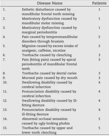
Currently, most disease names for prosthodontic treatment convey the patient’s present oral condition, without informa-tion on etiology. The JPS has recently proposed a new disease naming system for prosthodontic dentistry that conveys the specific disability as well as the cause of the disability. We consider that diseases names based on this new system should facilitate treatment planning for patients. The purpose Table 1 – List of reference disease names formulated by the clinical guideline committee, Japan Prosthodontic Society.
Disease Name Patients
1. Esthetic disturbance caused by mandibular frontal tooth missing
1 2. Masticatory dysfunction caused by
mandibular molar missing
1 3. Masticatory dysfunction caused by
marginal periodontitis
1 4. Pain caused by temporomandibular
disorders through bruxism
2 5. Migraine caused by excess intake of
analgesic, caffeine, nicotine
2
6. Toothache caused by clenching 3
7. Pain (biting pain) caused by apical periodontitis of mandibular frontal teeth
3
8. Toothache caused by dental caries 3
9. Mucosal pain caused by dry mouth 3
10. Swallowing disability caused by cerebral infarction
4 11. Pronunciation disability caused by
cerebral infarction
4 12. Swallowing disability caused by
ill-fitting denture
4 13. Pronunciation disability caused by
ill-fitting denture
4 14. Abnormal occlusal sensation
caused by ugly-looking phobia
5 15. Toothache caused by upper and
lower tooth clenching
5
Table 2 – Concordance rates of the prosthodontic disease names devised with the new prosthodontic diagnostic nomenclature system among validators.
Krippendorff’s a 95% CI
Specialist + Resident 0.378 0.256–0.494
Specialist 0.370 0.251–0.486
[(Fig._4)TD$FIG]
t
n
e
d
i
s
e
R
t
s
i
l
a
i
c
e
p
s
c
i
t
n
o
d
o
h
t
s
o
r
P
0 10 20 30 40 50 60 70 80 90 100 1 2 3 4 5 6 7 8 9 10 11 12 13 14 15Both causative factor A and disability B are concordant with the reference disease name
0 10 20 30 40 50 60 70 80 90 100 1 2 3 4 5 6 7 8 9 10 11 12 13 14 15 Concordance ratio (% )
Either factor A or disability are concordant Both factor A and disability B are not concordant
Concordance
ratio
(%
)
Fig. 4 – Concordance rates of the disease names between validators and the guideline committee. Details of the reference disease names are presented inTable 1andFig. 3.
[(Fig._5)TD$FIG]
0
5
10
15
20
25
Swallowing disability caused by cerebral infarction (patient 4) Pronunciation disability caused by cerebral infarction (patient 4) Abnormal occlusal sensation caused by ugly-looking phobia (patient 5) Swallowing disability caused by ill-fitting denture (patient 4) Pronunciation disability caused by ill-fitting denture (patient 4) Masticatory dysfunction caused by mandibular molar missing (patient 1) Pain caused by temporomandibular disorders through bruxism (patient 2) Esthetic disturbance caused by mandibular frontal tooth missing (patient 1) Masticatory dysfunction caused by marginal periodontitis (patient 1) Pain caused by apical periodontitis of mandibular frontal teeth (patient 3) Toothache caused by dental caries (patient 3) Migraine caused by excessive intake of analgesic, caffeine, nicotine (patient 2) Mucosal pain caused by dry mouth (patient 3) Toothache caused by upper and lower tooth clenching (patient 5) Toothache caused by clenching (patient 3)
Fig. 5 – Cluster analysis for the disease names.
[(Fig._6)TD$FIG]
0
2
4
6
8
10
12
14
0
20
40
60
Concordance
score
Time taken for diagnostic naming (min)
Full concordance score
0
2
4
6
8
10
12
14
0
10
20
30
40
Clinical experience (years)
Concordance
score
Fig. 6 – Relationship between concordance scores of the disease names and the time taken to devise the disease name or length of experience of the validators.
of this study was to evaluate the reliability and validity of this new DNS.
Statistical analysis using Krippendorff’s a revealed a lack of high consistency between disease names devised by the validators. Several methods, such as coincidence ratio, Scott’s p, Cohen’s k and Fleiss’s k, can evaluate the consistency of multiple observations. In this study, we chose Krippendorff’s a for the analyses. The advantages of Krippendorff’s a compared with other methods include its amenability for both numerical and categorical variables and its applicability for multiple observer evaluation. Krippendorff’s a for disease names calculated for all validators combined was low, suggesting lower consistency in disease naming. Krippendorff’s a values were higher for validators with higher concordance rates with reference disease names, indicating higher consistency.
In this study, the validity of the disease name was evaluated by determining the concordance rate between the disease name devised by the validator and the reference disease name formulated by the CGC. Common disease names encountered in daily clinical practice tended to exhibit higher concordance rates, while uncommon disease names showed lower concordance rates. For instance, ‘‘Swallowing disability caused by ill-fitting denture’’ and ‘‘Pronunciation disability caused by ill-fitting denture’’ are common disease names. Also, the present illness reported ‘‘When she wore the denture, these difficulties worsened. She also had a strange sensation in her mouth with the denture.’’ There might be the reasons that most of the validators could diagnose correctly even though the validators did not have clear message that the patient denture was ill-fitting. Some uncommon disease names, such as ‘‘swallowing disability caused by cerebral infarction’’, ‘‘pronunciation disability caused by cerebral infarction’’ and ‘‘abnormal occlusal sensation caused by ugly-looking phobia’’, exhibited higher concordance rates. These diseases were likely easy to diagnose from the patient charts, resulting in high concordance rates. In contrast, ‘‘pain (biting pain) caused by apical periodontitis of mandibular frontal teeth’’, ‘‘mucosal pain caused by dry mouth’’ and ‘‘toothache caused by upper and lower tooth clenching’’ exhibited low levels of validity.
In this study, mock patient charts were prepared from real patient data at Okayama University Hospital, and sufficient data for an accurate diagnosis were difficult to obtain from the charts. Therefore, incomplete patient chart records might account for the poor validity. Insufficient information on diagnostic criteria might also have contributed to the lack of concordance between validator-derived disease names and reference disease names. Although the instructions for the validators (Fig. 1) contained some examples of ‘‘disability B caused by factor A’’, detailed diagnostic criteria were not provided. Without such information, most validators would have been able to make correct diagnoses only for patients with symptoms that they were familiar with. Therefore, diagnostic criteria for both causative factor and disability need to be clearly defined and should provide information for standardized diagnosis and disease naming. Because the concordance rate of the disease names with reference disease names did not show significant differences between prostho-dontic specialists and dental residents, this new DNS should be amenable to use by non-prosthodontic specialists as well.
In this study, clinical experience of the validator or time taken to devise the disease name did not correlate with the full concordance score. Validators with either short or long time periods for diagnosis showed lower full concordance scores. This suggests that difficulty in under-standing patient charts may underlie the low concordance scores.
Taken together, our findings indicate that, at present, the new DNS for prosthodontic dentistry proposed by the JPS does not possess sufficiently high reliability. This system is acceptable for use by general dentists who are not prostho-dontic specialists, and it might have a high level of validity for common disease names. Our findings also show that higher validity for disease names is associated with higher reliability. A diagnostic standard for uncommon disease names with detailed criteria for determining disability and causative factor is required to improve this nomenclature system.
5.
Conclusion
To evaluate the new DNS for prosthodontic dentistry proposed by the JPS, the CGC of the JPS prepared mock patient charts. The validators, comprising prosthodontic specialists and dental residents, made diagnoses using these patient charts. Krippendorff’s a, an indicator of consistency in disease naming, was 0.378 among all validators, 0.370 among prosthodontics specialists and 0.401 among dental residents. Common disease names exhibited higher concordance rates with the reference disease names, while uncommon disease names showed lower concordance rates.
Acknowledgements
The authors would like to thank Dr. Naomi Tanoue, Graduate School of Biomedical Sciences, Nagasaki University. A Japa-nese-language version of this report has been published previously[4], and this is an English translation.
r e f e r e n c e s
[1] Ishibashi K, Fujii S, Hirai T, Sasaki K, Koyano K, Yatani H, et al. Prosthodontic disease name; 2012, <http://hotetsu. com/s/doc/cure_about.pdf>[accessed 05.10.15].
[2] Kuboki T, Ichikawa T, Baba K, Hideshima M, Sato Y, Wake H, et al. A multi-centered epidemiological study evaluating the reliability of the treatment difficulty indices developed by the Japan Prosthodontic Society. J Prosthodont Res 2012;56:71–86.
[3] Hayes AF, Krippendorff K. Answering the call for a standard reliability measure for coding data. Commun Methods Meas 2007;1:77–89.
[4] Matsuka Y, Hagiwara Y, Tamaki K, Takeuchi H, Fujisawa M, Ono T, et al. Reliability and validity of diagnostic
nomenclature system of prosthetic dentistry. Ann Jpn Prosthodont Soc 2013;5:281–90.