Gut microbial metabolites of linoleic acid are metabolized by accelerated peroxisomal
β-oxidation in mammalian cells
Katsuya Morito1, Ryota Shimizu1, Nahoko Kitamura2, Si-Bum Park2, Shigenobu Kishino2, Jun Ogawa2,3, Tatsuya Fukuta1, Kentaro Kogure1, Tamotsu Tanaka1,4*
1Graduate School of Biomedical Sciences, Tokushima University, Tokushima, 770-8505, Japan
2Division of Applied Life Sciences, Graduate School of Agriculture, Kyoto University, Kyoto,
606-8502, Japan
3Center for the Promotion of Interdisciplinary Education and Research, Kyoto University, Kyoto,
606-8501, Japan
4Graduate School of Technology, Industrial and Social Sciences, Tokushima University, Tokushima,
770-8502, Japan
*To whom correspondence should be addressed e-mail: tanaka.tamotsu@tokushima-u.ac.jp
© 2019. This manuscript version is made available under the CC-BY-NC-ND 4.0 license http://creativecommons.org/licenses/by-nc-nd/4.0/ The published version is available via https://doi.org/10.1016/j.bbalip.2019.07.010.
Abstract
Microorganisms in animal gut produce unusual fatty acids from the ingested diet. Two types of hydroxy fatty acids (HFAs), cis-12-octadecenoic acid (HYA) and 10-hydroxy-octadecanoic acid (HYB), are linoleic acid (LA) metabolites produced by Lactobacillus plantarum. In this study, we investigated the metabolism of these HFAs in mammalian cells. When Chinese hamster ovary (CHO) cells were cultured with HYA, approximately 50% of the supplemented HYA disappeared from the dish within 24 hours. On the other hand, the amount of HYA that disappeared from the dish of peroxisome (PEX)-deficient CHO cells was lower than 20%. Significant amounts of C2- and C4-chain-shortened metabolites of HYA were detected in culture medium of HYA-supplemented CHO cells, but not in medium of PEX-deficient cells. These results suggested that peroxisomal β-oxidation is involved in the disappearance of HYA. The PEX-dependent disappearance was observed in the experiment with HYB, but not with LA. We also found that HYA treatment up-regulates peroxisomal -oxidation activity of human gastric MKN74 cells and intestinal Caco-2 cells. These results indicate a possibility that HFAs produced from gut bacteria affect lipid metabolism of host via modulation of peroxisomal -oxidation activity.
Keywords: Peroxisomal -oxidation, Hydroxy fatty acid, Linoleic acid, Fatty acid metabolism, Gut
Abbreviations: ACOX, acyl-CoA oxidase; CHO, Chinese hamster ovary; DAPC,
1,2-diarachidoyl-sn-glycero-3-phosphocholine; DAPI, 4’,6’-diamidino-2-phenylindole; FA, fatty acid; FAME, fatty acid methyl ester; GAPDH, glyceraldehyde-3-phosphate dehydrogenase; GC, gas chromatography; GI, gastrointestinal; GPR, G-protein-coupled receptor; HFA, hydroxy fatty acid; HRP, horseradish peroxidase; HYA, 10-hydroxy-cis-12-octadecenoic acid; HYB, 10-hydroxy-octadecanoic acid; KetoA, 10-oxo-cis-12-octadecenoic acid; LA, linoleic acid; MS, mass spectrometry; PC, phosphatidylcholine; PE, phosphatidylethanolamine; PEX, peroxisome; PMI, polymethylene-interrupted; PMP, peroxisomal membrane protein; PUFA, polyunsaturated fatty acid; TG, triacylglycerol; THF-DA, tetrahydro-2,5-furan-diacetic acid; TRPV, transient receptor potential vanilloid; TBS-BT, Tris-buffered saline containing 3% BSA and 0.1% Tween 20
1. Introduction
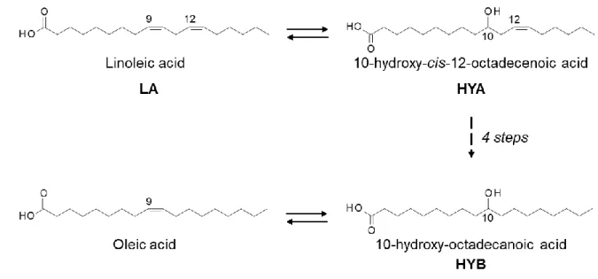
Gastrointestinal microbes have been shown to produce various compounds via fermentation of ingested foods. Some of them are known to affect the cellular functions of the host [1], [2]. For instance, short-chain fatty acids produced through fermentation of dietary fiber in the colon are reported to affect host immune response [3] and obesity [4] via activation of the fatty acid (FA)-specific G-protein-coupled receptor (GPR) 43. Previously, our group showed that a strain of lactic acid bacteria, Lactobacillus plantarum AKU1009a, converts linoleic acid (LA) to oleic acid. Our group also showed that significant amounts of “hydroxy” fatty acids (HFAs) such as hydroxy-cis-12-octadecenoic acid (HYA) and hydroxy-octadecanoic acid (HYB), and “oxo” FAs such as 10-oxo-cis-12-octadecenoic acid (KetoA) are produced during this conversion process (Fig. 1). HYB is also generated when oleic acid is used as a substrate of this conversion. These unusual FAs have been shown to be absorbed from the gastrointestinal (GI) tract and circulate in host animals [5].
Recently, our group revealed that HYA ameliorates intestinal epithelial barrier impairment partially via the GPR40 pathway [6], and that KetoA enhances energy expenditure by activation of transient receptor potential vanilloid (TRPV) 1 in mice [7]. Our group also reported that KetoA induces adipocyte differentiation by activating peroxisome proliferator-activated receptor (PPAR)
sterol regulatory element binding protein-1 mRNA expression [9]. Although much information on the biological effects of gut bacteria-derived FAs on hosts has accumulated so far, little is known about their metabolism in mammalian cells.
Peroxisome (PEX) is an organelle present in virtually all eukaryotic cells. One of roles of PEX is oxidation of FAs that are not metabolized in mitochondria. These include very-long-chain FAs, eicosanoids, dicarboxylic acids, branched-chain FAs, and certain xenobiotics [10]. Recently, we found that peroxisomal -oxidation and chain elongation in the endoplasmic reticulum cooperatively remodel FA. In this process, gymnosperm derived C20 polymethylene-interrupted PUFA (PMI-PUFA) is chain-shortened in PEXs. The resulting FA was then chain-elongated to a C18 FA, such as LA or -linolenic acid [11], [12]. During the course of this study, we learned that PEX-deficient cells are a useful tool to find PEX-intrinsic metabolic pathways.
In this study, we examined metabolisms of two kinds of HFAs, HYA and HYB, produced by gut lactic acid bacteria (Fig. 1), and found that these HFAs are exclusively metabolized by peroxisomal
-oxidation in gastrointestinal cells. We also showed that the HYA treatment potentiates oxidative metabolism of FA by up-regulating PEX function.
2. Materials & Methods 2.1. Materials
HYA and HYB were synthesized enzymatically as described previously [5]. LA was from Wako Pure Chemical Industries, Ltd. (Osaka, Japan). Fatty acid-free bovine serum albumin (BSA) and heptadecanoic acid were obtained from Sigma-Aldrich (St. Louis, MO, USA). 1,2-Diarachidoyl-sn-glycero-3-phosphocholine (DAPC) was purchased from Avanti Polar Lipids (Alabaster, AL, USA). Anti-peroxisomal membrane protein (PMP) 70 antibody (ab3421), anti-ACOX1 antibody [EPR19038] (ab184032), and goat anti-rabbit IgG (Alexa Fluor® 488) (ab150077) were from Abcam Inc. (Cambridge, UK). Mouse anti-glyceraldehyde-3-phoshate dehydrogenase (GAPDH) (5174) and horseradish peroxidase (HRP)-conjugated goat anti-rabbit IgG (A24531) were from Cell Signaling Technologies (Danvers, MA, USA) and Novex® by Life Technologies (Rockville, MD, USA), respectively. All other reagents were of reagent grade.
2.2. Cell culture and fatty acid supplementation
CHO-K1 (wild-type) cells, MKN74 stomach cancer cells, and Caco-2 colon cancer cells were obtained from RIKEN Cell Bank (Tsukuba Japan). CHO-zp102 cells were constructed by deletion of Pex5, coding a peroxisome-targeting signal-1 receptor, and used as described previously [11]-[14].
We used Ham’s F-12 medium for CHO, RPMI1640 medium for MKN74, and MEM for Caco-2. The cells were suspended in the medium containing 10% fetal bovine serum (FBS) (Biowest (Noaillé, France)) and 1% penicillin-streptomycin (Gibco BRL, Life Technologies, Inc. (Rockville, MD, USA)), seeded in 60-mm plastic dishes at 5 × 10⁵ cells, and maintained in humidified air with 5% CO2 at 37°C. After cells were attached to the dish, various concentrations of HFAs or LA were added to the cell cultures as an FA/BSA complex [14]. The molar ratio of FA and BSA of the complex is adjusted to 3:1.
2.3. Extraction and analyses of lipids
Lipids in the culture media and cells were separately extracted by the method of Bligh and Dyer [15] after addition of a known amount of DAPC as an internal standard. The extracted lipids were subjected to methanolysis with 5% HCl-methanol at 100°C for 1 h. The resultant FA methyl esters (FAMEs) were analyzed by gas chromatography (GC) (Shimadzu GC-15A; Shimadzu, Kyoto, Japan) equipped with a capillary column (DB-225, 0.25 μm film thickness, 30 m length, 0.25 mm ID; Agilent Technologies, Santa Clara, CA, USA). The oven temperature was kept at 100°C for 0.5 min and increased to 195°C at rate of 25°C/min. Then, it was increased to 205°C at 3°C/min followed by 240°C at 8°C/min, and kept 240°C for 10 min. The oven temperature was then decreased to 100°C
prior to injection of the next sample. Cellular lipids were separated by TLC. The solvent systems used are petroleum ether:diethyl ether:acetic acid = 80:20:1 (v/v/v) for isolation of triacylglycerol (TG) and free fatty acids. For isolation of phosphatidylethanolamine (PE) and phosphatidylcholine (PC), chloroform:methanol:28% ammonia = 60:35:8 (v/v/v) was used. Isolated lipids were subjected to methanolysis and analyzed with GC as described above. The amount of each FA was calculated based on the ratio of the peak areas between the objective peak and DAPC-derived 20:0 as the internal standard.
2.4. Determination of disappearance of HFAs from cells
After incubation of the cells with 50 μM HFAs or LA for 3 h, culture media were replaced with fresh medium without FAs and further incubated for the indicated period. The cells were harvested using a cell scraper after washing with 0.3% BSA in phosphate-buffered saline (PBS). Lipids were extracted, subjected to methanolysis, and analyzed by GC as described above. The amount of the HFAs or LA in the cellular lipid at initial 3 h incubation was set as 100%. Disappearance of the FAs was expressed as a percentage of the initial amount of FAs.
CHO cells were supplemented with 50 μM HYA. After 24 h incubation, lipids of the cells and culture media were separately extracted and subjected to methanolysis as described above. FAMEs from lipids were separated by TLC with petroleum ether:diethyl ether:acetic acid = 80:20:1 (v/v/v) as the developing solvent. The plate was sprayed with 0.01% primulin to visualize the lipids on the plate under a UV lamp. The FAME in each TLC band was extracted with methanol. After evaporation of the solvent under a stream of nitrogen gas, the FAMEs were trimethylsilylated with Sylon BTZ (Sigma-Aldrich). The derivatization was performed based on the instructions with this reagent. The resultant derivatives were analyzed by GC-mass spectrometry (MS) using a GC-MS QP5050 (Shimadzu) with a GC-17A gas chromatograph for mass spectral analyses. The condition of GC was the same as described above. MS was used in the electron impact mode at 70 eV with a source temperature of 250°C. Split injection was employed with the injector port at 250°C.
2.6. Immunofluorescent staining and microscopic observation
Cells were seeded in 35-mm glass-bottom dishes at 2 × 10⁵ cells and maintained in humidified air with 5% CO2 at 37°C. After they were attached to the dish, cells were incubated with the vehicle, 50 μM HYA, or LA for 3, 6, or 24 h. Cells were fixed with 4% paraformaldehyde for 20 min at 37°C. The fixed cells were washed three times with PBS containing 1% BSA and permeabilized with PBS
containing 0.1% Triton X-100 and 1% BSA for 20 min at 37°C. After washing with PBS containing 1% BSA, cells were treated with rabbit anti-rat PMP 70 antibody diluted 600-fold with PBS containing 1% BSA for 1 h at 37°C. After washing with PBS containing 1% BSA three times, the samples were incubated for 1 h at 37°C with Alexa Fluor488-labeled goat antibody to rabbit IgG diluted 500-fold in PBS containing 1% BSA, then washed with PBS containing 1% BSA three times. Nuclei were counterstained with 4’,6’-diamidino-2-phenylindole (DAPI, 1 μg/mL in PBS) for 15 min at 37°C. After washing with PBS, the stained cells were observed using a confocal laser scanning microscope (Zeiss LSM 700) with a 63× oil-immersion objective.
2.7. Western blotting
Cells were incubated with the vehicle, 50 μM HYA, or 50 μM LA for 24 h. After washing the cells with PBS, they were lysed with lysis buffer (10 mM Tris-HCl (pH 7.4), 0.1% Triton X-100, 2 μg/mL aprotinin, 2 μg/mL leupeptin, 2 μg/mL pepstatin A, and 1 mM phenylmethylsulfonyl fluoride). The protein concentration was determined using a bicinchoninic acid protein assay kit (Thermo Fisher Scientific Inc., Waltham, MA), and cell lysates were subjected to 10% SDS-PAGE. After protein transfer, polyvinylidene difluoride membranes were blocked with 20 mM tris-buffered saline (pH 7.4) containing 3% BSA and 0.1% Tween 20 (TBS-BT) for 1 h. The membranes were incubated with
anti-ACOX1 antibody (diluted 1:600) at 4°C overnight and then washed three times with TBS-BT. The membranes were incubated with anti-GAPDH antibody at room temperature for 1 h, washed three times with TBS-BT, and incubated with HRP-conjugated secondary antibody (diluted 1:20,000) at room temperature for 1 h. The positive bands were detected using the ECL Western Blotting Detection Reagent (GE Healthcare, Waukesha, WI) with a Fuji LAS-4000 imaging system (Fuji Film, Tokyo, Japan). Densitometric analysis was conducted using ImageJ. GAPDH protein was used as an internal control.
2.8. Effect of HYA on oxidative metabolism of FA of the cells
MKN74 cells were incubated with 50 µM HYA or LA for 24 h. After replacement of the culture medium with a medium containing 50 µM 17:0, cells were further incubated for 0, 3, 18, and 24 h. Lipids of the cells and media were extracted separately, subjected to methanolysis, and analyzed using GC for determination of metabolism of 17:0 as described above.
2.9. Statistical analysis
Statistical analyses were performed by Student’s t-test (to compare two groups) and one-way ANOVA with post-hoc Tukey test (to compare more than two groups).
3. Results
3.1. Cellular levels of HFAs in HFA-supplemented CHO cells
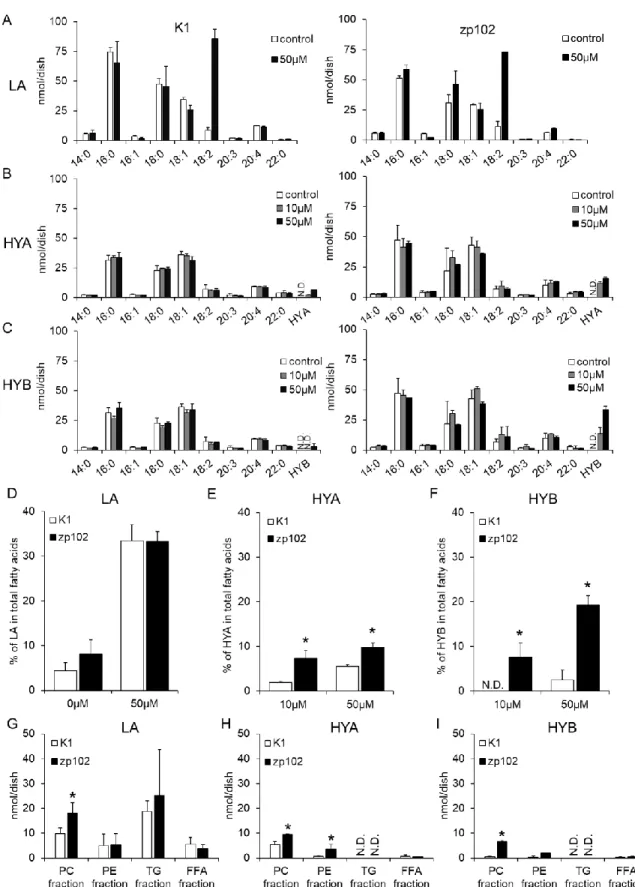
Supplementation of 50 µM LA (250 nmol/dish) resulted in prominent accumulation of LA in cellular lipids in both CHO-K1 (wild-type) and CHO-zp102 (PEX-deficient) cells (Fig. 2A). There was no significant difference in the levels of cellular accumulation of LA between wild-type and PEX-deficient cells (Fig. 2D). In both cell types, accumulations of HFAs were considerably lower than those of LA (Fig. 2B, C). Interestingly, PEX-deficient cells accumulated HYA at a significantly higher level than wild-type cells did (Fig. 2E). This was also the case in the experiments with HYB (Fig. 2F). Distribution of incorporated LA or HFAs in CHO cells was examined. In both CHO cells, LA was acylated to PC, PE, and TG with TG having the highest LA content among these lipid classes (Fig. 2G). On the other hand, HYA was acylated to PC and PE, but not in TG or free fatty acid fraction. It should be noted that the TG band was detectable on the TLC plate of cellular lipids of LA-supplemented cells, whereas it was scarcely detected in cellular lipids of HFA-LA-supplemented cells. Amounts of HYA in PC and PE of CHO-zp102 cells were significantly higher than those of corresponding fraction of wild-type CHO cells (Fig. 2H). Similar results were obtained in the experiments with HYB (Fig. 2I). These observations imply that the metabolism of HFA is different from that of LA in CHO cells.
CHO-K1 (wild-type) and CHO-zp102 (PEX-deficient) cells were incubated with the indicated concentration of LA (A), HYA (B), or HYB (C) for 24 h. Fatty acids of the cellular lipids were determined by GC using DAPC as an internal standard. Percentages of LA (D), HYA (E), and HYB (F) in total fatty acids of cellular lipids are shown. Amounts of LA (G), HYA (H), and HYB (I) in PC, PE, TG, and FFA fraction were analyzed after separation of each lipid class by TLC. Values shown are the means ± S.D. PC: phosphatidylcholine, PE: phosphatidylethanolamine, TG: triacylglycerol, FFA: free fatty acid. n = 3 for each cell. *p<0.05 compared with corresponding CHO-K1 cells (Student’s t test). N.D.: not detected.
3.2. PEX-dependent disappearance of HFAs in CHO cells
The disappearance of incorporated FA was examined in wild-type and PEX-deficient cells. When wild-type cells were incubated with 50 µM LA (250 nmol/dish) for 3 h, the level of LA accumulated in the cells was approximately 15 nmol/dish. These LA-repleted wild-type cells maintained the LA level throughout 24 h of additional incubation without LA supplementation. Similarly, the LA level in LA-repleted PEX-deficient cells did not change much throughout the incubation (Fig. 3A). In contrast, around 80% of HYA incorporated into wild-type cells (2.2 nmol/dish) disappeared from the cells within 18 h of incubation, whereas PEX-deficient cells held the initial level of HYA (2.5 nmol/dish) throughout the incubation (Fig. 3B). Similar results were obtained in the experiments with HYB (Fig. 3C). These results suggest that incorporated HFAs were metabolized in a PEX-dependent manner, whereas LA was not so eliminated in this condition.
Fig. 3. Change in the amounts of HFAs incorporated in CHO cells.
CHO-K1cells and CHO-zp102 cells were incubated with 50 M LA (A), HYA (B), or HYB (C) for 3 h. The cells were further incubated in FA-free media for indicated times. Amounts of each fatty acid were quantified and expressed as a percentage of those obtained at initial 3 h incubation. Values shown are means ± S.D. n = 3 for each cell at every time point. *p<0.05
3.3. PEX-dependent disappearance of HFAs in culture dishes of CHO cells
We examined the disappearance of supplemented FAs from culture dishes by measuring the amounts of FAs in the media and cells. In experiments with CHO-K1 cells, LA in the medium disappeared in proportion to incubation time. The decrements of LA in the medium were balanced by increments of LA in the cells (Fig. 4A). As a result, the total decrement of LA in culture dishes in 24 h was 5% of supplemented LA (250 nmol). Similar results were acquired in the experiments with PEX-deficient cells. There was no significant difference in the fluctuations of LA between culture dishes of wild-type and PEX-deficient cells. Together with results in Fig. 2, these results suggest that acylation to cellular lipids is the predominant metabolic route of LA. In contrast, decrements of HFAs in a culture dish were remarkably different between wild-type CHO cells and PEX-deficient cells. The decrements of HYA in the medium and increments of HYA in the cells were 40 and 15 nmol, respectively, in PEX-deficient cells at 24 h. Thus, the total decrement of HYA in a culture dish in 24 h was 25 nmol, which was 10% of supplemented HYA (Fig. 4B; closed squares). In contrast, 110 nmol of HYA, which corresponds to 44% of supplemented HYA, disappeared in the dish of CHO-K1 cells. Such a large disappearance resulted from the imbalance between decrements of HYA in the media (120 nmol) and increments of HYA in cells (less than 10 nmol) (Fig. 4B; open circles). These results indicate that the disappearance of most of the supplemented HYA observed in CHO-K1 cells
is PEX-dependent. Surprisingly, the amount of HYA that disappeared from the culture dish (120 nmol) was equivalent to the total amount of FAs in cellular lipids (118 nmol, Fig. 2B). Similar results were obtained in the experiments with HYB (Fig. 4C). Again, these results suggest that HFAs produced by gut microbiota are metabolized in animal cells in a PEX-dependent manner.
Fig. 4. Time-dependent changes in the amounts of HFAs in the cells and media of CHO cells.
CHO-K1cells and CHO-zp102 cells were incubated with 50 M LA (A), HYA (B), or HYB (C). The cells and media were collected separately in the indicated time, and subjected to lipid extraction. The amount of HFA in the whole dish is the sum of HFA in the cells and that in the medium. Values shown are means ± S.D. n = 3 for each cell on every time point. *p<0.05
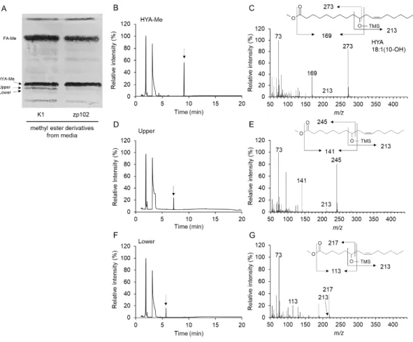
3.4. Identification of HYA metabolites in CHO cells
HYA has been shown to be primary HFAs produced during LA-saturating process in gut lactic acid bacteria [5]. In subsequent experiments, we used HYA as a representative of HFAs. FAMEs prepared from medium lipids and cellular lipids were analyzed by TLC. Two bands located just below HYA methyl ester on TLC were found in lipids prepared from the medium of wild-type CHO cells (Fig. 5A). Lipids recovered from these two bands were analyzed by GC-MS after trimethylsilyl derivatization. Fragment ions containing the carboxyl terminus indicated that they are C2- and C4-methylene unit-shortened products of HYA (Fig. 5C, E, G). Possible assignments of upper and lower bands were 8-hydroxy-cis-10-hexadecenoic acid and 6-hydroxy-cis-8-tetradecenoic acid, respectively (Fig. 5B-E). These two compounds are considered to be formed by one or two cycles of
-oxidation of HYA. Interestingly, the chain-shortened metabolites were detected only in culture media but not in cellular lipids of CHO-K1. We also analyzed the FAMEs from lipids of CHO-zp102, and found that the chain-shortened metabolites of HYA were under the detection level both in medium lipids and cellular lipids (data not shown). These results suggest that HYAs are -oxidized in PEXs, and that a portion of the chain-shortened metabolites are released to extracellular space.
Fig. 5. TLC and GC-MS of metabolites of HYA.
Fatty acid methyl esters were prepared from HYA-supplemented media of CHO-K1 and CHO-zp102 cells, and separated by TLC for fractionation (A). Silica gel corresponding to the methyl ester of HYA, and its metabolites (indicated as Upper and Lower) were scraped off the plate. FAs extracted from the silica gel were trimethylsilylated for analysis by GC-MS. Mass spectra were obtained at the time points indicated by arrows on the total ion chromatogram of methyl HYA fraction (B, C), Upper fraction (D, E) and Lower fraction (F, G). Note that peaks earlier than 5 min are derived from trimethylsilylation reagents.
3.5. PEX-biogenerating effect of HFA in CHO cells
As shown in Fig. 4, the disappearance rate of HFAs from 6 to 24 h was higher than that of the initial incubation phase, suggesting accelerated disappearance of HFAs in the later incubation phase. Based on this observation, we examined the effect of HFAs on the biogenesis of PEX. Immunocytochemical staining was performed with an antibody against PMP 70, a PEX-specific membrane protein. The number and density of PEXs, as shown by green dots, seemed to be increased by HYA treatment in wild-type cells, especially at the perinuclear area (Fig. 6A-D). CHO-zp102 cells are known to have peroxisomal ghosts containing PMP 70 [16]. Consistent with this report, PMP 70 staining in CHO-zp102 cells was positive. The number of peroxisomal ghosts in CHO-zp102 cells was constant throughout the incubation with HYA (Fig. 6F-I). The effect of LA on PEX biogenesis in CHO-K1 cells seemed to be weaker than that observed in HYA-treated cells (Fig. 6E). The number of peroxisomal ghosts in CHO-zp102 cells was unchanged by LA treatment (Fig. 6J). These results suggest that HFAs, but not LA, induce biogenesis of PEX.
Fig. 6. Peroxisome proliferating effect of fatty acids in CHO cells.
CHO-K1 and CHO-zp102 cells were incubated with vehicle (A, F), 50 M HYA (B-D, G-I), or 50
M LA (E, J) for indicated times. PMP70, a peroxisomal marker, was stained with anti-PMP70 antibody (green). Nuclei were stained with DAPI (blue). Scale bar, 20 µm.
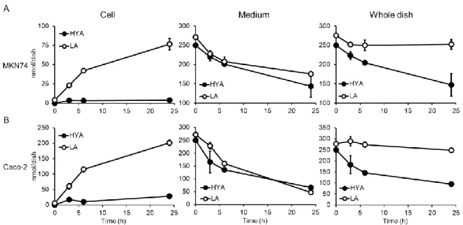
3.6. Disappearance of HFAs in culture dish of human GI tract cells
GI cells are the first cells exposed to HFAs released from gut microbiota. To investigate the metabolism of HFA in human GI cells, experiments were performed with cells that originated from the human GI tract. We used MKN74 cells, a moderately differentiated tubular adenocarcinoma from human stomach, and Caco-2 cells, which are derived from human colon carcinoma. In MKN74 cells, most of the decrement of LA supplemented in media was balanced by the increment of LA in the cells. The total decrement of LA in culture dishes of MKN74 cells in 24 h was 8% of supplemented LA (Fig. 7A; open circles). On the other hand, the total decrement of HYA in culture dishes of MKN74 cells was 41% of supplemented HYA at 24 h. The cells seemed to incorporate HYA at similar level to LA, but eliminated HYA much faster than LA (Fig. 7A; closed circles). Similar results were obtained in the experiments with Caco-2 cells (Fig. 7B). These results indicate that peroxisomal fatty acid -oxidation mainly contributes to the metabolism of HFAs in human GI tract cells, as observed in the CHO-K1 cells.
Fig. 7 Time-dependent change in the amounts of HFAs supplemented to GI tract-derived
cells.
MKN74 (A) and Caco-2 (B) cells were supplemented with 50 M LA or HYA. The cells and media were collected separately at the indicated times and subjected to lipid extraction. The amount of HFA in a whole dish is the sum of HFA in the cells and that in the medium. Values shown are means ± S.D. n = 3 for each cell on every time point. *p<0.05
3.7. PEX-biogenerating effect of HFA in human GI tract cells
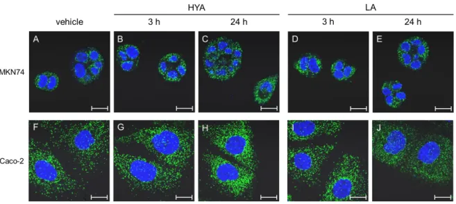
We immunocytochemically stained PMP 70 as a peroxisomal marker in MKN74 and Caco-2 cells. Consistent with the results in CHO-K1 cells, HYA treatment increased the peroxisomal signal in these GI-derived cells in a time-dependent manner. Again, LA treatment did not increase the PEX signal in either cell line (Fig. 8). These results suggest that HFAs induce the biogenesis of PEX in human GI tract cells.
Fig. 8. Peroxisome-proliferating effect of fatty acids in human GI tract-derived cells.
MKN74 and Caco-2 cells were treated with vehicle (A, F), HYA (B, C, G, H), or LA (D, E, I, J) for the indicated times. PMP70, a peroxisomal marker, was stained with anti-PMP70 antibody (green). Nuclei were stained with DAPI (blue). Scale bar, 20 µm.
3.8. Effect of HFAs on peroxisomal fatty acid -oxidation in human GI tract cells
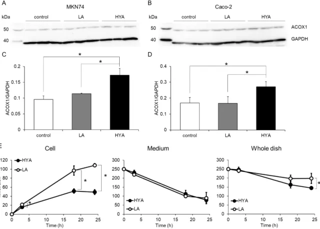
Acyl-CoA oxidase (ACOX) 1 is the rate-limiting enzyme in the peroxisomal -oxidation. We evaluated the effect of HYA on the protein level of ACOX1 in human GI tract cells by Western blotting. Treatment with HYA for 24 h significantly increased the ACOX1 protein in both MKN74 cells and Caco-2 cells (Fig. 9A and B). On the other hand, there was no significant difference in the levels of ACOX1 protein between vehicle-treated cells and LA-treated cells (Fig. 9C and D). We examined whether HYA treatment affected FA metabolism using HYA- and LA-treated MKN74 cells. Judging from the decrements of 17:0 from the media, HYA-pretreated cells incorporated 17:0 at a similar level to LA-pretreated cells. However, accumulated 17:0 in HYA-treated cells was around half the level of that in LA-treated cells (Fig. 9E). Chain-elongation metabolites of 17:0 were modestly detected, but there was no significant difference between HYA- and LA-treated cells (data not shown). These results indicated that pretreatment of HYA enhances oxidation of incorporated 17:0 by inducing PEX biogenesis and up-regulating the peroxisomal -oxidation activity.
Fig. 9. Effects of HYA on expression level of acyl-CoA oxidase and FA oxidation activity of
GI tract-derived cells.
MKN74 and Caco-2 cells were incubated with vehicle, 50 M LA, or HYA for 24 h. The cells were harvested and subjected to Western blotting. Representative blots for acyl-CoA oxidase 1 (ACOX1) and GAPDH of MKN74 (A) and Caco-2 (B) are shown. Protein levels of ACOX1 in MKN74 (C) and Caco-2 (D) cells were quantified by image analysis and normalized to GAPDH. Values shown are means ± S.D. n = 3 for MKN74 experiment and n = 6 for Caco-2 experiment. *p<0.05 using one-way ANOVA with post-hoc Tukey test. Time-dependent changes in the amounts of 17:0 supplemented to
MKN74 cells were analyzed in the cellular fraction and medium fraction (E). The cells that had been incubated with 50 µM HYA or LA for 24 h were incubated with 50 µM 17:0 for 0, 3, 18, and 24 h. Cells and media were collected separately, and subjected to lipid extraction. The amount of HFA in whole dish is sum of HFA in the cells and that in the medium. Values shown are means ± S.D. n = 3 at every time point. *p<0.05.
4. Discussion
Dietary PUFA affects not only FA composition of cellular membrane [17], but also gut microbiome [18]. It has been reported that dietary LA influence gut microbial diversity and affect tendency of obesity [19]. Previously, our group revealed that a gut lactic acid bacterium, Lactobacillus plantarum, possesses a PUFA-saturating metabolism that generates HFAs and oxo FAs as intermediates [5]. These gut bacteria-derived FAs have been shown to affect mammalian cells by acting as ligands of GPR40 [6], [20], TRPV1 [7], and PPAR [8]. In this study, we investigated the metabolism of gut bacteria-derived FAs in animal cells.
We found that HFAs are metabolized exclusively by peroxisomal-oxidation. This conclusion was led by the following observations. Firstly, PEX-deficient CHO-zp102 cells retained incorporated HFAs within cells, whereas wild-type cells did not retain the HFAs (Figs. 2, 3). Secondly, the clearance rate of HYA in a dish of PEX-deficient cells was much lower than that in a dish of type cells (Fig. 4). Thirdly, C2- and C4-chain-shortened metabolites of HYA were detected in wild-type but not in PEX-deficient CHO cells (Fig. 5). PEX has been shown to oxidize very-long-chain FAs, dicarboxylic acids, branched-chain FAs, and certain xenobiotics [10]. Furthermore, bioactive lipids such as prostaglandins [21], [22], thromboxane B2 [23], leukotrienes [24], [25], hydroxyeicosatetraenoic acids [26], and PMI-PUFA [11], [12] were shown to be metabolized in a
PEX-dependent manner. We showed, for the first time, that HFAs from gut bacteria are exclusively chain-shortened by peroxisomal -oxidation.
An interesting phenomenon observed here is that HFAs are acylated to PC and PE, but not in TG or free fatty acid fraction. This is distinct property of LA which is accumulated in TG. It is worthwhile to examine whether HFAs influence the cellular membrane property and affect enzyme activity in cellular membrane.
Alpha-oxidation and -oxidation pathways are known to be present in PEX for degradation of FA. In this study, we detected C2 and C4 chain-shortened metabolites of HYA in wild-type CHO cells. HYA metabolites with odd chain length were not detected. Based on these results, it is considered that HFAs are exclusively -oxidized in a PEX-dependent manner. The C2 and C4 chain-shortened metabolites of HYA detected in wild-type CHO cells accounted for only 20% of total disappearance of HYA from the medium (data not shown). The structures of other metabolites of HYA are unknown. They could be further chain-shortened by peroxisomal -oxidation. It has been reported that tetrahydro-2,5-furan-diacetic acid (THF-DA) is one of the metabolites of ricinoleic acid in mammals [27]. Because ricinoleic acid (12-hydroxy-cis-9-octadecenoic acid) is a regioisomer of HYA, we speculated that such metabolites could be formed from HYA. However, attempts to detect THF-DA or its homologues by mass spectrometry were unsuccessful. Further studies are required for
elucidation of the metabolic fate of HYA in animal cells.
The amount of FA oxidized by peroxisomal -oxidation can be determined by our experimental design using PEX-deficient cells and their wild-type cells. Here, we found that the amounts of HFAs eliminated by peroxisomal -oxidation in 24 h were equivalent to FAs in cellular lipids (Fig. 4). Such a vast and rapid elimination of HFAs is considered to be attained by up-regulation of peroxisomal -oxidation activity. This conclusion was led by following observations: 1) incubation of mammalian cells with HYA induced biogenesis of PEXs (Figs. 6 and 8), and 2) expression levels of ACOX1 and FA-oxidation were enhanced in HYA-treated cells compared to LA-treated cells (Fig. 9). Although we have not clarified the mechanisms underlying the up-regulation of PEX, our group has reported that HYA acts as ligands of PPARs [8] and TRPV1 [7]. It is known that activation of these receptors enhances peroxisomal -oxidation [28], [29].
HYA concentration in plasma of mouse maintained under specific pathogen free condition with a sterile diet has been reported to be around 50 nM [5]. Thus, HFA concentrations used in this study (10 M and 50 M) will be unusually high. However, stomach or intestinal epithelial cells which are facing to digestions and microbial metabolites are considered to be exposed to such high concentration of HFA. Approximately 13 g/day of LA are reported to be ingested from the diet [30] and 25% of ingested LA are converted to HFAs [31]. Considering that sum of volume of exocrine
gland secretions into the gastrointestinal lumen and the water intake from diet is estimated to be approximately 10 L/day [32], it is likely that concentrations of LA and HFAs could reach to sub-mM order in GI tract.
Hyperlipidemia is major risk factor of cardiovascular disease. One of strategies to reduce TG in blood is up-regulation of PEX function. Chemicals such as fibrates [28] and fish oil containing EPA [33] and DHA [34] have been shown to enhance peroxisomal -oxidation and are widely used for treatment of hyperlipidemia. In the current study, we showed that HFAs released from gut bacteria enhanced FA oxidation activity of PEX in mammalian cells including intestinal cells. Gastrointestinal microbes have a possibility to affect the lipid metabolism of our bodies by modulating peroxisomal β-oxidation activity.
In summary, we revealed that HFAs produced by gut bacteria are -oxidized in animal and human GI tract cells in a PEX-dependent manner. We also demonstrated that HYA up-regulates peroxisomal
-oxidation activity. These results indicate a possibility that FAs from gut microbiota affect the lipid metabolism of the host.
Acknowledgments/grant support
This study was partly supported by the Mishima Kaiun Memorial Foundation (to K.M.), JSPS Grant-in-Aid for challenging Exploratory Research Grant Numbers JP 18K19175 (to S.K.), JSPS KAKENHI Grant Numbers JP 15H02441 (to J.O.), and research program for development of intelligent Tokushima artificial exosome (iTEX) from Tokushima University.
References
[1] Tremaroli, V., and F., Bäckhed. 2012. Functional interactions between the gut microbiota and
host metabolism. Nature 489: 242-249. https://doi.org/10.1038/nature11552
[2] Gibson, G. R., and M. B. Roberfroid. 1995. Dietary modulation of the human colonic
microbiota: Introducing the concept of prebiotics. J Nutr 125: 1401-1412.
https://doi.org/10.1093/jn/125.6.1401
[3] Maslowski K. M., and C. R. Mackay. 2011. Diet, gut microbiota and immune responses. Nat
Immunol 12: 5-9. https://doi.org/10.1038/ni0111-5
[4] Carding, S., K. Verbeke, D. T. Vipond, B. M. Corfe, and L. J. Owen. 2015. Dysbiosis of the gut
microbiota in disease. Microb Ecol Health Dis 26: 26191. https://doi.org/10.3402/mehd.v26.26191 [5] Kishino, S., M. Takeuchi, S. B. Park, A. Hirata, N. Kitamura, J. Kunisawa, H. Kiyono, R.
Iwamoto, Y. Isobe, M. Arita, H. Arai, K. Ueda, J. Shima, S. Takahashi, K. Yokozeki, S. Shimizu,
and J. Ogawa. 2013. Polyunsaturated fatty acid saturation by gut lactic acid bacteria affecting host
lipid composition. Proc Natl Acad Sci U S A 110: 17808-17813.
https://doi.org/10.1073/pnas.1312937110
[6] Miyamoto, J., T. Mizukure, S. B. Park, S. Kishino, I. Kimura, K. Hirano, P. Bergamo, M. Rossi,
10-hydroxy-cis-12-octadecenoic acid, ameliorates intestinal epithelial barrier impairment partially via
GPR40-MEK-ERK pathway. J Biol Chem 290: 2902-2918. https://doi.org/10.1074/jbc.M114.610733
[7] Kim, M., T. Furuzono, K. Yamakuni, Y. Li, Y. I. Kim, H. Takahashi, R. Ohue-Kitano, H. F.
Jheng, N. Takahashi, Y. Kano, R. Yu, S. Kishino, J. Ogawa, K. Uchida, J. Yamazaki, M. Tominaga,
T. Kawada, and T. Goto. 2017. 10-Oxo-12(Z)-octadecenoic acid, a linoleic acid metabolite produced
by gut lactic acid bacteria, enhances energy metabolism by activation of TRPV1. FASEB J 31:
5036-5048. https://doi.org/10.1096/fj.201700151R
[8] Goto, T., Y. I. Kim, T. Furuzono, N. Takahashi, K. Yamakuni, H. E. Yang, Y. Li, R. Ohue, W.
Nomura, T. Sugawara, R. Yu, N. Kitamura, S. B. Park, S. Kishino, J. Ogawa, and T. Kawada. 2015. 10-Oxo-12(Z)-octadecenoic acid, a linoleic acid metabolite produced by gut lactic acid bacteria, potently activates PPAR and stimulates adipogenesis. Biochem Biophys Res Commun 459: 597-603. https://doi.org/10.1016/j.bbrc.2015.02.154
[9] Nanthirudjanar, T., H. Furumoto, J. Zheng, Y. I. Kim, T. Goto, N. Takahashi, T. Kawada, S. B.
Park, A. Hirata, N. Kitamura, S. Kishino, J. Ogawa, T. Hirata, and T. Sugawara. 2015. Gut microbial fatty acid metabolites reduce triacylglycerol levels in hepatocytes. Lipids 50: 1093-1102. https://doi.org/10.1007/s11745-015-4067-z
[10] van Veldhoven, P. P. 2010. Biochemistry and genetics of inherited disorders of peroxisomal
fatty acid metabolism. J Lipid Res 51: 2863-2895. https://doi.org/10.1194/jlr.R005959.
[11] Tanaka T, J. Morishige, D. Iwawaki, T. Fukuhara, N. Hamamura, K. Hirano, T. Osumi and K.
Satouchi. 2007. Metabolic pathway that produces essential fatty acids from polymethylene-interrupted polyunsaturated fatty acids in animal cells. FEBS J 274: 2728-2737. https://doi.org/10.1111/j.1742-4658.2007.05807.x
[12] Tanaka, T., S. Uozumi, K. Morito, T. Osumi, and A. Tokumura. 2014. Metabolic conversion
of C20 polymethylene-interrupted polyunsaturated fatty acids to essential fatty acids. Lipids 49: 423-429. https://doi.org/10.1007/s11745-014-3896-5
[13] Tsukamoto, T., A. Bogaki, K. Okumoto, K. Tateishi, Y. Fujiki, N. Shimozawa, Y. Suzuki, N.
Kondo, and T. Osumi. 1997. Isolation of a new peroxisome-deficient CHO cell mutant defective in peroxisome targeting signal-1 receptor. Biochem Biophys Res Commun 230: 402–406. https://doi.org/10.1006/bbrc.1996.5971
[14] Tanaka, T., J. Morishige, T. Takimoto, Y. Takai, and K. Satouchi. 2001. Metabolic
characterization of sciadonic acid (5c,11c,14c-eicosatrienoic acid) as an effective substitute for arachidonate of phosphatidylinositol. Eur J Biochem 268: 4928–4939. https://doi.org/10.1046/j.0014-2956.2001.02423.x
[15] Bligh, E. G., and W. J. Dyer. 1959. A rapid method of total lipid extraction and purification.
Can J Biochem Physiol 37: 911–917. https://doi.org/10.1139/o59-099
[16] Yamasaki, M., N. Hashiguchi, C. Fujiwara, T. Imanaka, T. Tsukamoto, and T. Osumi. 1999.
Formation of peroxisomes from peroxisomal ghosts in a peroxisome-deficient mammalian cell mutant upon complementation by protein microinjection. J Biol Chem 274: 35293-35296. https://doi.org/10.1074/jbc.274.50.35293
[17] Abbott, S. K., P. L. Else, T. A. Atkins, and A. J. Hulbert. 2012. Fatty acid composition of
membrane bilayers: Importance of diet polyunsaturated fat balance. Biochim Biophys Acta 1818:
1309-1317. https://doi.org/10.1016/j.bbamem.2012.01.011.
[18] Watson, H., S. Mitra, F. C. Croden, M. Taylor, H. M. Wood, S. L. Perry, J. A. Spencer, P. Quirke,
G. J. Toogood, C. L. Lawton, L. Dye, P. M. Loadman, and M. A. Hull. 2018. A randomised trial of the effect of omega-3 polyunsaturated fatty acid supplements on the human intestinal microbiota. Gut 67: 1974-1983. https://doi.org/10.1136/gutjnl-2017-314968.
[19] Zhuang, P., Q. Shou, W. Wang, L. He, J. Wang, J. Chen, Y. Zhang, and J. Jiao. 2018. Essential
fatty acids linoleic acid and -linolenic acid sex-dependently regulate glucose homeostasis in obesity. Mol Nutr Food Res 62: e1800448. https://doi.org/10.1002/mnfr.201800448
[20] Ohue-Kitano, R., Y. Yasuoka, T. Goto, N. Kitamura, S. B. Park, S. Kishino, I. Kimura, M.
Kasubuchi, H. Takahashi, Y. Li, Y. S. Yeh, H. F. Jheng, M. Iwase, M. Tanaka, S. Masuda, T. Inoue,
H. Yamakage, T. Kusakabe, F. Tani, A. Shimatsu, N. Takahashi, J. Ogawa, N. Satoh-Asahara, and T.
Kawada. 2018. -Linolenic acid–derived metabolites from gut lactic acid bacteria induce
differentiation of anti-inflammatory M2 macrophages through G protein-coupled receptor 40. FASEB
J 32: 304-318. https://doi.org/10.1096/fj.201700273R
[21] Fauler, J., D. Tsikas, E. Mayatepek, D. Keppler, and J. C. Frölich. 1994. Impaired degradation
of prostaglandins and thromboxane in Zellweger syndrome. Pediatr Res 36: 449-455. https://doi.org/10.1203/00006450-199410000-00006
[22] Diczfalusy, U., and S. E. H. Alexson. 1988. Peroxisomal chain-shortening of prostaglandin F2.
J lipid Res 29: 1629-1636.
[23] Diczfalusy, U., O. Vesterqvist, B. F. Kase, E. Lund, and S. E. H. Alexson. 1993. Peroxisomal
chain-shortening of thromboxane B2: evidence for impaired degradation of thromboxane B2 in Zellweger syndrome. J lipid Res 34: 1107-1113.
[24] Jedlitschky, G., M. Huber, A. Völkl, M. Müller, I. Leier, J. Müller, W. D. Lehmann, H. D.
Fahimi, and D. Keppler. Peroxisomal degradation of leukotrienes by -oxidation from the -end. J Biol Chem 266: 24763-24772.
[25] Mayatepek, E., W. D. Lehmann, J. Fauler, D. Tsikas, J. C. Frölich, R. B. Schutgens, R. J.
Wanders, and D. Keppler. 1993. Impaired degradation of leukotrienes in patients with peroxisome deficiency disorders. J Clin Invest 91: 881-888. https://doi.org/10.1172/JCI116309
[26] Gordon, J. A., P. H. Figard, and A. A. Spector. 1990. Hydroxyeicosatetraenoic acid metabolism
in cultured human skin fibroblasts. Evidence for peroxisomal -oxidation. J Clin Invest 85: 1173-1181. https://doi.org/10.1172/JCI114550
[27] Hagenfeldt, L., L. Blomquist, and T. Midtvedt. 1986. Epoxydicarboxylic aciduria resulting
from the ingestion of castor oil. Clin Chim Acta 161: 157-163. https://doi.org/10.1016/0009-8981(86)90209-3
[28] Lakhia, R., M. Yheskel, A. Flaten, E. B. Quittner-Strom, W. L. Holland, and V. Patel. 2018.
PPAR agonist fenofibrate enhances fatty acid -oxidation and attenuates polycystic kidney and liver disease in mice. Am J Physiol Renal Physiol 314: F122-F131.
https://doi.org/10.1152/ajprenal.00352.2017
[29] Baboota, R. K., N. Murtaza, S. Jagtap, D. P. Singh, A. Karmase, J. Kaur, K. K. Bhutani, R. K.
Boparai, L. S. Premkumar, K. K. Kondepudi, and M. Bishnoi. 2014. Capsaicin-induced transcriptional changes in hypothalamus and alterations in gut microbial count in high fat diet fed mice. J Nutr Biochem 25: 893-902. https://doi.org/10.1016/j.jnutbio.2014.04.004
[30] Kalmijn, S., M. P. van Boxtel, M. Ocké, W. M. Verschuren, D. Kromhout, and L. J. Launer.
2004. Dietary intake of fatty acids and fish in relation to cognitive performance at middle age.
Neurology 62: 275-280. https://doi.org/10.1212/01.WNL.0000103860.75218.A5
[31] Ogawa, J., K. Matsumura, S. Kishino, Y. Omura, and S. Shimizu. 2001. Conjugated linoleic
acid accumulation via 10-hydroxy-12-octadecaenoic acid during microaerobic transformation of
linoleic acid by Lactobacillus acidophilus. Appl Environ Microbiol 67: 1246-1252.
https://doi.org/10.1128/AEM.67.3.1246-1252.2001
[32] Cheng, H. M., Secretion of Digestive Juices, in: H. M. Cheng (Eds.), Physiology
Question-Based Learning, Springer, Singapore, 2016, pp. 137-148.
[33] Aarsland, A., M. Lundquist, B. Børretsen, and R. K. Berge. 1990. On the effect of peroxisomal
-oxidation and carnitine palmitoyltransferase activity by eicosapentaenoic acid in liver and heart from rats. Lipids 25: 546-548. https://doi.org/10.1007/BF02537162
[34] Willumsen, N., S. Hexeberg, J. Skorve, M. Lundquist, and R. K. Berge. 1993.
Docosahexaenoic acid shows no triglyceride-lowering effects but increases the peroxisomal fatty acid oxidation in liver of rats. J Lipid Res 34: 13-22.