INTRODUCTION
The recent advances in the establishment and controlled differentiation of embryonic stem cells (ESCs) have produced new model systems to access the biology of early mammalian development includ-ing the human development, and new approaches
to regenerative medicine (1-2) . Herein, we developed a novel protocol for generation and selective amplification of neural progenitor cells regionally specified to the rostral brain from the mouse ESCs. The neural progenitors could differentiate into cholinergic and GABA ergic neurons both in vitro and in vivo. Transplantation of the neural progenitors
ORIGINAL
Differences in the neuronal stem cells survival, neuronal
differ-entiation and neurological improvement after
transplan-tation of neural stem cells between mild and severe
experimental traumatic brain injury
Tokuhisa Shindo, Yoshihito Matsumoto, Qinghua Wang, Nobuyuki Kawai,Takashi Tamiya,
and Seigo Nagao
Department of Neurological Surgery, Faculty of Medicine, Kagawa University, Kagawa, Japan
Abstract : We developed a novel protocol for generation and selective amplification of neural progenitor cells regionally specified to the rostral brain but not the spinal cord from mouse embryonic stem cells (ESCs) . The neural progenitors could differentiate in vitro and in vivo into many cholinergic and a few GABAergic neurons but rarely into astrocytes. The transplanted neurospheres could survive in the hippocampus (CA3) of animals with mild traumatic brain injury (TBI) . Twelve weeks after transplantation (a week after the behavioral test) , we found significant cholinergic differentiation recognized as ChAT immunoreactivity in the eGFP+ transplanted cells. Moreover, the grafts contained a few GAD67+cells. However, we barely found GFAP+ astrocytes within the grafts. Furthermore, presynaptic formations of graft-derived neurons were recognized by immunohistochemistry of near the grafts around CA3. However, these findings were not observed in severe TBI group. So, we examined NGF, BDNF, and FGF-2 mRNA by RT-PCR in 12 mice including normal, mild TBI and severe TBI group. Increases in the neurotrophic factors’ mRNA were evident in the hippocampus on the ipsilateral side in the mild TBI group. Statistical analysis revealed significant differences between the mild and severe TBI groups. The data also revealed significant differences between the mild TBI and normal groups. The transplanted neurospheres could survive in the mild TBI animals, but not in the severe TBI group. J. Med. Invest. 53 : 42-51, February, 2006
Keywords : embryonic stem cell, neural stem cell, transplantation, cholinergic, traumatic brain injury
Abbreviations used in this paper:AD = Alzheimer’s disease;ChAT= choline acetyltransferase ; ANOVA = analysis of variance ; AP =amyloid-βprotein ; ESCs=embryonic stem cells ; GAD=glutamate decarboxylase ; GFAP=glial fibrillary acidic protein ; GFP=green fluorescent protein;GMEM = Glasgow minimal essential medium; HE=hematoxylin and eosin;LIF=leukemia inhibitory factor;LSM= laser scanning microscope;NBM = nucleus basalis of Meynert; NPCs = neural progenitor cells ; NSCs = neural stem cells,
Received for publication October 11, 2005 ; accepted August 17, 2005.
Address correspondence and reprint requests to Yoshihito Matsumoto, Department of Neurological Surgery, Faculty of Medicine, Kagawa University, Miki-cho, Kita-gun, Kagawa 76 1-0793, Japan and Fax : +81-87-891-2208.
The Journal of Medical Investigation Vol. 53 2006 42
into the hippocampus of mouse model of traumatic brain injury (TBI) resulted in its functional recovery (3-6) . This could be one of the critical steps for ap-plication of ESCs in understanding of their biology and treatment of neurodegenerative diseases. The identification of the mammalian neural stem cells (NSCs) and the establishment of protocols for their amplification and differentiation in vitro and in vivo have brought high expectations for the clinical application of NSCs in the cell replacement thera-peutics to treat the damaged central nervous system (CNS). Some studies on transplantation of in vitro expanded neural progenitor cells (NPCs) including NSCs and mobilization of latent NPCs in vivo in the animal models of several neurological disorders revealed functional recovery of the animals to some extent (7-10) The ideal cell replacement therapeutics using NSCs to various neurological disorders can include replacement of specific types of neurons. Herein, we report a novel differentiation protocol of the mouse ESCs to generate NPCs which can generate many functional cholinergic neurons in vitro and
in vivo(11-13) .
Neurotrophins are a family of structurally related polypeptides that play a critical role during neuro-nal development and appear to mediate a protective response in mature animals. The members of the neurotrophin family include nerve growth factor (NGF), brain-derived neurotrophic factor (BDNF), and fibroblast growth factor (FGF)(11-12). Neu-rotrophins are believed to promote cell survival, growth, and differentiation effects through interac-tions primarily with specific high affinity tyrosine kinase receptors and subsequent activation of the intracellular signal transduction pathway.
In this study, we evaluated the differences in the NSCs survival, neuronal differentiation, and spatial memory improvement after transplantation of neu-rospheres between mild and severe experimental models of TBI. Moreover, much less is known about modulation of neurotrophin expression fol-lowing TBI. We also examined the mRNA levels of NGF, BDNF, and FGF in the hippocampus follow-ing mild and severe TBI.
MATERIALS AND METHODS
Animals
The experimental protocol was approved by the Institutional Animal Care and Use Committee, and conformed to the National Institutes of Health
(NIH) guidelines for the care and use of laboratory animals. We used 9-week-old adult male C 57 BL/6 mice weighing 20 to 22 g in all experiments.
ESC culture
Mouse ES cell lines transfected with GFP gene were cultivated as described previously (14) . Briefly, the cells were maintained in the absence of feeder cells in Glasgow minimal essential medium (GMEM; Sigma, Colorado, USA) supplemented with 10% FCS (Equitech-Bio Inc., Kerrville, TX, USA), 1 mM sodium pyruvate, 10-4
M 2-mercaptoethanol, 1 x non-essential amino acids, and 1,000 U of leukemia inhibitory factor (LIF) per ml on gelatin-coated dishes. Then, we added 5µg/ml of blastcydine S (Funakoshi, Tokyo, Japan) for stem cell selection.
Differentiation of ESCs and selective amplification and differentiation of neural progenitor cells
To induce differentiation of ESCs, a single cell suspension of ESCs was plated onto non-adherent bacterial culture dishes at a density of 5×10⁴
cell/ ml inαMEM (GIBCO/Invitrogen, California, USA) supplemented with 10% FCS, 10-4
M2-mercaptoethanol and Noggin (inhibitor of bone morphogenic factor). After EBs formation 6 days later , a single suspension was obtained by trypsinization for 5 min with 0.25% trypsin in 1 mM EDTA (GIBCO/Invitrogen) , followed by quenching with10% FCS. The cells were washed twice with basic neurosphere medium composed of DMEM : F 12 (1 : 1) , 0.6% glucose, 2 mM glutamine, 3 mM sodium bicarbonate, 5 mM HEPES buffer, 25µg/ml insulin, 100µg/ml transferring, 20 nM progesterone, 60µm putrescine, 30 nM selenium chloride (all from Sigma except glutamine from GIBCO/Invitrogen) and plated onto T 75 flasks (Greiner, city, country) at a density of 2×10⁴
or 1×10⁵
cells/ml in the neurosphere medium-basic neuro-sphere medium supplemented with 20 ng/ml FGF 2 (Peprotech, Rocky Hill, NJ, USA), 2µg/ml heparin sulfate (Sigma) and sonic hedgehog (Shh) (15-17). Shh is a secreted protein known to be expressed in the notochord and floor plate, and induces ventralization of the developing CNS (18) . The cells were grown for 5 to 7 days and formed neurospheres. For in vitro differentiation of NPCs, the neurospheres were plated onto poly-L-ornithin coated dishes in the basic neurosphere medium and cultured for 5 to 14 days. Neurospheres were collected by settling down for 15 min and re-suspended in the neurospheres culture medium. Cell suspensions were made at a density of 1×10⁴
cells/µl and were kept on ice to optimize the
cell viability.
Brain Injury and Cell Transplantation
C57BL/6 mice were subjected to mild (2atm) or severe (4 atm) lateral controlled cortical impact brain injury (by fluid percussion). Animals were anesthetized using a combination of ketamine (60 mg/kg body weight; Bayer, Germany) and xylazine (20mg/kg body weight; Bayer, Germany) in saline administered intraperitoneally. A1-cm midline scalp incision was made and a 5-mm craniectomy was made in the left parietal bone centered between the coronal, sagittal, and lambdoid sutures. A hollow plastic Luer-loc fitting was cemented into the cra-niectomy site. The Luer-loc cap was fitted to the fluid percussion device, and a lateral fluid percussion injury of mild (2atm) or severe (4atm) severity was delivered by a rapid percussion? of saline that struck the exposed dura mater through the sealed fitting (40 animals) . After the injury, the caps were removed and the scalps were sutured. Eight animals (control group) were subjected to the same anesthesia and surgical procedures but without TBI (2, 3, 9).
On day 7 post-injury, all animals were anesthe-tized with intraperitoneal injection of 60 mg/kg sodium pentobarbital and placed in a stereotactic frame. In the animals assigned to receive trans-plants, separate injections of 2 ul of cell suspension (a total 1×10⁶ cells/brain) were stereotactically delivered through a 10-ul Hamilton syringe into each injured site by using the following coordinates cal-culated from the bregma (A and L) and dura mater (V) according to the atlas of Paxinos and Watson : A=2.5 mm, L=2.5 mm, V=2 mm. In all animals that received injections of cells or vehicle, the syringe was slowly advanced through the dura mater and cortex. The cell suspension was injected over 10 min into the injured site. The needle was left in place for 5 min, and then gradually withdrawn over 5 min. The injured animals and uninjured controls were subjected to similar procedures and injection of vehicle to control the effects of cannula microin-jection.
Behavioral Assessment
Eight-direction maze test was initiated 8 weeks after transplantation and conducted as described elsewhere (19). The radial maze consisted of 8 sym-metrical arms (25cm long, and 6cm wide, with side walls 6 cm high), radiating from an octagonal central platform (10.5cm in diameter) . The platform was enclosed by a wall (6cm high) . The floor of the
maze was made of wood painted white, and the wall consisted of transparent Plexiglas. The apparatus was elevated 50 cm above the floor and placed in a closed room where all visual cues were kept unchanged throughout the test trial. During the experiment, the maze was maintained in a constant orientation, and the investigator stayed in a constant position beside the maze and observed the performances of the mice. Completion of the radial maze entailed mouse running down and retrieving a food pellet of 15 mg at the end of each arm. An arm entry was defined as crossing of the four paws into an arm. Before each session of the test, the mice were deprived of food until their body weight was reduced to 80-85% of the initial level. Pretraining started three days before the test, in which each animal was placed in the central platform and allowed to explore and consume a food pellet at the end of each arm. This was repeated daily for three con-secutive sessions. During the test trail (3 weeks), only 4 arms were baited with food pellets, and each mouse was given one daily trail for 6 days/week. For each trial, two parameters ; namely, reference memory error (entry into the unbaited arms) and working memory error (repeated entry into the arms that had already been visited within a trial), were measured.
Histology and Immunohistochemistry
One week after completion of the behavioral testing (12 weeks after transplantation) , all mice were deeply anesthetized with ketamine and xylazine and perfused transcardially with saline for 3 min and then with ice-cold 4% paraformaldehyde (PFA) in 0.1 M PBS (pH 7.4) for 30min. Then, the brains were removed and placed in paraformaldehyde? (PFA) overnight. Then, 30-µm thick coronal or sagittal sections were cut on a vibratome (D. S. K microslicer, Japan) into series and placed free floating in PBS at 4!. The primary antibodies (final dilution and source) used in this study were as follows : rabbit polyclonal anti-human choline acetyl transferase (ChAT) (1 : 200 ; Chemicon, Temecula, CA, USA), rabbit polyclonal anti-humanβ-III-tubulin (1:200;DAKO, Carpinteria,
CA, USA) , rabbit polyclonal anti-feline glutamic acid decarboxylase (GAD67) (1:200;Chemicon) , mouse monoclonal anti-human GFAP IgG (1:100, Sigma) , rabbit polyclonal anti-human glial fibrillary acidic protein (GFAP) IgG (1:200;DAKO) , rabbit polyclonal anti-green fluorescence protein (GFP) (1 : 1000 ; MBL, Nagoya, Japan), mouse monoclonal anti-GFP (Molecular Probes, Eugene, OR, USA), mouse
T. Shindo, et al. Neural transplantation in injured brain
monoclonal anti-Hu C/D (Molecular Probes) . Guinea pig polyclonal antibodies to VAChT (1µg/ml) were raised against amino acid residues 472-530 of mouse VAChT (GenBank, AF 019045) as described previously (20). The specificity of the VAChT antibodies was confirmed by the Western blotting analysis of adult mouse brain. Goat polyclonal anti-mouse, rabbit and guinea pig IgG conjugated with Alexa 350,488 and 568(Molecular Probes) were used as secondary antibodies. For immuno-labeling, fluorescent signals were amplified using TSATM
fluorescein system (Perkin Elmer Life Science, Boston, MA, USA) using biotinylated secondary antibodies according to the manufacturer's protocol.
For the in vitro studies, cells on coverslips were fixed with 4% PFA in PBS in the presence or absence of 0.1% gluteraldehyde and processed for indirect immunocytochemistry. For the in vivo studies, mice were killed by anesthetic overdose and perfused transcardially with 4% PFA in PBS in the presence or absence of 0.1%gluteraldehyde. The brains were post-fixed in the perfusion solution for 2-12 hr at 4!. Free-floating sections from the brains were prepared as 40-µm coronal cryosections (-20!) using a mi-croslicer (DTK-2000, Dosaka EM, Osaka, Japan). The sections were rinsed with 0.1% Triton X-100/ PBS, then incubated for 24 hr (4!) in the primary antibody diluted in 10% normal goat serum /PBS. After incubation in the primary antibody, the sec-tions were washed and incubated with the regular secondary antibodies for 2 hr or with the bioti-nylated secondary antibodies 1 hr at room tem-perature. Fluorescent signals were detected and analyzed using a fluorescence microscope(Axio-Photo 2 ; manufacturer, city, country)and a confocal laser scanning microscope (LSM 510 ; Zeiss, Jena, Germany).
Reverse Transcription-Polymerase Chain Reaction (RT-PCR) Analysis
The hippocampus tissue was collected under a stereotactic microscope. RNA was extracted from the frozen tissue sample by using ISOGEN reagent (Nippon Gene, Toyama, Japan). Briefly, 10 mg of tissue were homogenized in 0.5 ml of ISOGEN. Subsequently, 0.1 ml of chloroform was added and the mix was centrifuged. This separated the solution into an aqueous phase containing RNA, an interphase containing DNA, and an organic phase containing protein. The aqueous layer was aspirated and added to 0.5ml of isopropanol for RNA precipitation. Next, the solution was centrifuged and the pellet was washed
with 70% ethanol and centrifuged. Subsequently, RNA was collected into 50µl of diethylpyrocarbon-ate (DEPC) -trediethylpyrocarbon-ated wdiethylpyrocarbon-ater. RT-PCR was done using a First-Strand cDNA Synthesis Kit (Amersham Pharmacia Biotech Limited, Buckinghamshire, UK). Two microliters of total RNA (2µg) were added to 5.5µl of the RT mixture. After mixing, the samples were incubated at 37! for 45 min, 95! for 5 min, and 4! for at least 5 min. Then, 17.5µl of PCR-mixture containing 12.5µl primers and Taq DNA polymerase (Amersham Pharmacia Biotech Limited) were added to the RT products. Initial denaturation for 2 min at 94! was followed by 30 cycles of 1 min at 94!, 1min at 55!, and 2min at 72!, and a final extension for 6 min at 72!. The PCR products were separated on 2% agarose gels, and ethidium bromide-stained bands were recorded by Mupid-2 R (Cosmo-Bio, Tokyo, Japan) . The primer sequences were as follows :
NGF : forward primer : 5’primer :
5’-TAAAAAGCGGCGACTCCGTT-3’, reverse primer:5’-ATTCGCCCCTGTGGAAGATG-3’.
BDNF : forward primer : 5’primer : 5’-AGCCTCCTCTGCTCTTTCTGCTGGA-3’, reverse primer :
5’-CTTTTGTCTATGCCCCTGCAGCCTT-3’. (15) FGF-2 : forward primer : 5’primer : 5’-TTTCTCGCTTATCTCCGTGGCATCC-3’, reverse primer :
5’-GGCAGGGTGCTCTGGTAATTTTCCT-3’. ββ-actin : forward primer : 5’primer :
5’-ATCACCATTGGCAATGAGGG-3’,reverse primer:5’-TTGAAGGTAGTTTCGTGGAT-3’.
The percentages of NGF, BDNF, and FGF-2 mRNA/β-actin
mRNA were assessed by NIH software using computer.
Statistical Analysis
The behavioral scores were analyzed using one-way analysis of variance (ANOVA) and the Student’s t-test. P values of less than 0.05 were considered significant. The values in the graphs are expressed as mean±SEM. The Student’s t-test was used to evaluate the relationships among various neu-rotrophins mRNA expression. The differences in the percentages of NGF mRNA/β-actin mRNA,
BDNF mRNA/β-actin mRNA, and FGF-2 mRNA/
β-actin mRNA were assessed, using the StatView
software for windows.
RESULTS
Derivation and selective amplification of neural progenitor cells from embryonic stem cells
Clonally derived spheres were allowed to differentiate on the adhesive substrate in the absence of mitogen for five days followed by β-III-tubulin (neurons),
GFAP (astrocytes) and O4 (oligodendrocytes) im-munocytochemistry. Virtually, all the clones were neuronal but no sign of glial differentiation (Fig.1a) (Fig.1 b: positive control forβ-III-tubulin:red, GFAP:
blue, and O4:green) . To further characterize the ES-derived NPCs, we analyzed the neurotransmitter phenotypes of the differentiated neurons from the neurospheres. As shown in Fig.1c, most of the neurons that differentiated from neurospheres were ChAT-immunoreactive (green) cholinergic neurons in Hu C/D+ (red) neuron. GAD67-immunoreactive GABAergic or GFAP+ astrocytes were barely observed (data not shown) in the neurons derived from the neuro-spheres.
Neurospheres survival and differentiation mainly into cholinergic neurons after transplantation into the cortex
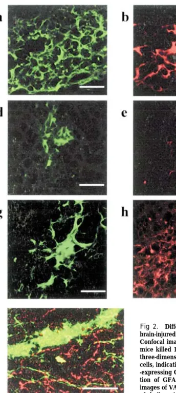
At 12 weeks (a week after the behavioral test) post-injury, surviving grafts could be identified in all mild TBI animals that received transplantation of neurospheres. Initial evaluation of the graft survival was conducted using eGFP-stained sections. None-theless, no surviving grafts could be identified in the severe TBI mice that received transplantation of neurospheres. To know whether the grafted ES-derived NPCs generated cholinergic neurons, we carried out immunohistochemical analysis of the brains of the transplanted mice. We found significant cholinergic differentiation recognized as ChAT im-munoreactivity in the eGFP+ transplanted cells in the mild TBI mice (Fig.2a, b, c). Moreover, the grafts contained little amount of GAD 67+cells (Fig.2 d, e, f). However, we barely found GFAP+ astrocytes within the grafts (Fig.2 g, h, i). Furthermore,
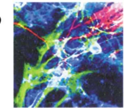
pre-Fig1. Immunocytochemistry of differenti-ated progeny of ES-derived NPCs.
Neurospheres (a) and positive control (b) were differentiated as described in the text. Scale bars = 50µm. Triple labeling (β-III-tubulin : red, GFAP : blue, and O4 : green) immuno-cytochemistry showed unipotent neuronal differentiation of clonally derived and mul-tipotent (neurons and glia) differentiation of positive control neurospheres. c) Differen-tiation of neurospheres into ChAT+ green) expressions in HuC/D+ (red) neurons.
T. Shindo, et al. Neural transplantation in injured brain
synaptic formations of graft-derived neurons were recognized by immunohistochemistry of vesicular acetylcholine transporter (VAChT) which is spe-cifically expressed on the axons of the cholinergic neurons, near the grafts around CA 3 (Fig.2 j, eGFP ; green, VAChT ; red).
mRNA Expression of neurotrophins
We examined NGF, BDNF, and FGF-2 mRNA by RT-PCR from 12 mouse including normal, mild TBI and severe TBI groups. Increases in the neurotrophic factors mRNA were evident in the
hippocampus on the ipsilateral side in the mild TBI group. Statistical analysis revealed significant differences between the mild and severe TBI groups. The data also revealed significant differences be-tween the mild TBI and normal groups (Fig. 3). The percentages of NGF, BDNF, and FGF-2 mRNA/β-actin mRNA were significantly higher in
the mild TBI group (68.2±15.3%, 94.7±45.6%, 48.4 ±15.4%)than in the severe TBI group (5.4±2.2%, 2.8±1.5%,1.8±1.9% ; p<0.01, p<0.01, p<0.01). The percentages of NGF, BDNF, and FGF-2 mRNA/β
-actin mRNA were significantly higher in the mild
Fig 2. Differentiation of ES-derived NPCs in the hippocampus of the brain-injured mice.
Confocal images of fluorescent immunohistochemistry obtained from the mice killed 12 weeks after transplantation. Scale bars=50µm. (a, b, c) A three-dimensional confocal image showing GFP+ (green)/ChAT+ (red) cells, indicating neuronal differentiation. (d, e, f) Differentiation of GAD 67 -expressing GABAergic neurons was barely observed. (g, h, i) Differentia-tion of GFAP-expressing astrocytes was barely observed. (j) Confocal images of VAChT immunohistochemistry showing presynaptic formations of cholinergic neurons derived from the grafted NPCs around CA 3.
TBI group than in the normal group (21.5±8.3%, 18.3±7.8%, 13.5±6.2% ; p<0.05, p<0.05, p<0.05).
Eight-arm radial maze testing
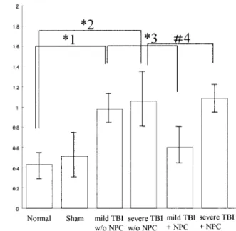
The mild TBI animals that received transplantation of neurospheres showed significantly improved learn-ing ability as compared with the severe TBI animals and no transplantation of neurospheres. Fig. 4 shows the mean scores of working memory errors and reference memory errors on the eight-arm radial maze performance in all 5 groups of mice, with data averaged from 18 trials. In the NBM-lesioned mice,
the working memory errors increased signifi-cantly (t=6.13, p<0.001 vs. the control group). In the mice transplanted with neurospheres, the scores were restored (t=0.307, p<0.05, vs. the control group). With respect to the scores of mice in the non-treated group with NPC transplants, the working memory deteriorated remarkably (t=5.95, p<0.001 vs. the normal group; t=6.46, p<0.001 vs. the NBM-lesioned group). There were no differences in the working memory errors between the NPC trans-plants group and non-treated group in the severe TBI (p>0.05).
DISCUSSION
One of major the findings in this study is that ES-derived NPCs exhibited similar differentiation potential both in vitro and in vivo. This is in contrast to the study by Wu et al. (21) who found” primed” human neural progenitors capable of generating multiple types of neurons including cholinergic neurons in vitro differentiated into neu-rons specific to the site of transplantation in the adult rat CNS. For example, the primed human NPCs generated only GABAergic neurons in the adult hippocampus (1) . In our present study, however, NPCs derived from mouse ESCs transplanted into the adult hippocampus generated many cholinergic and a few of GABAergic neurons but rarely astrocytes, like their in vitro differentiation. Strikingly, inner-vations of the graft-derived cholinergic axons into CA 3 regions which are the target sites of the septal cholinergic projections, synaptic formations and functional recovery from the TBI-induced learning Fig.3. mRNA Expression of neurotrophins.
We examined the expressions of NGF, BDNF, and FGF-2 mRNA by RT-PCR from 12 mice including normal, mild TBI and severe TBI groups. Increases in the neurotrophic factors mRNA were evident in the hippocampus on the ipsilateral side in the mild TBI group. Statistical analysis revealed significant differences between the mild and severe TBI groups. The data also revealed significant differences between the mild TBI and normal groups. The percentages of NGF, BDNF, and FGF-2 mRNA/β-actin mRNA were significantly higher in the mild TBI group (68.2±15.3 %, 94.7±45.6 %, 48.4±15.4 %) than in the severe TBI group (5.4±2.2 %, 2.8±1.5 %, 1.8±1.9 % ; p<0.01, p <0.01, p<0.01). The percentages of NGF, BDNF, and FGF-2 mRNA/β-actin mRNA were significantly higher in the mild TBI group than in the normal group (21.5±8.3 %, 18.3±7.8 %, 13.5 ±6.2 % ; p<0.05, p<0.05, p<0.05).
Fig.4. Effects of neurospheres and ESCs on the eight-arm radial maze performance of mice.
The mild TBI animals that received transplantation of neuro-spheres showed significantly improved learning ability as com-pared with the severe TBI animals and no transplantation of neurospheres. The mean scores of the working memory errors and reference memory errors on the 8-arm radial maze per-formance of all 6 groups of mice are shown, with data averaged from 18 trials. With respect to the scores of mice in the no-treatment group with NPC transplants, the working memory of TBI group mice deteriorated remarkably (*1,*2 : t=5.95, p< 0.001 vs. normal group ; t =6.46, p<0.001 vs the TBI group). In the mild TBI group mice transplanted with neurospheres, the scores were restored (*3 : t=0.307, p>0.05, vs. the NPC group). There were no differences in the working memory errors be-tween NPC transplants group and no-treatment group in severe TBI (#4 : p>0.05, F=0.1439, d. f. = 4.62)
T. Shindo, et al. Neural transplantation in injured brain
and memory deficits were observed. Thus, the ESCs can be excellent source for production of functional cholinergic neurons. However, we could not exclude the possibility that trophic effects from the grafts enhanced the function of the remaining septal cholinergic neurons and ameliorated the deficits.
Another important finding is that the neurosphere
-transplanted animals with mild TBI survived in
the hippocampus (CA 3). At 12 weeks after trans-plantation (a week after the behavioral test) , we found significant cholinergic differentiation recognized as ChAT immunoreactivity in the eGFP+ trans-planted cells. Moreover, the grafts contained few GAD 67+cells. However, we barely found GFAP+ astrocytes within the grafts (Fig. 2) . Moreover, pre-synaptic formations of graft-derived neurons were recognized by VAChT immunohistochemistry which is specifically expressed on the axons of the cholinergic neurons, near the grafts around CA 3 (Fig.3). However, these findings were not observed in the severe TBI group. These results might be due to differences in the microenvironment between the mild and severe TBI groups. So, we examined NGF, BDNF, and FGF-2 mRNA by RT-PCR from 12 mice including normal, mild TBI and severe TBI groups. Increases in the neurotrophic factors mRNA were evident in the hippocampus on the ipsilateral side in the mild TBI group. Statistical analysis revealed significant differences between the mild and severe TBI groups. The data also revealed significant dif-ferences between the mild TBI and normal groups (Fig. 3). The percentages of NGF, BDNF, and FGF-2 mRNA/β-actin mRNA were significantly
higher in the mild TBI group than in the severe TBI group. The percentages of NGF, BDNF, and FGF-2mRNA/β-actin mRNA were significantly higher
in the mild TBI group than in the normal group. Most of the surviving grafts in CA3 differentiated into cholinergic neurons. A few cells around the surviving grafts in CA 3 differentiated into GABAergic neurons. The surviving grafts might promote differentiation of the cells around them into GABAergic neurons.
It has been reported that transplantation of BDNF promoted differentiation into neurons after TBI (22) . Accordingly, BDNF might activate the NSCs and then lead to differentiation of NSCs into neurons. Hicks et al. noticed a moderate increase in BDNF mRNA expression in CA3 region of the hippocam-pus up to 12 hr post-injury and then remained above the control levels for 72 hr post-injury after mild FP injury (23) . A significant increase in BDNF mRNA expression was observed in CA 3 region and then
remained above the control levels after severe FP injury. Nonetheless, they did notice BDNF mRNA expression after 7 days post-injury. Our study showed that the transplanted neurosphere survived in the mild TBI animals, but not in the severe TBI group. These findings might correlate with the absence of neurotrophic factors in the hippocampus of the severe TBI animals. Several previous studies on ES cell transplantation examined the functional recovery following injury. It has been showed that minced fetal cortical grafts (E16) implanted after TBI in the rat significantly improved the neuromotor function and cognitive function in MWM as compared to rats injected with saline (5, 24). Significant improve-ments in the cognitive performance and neuromotor function were demonstrated with immortalized neural progenitors derived from the embryonic rat hip-pocampus (HIB5) , that were subsequently transfected to secrete NGF transplanted into the traumatically injured rat brain (1) . It has also been shown that transplantation of NT 2 N human neuronal cells transfected to secrete NGF can improve the cognitive function when transplanted into the striatum of brain-injured mice (25). On the contrary, trans-plantation of neuronal and glial precursors reportedly improves the sensorimotor function dramatically but not the cognitive function in the traumatically injured brain (26) . The latter investigators stated that the ESCs migrating to the hippocampus or striatum might have produced significant improve-ments in the cognitive function if testing was delayed for another couple of months. Thus, the improve-ments in the cognitive function following injury in the present study are consistent with the findings in other transplantation studies.
An involvement of the basal forebrain cholinergic system in the cognitive functions is evident. Nu-merous studies analyzed the functional roles of the basal forebrain cholinergic neurons and indicated their contribution to the learning and memory functions ; supporting the cholinergic hypothesis of memory dysfunction in the aged and Alzheimer’s disease (AD) patients, since loss of basal the forebrain cholinergic neurons or acetylcholine synthesis is often observed in the brains of patients with advanced dementia (27, 28). On the other hand, however, recent sensitive and reliable neurological analyses of AD patients in the early stages suggest that the cholin-ergic deficit occurs only in the late stage of the disease (29). The in vitro generation of functioning cholinergic neurons and their precursors presented here should facilitate the development of efficient
systems for drug development and screening. For further understanding of those diseases and devel-opment of medications, application of our system into the human ESCs, which provides another access to the biology of the human cholinergic neurons, could be an important step.
REFERENCES
1. Philips MF, Mattiasson G, Wieloch T, Bjorklund A, Johansson BB, Tomasevic G, Martinez-Serrano A, Lenzlinger PM, Sinson G, Grady MS, Mcintosh TK: Neuroprotective and behavioral efficacy of nerve growth-transfected hippocampal progenitor cell transplants after experimental traumatic brain injury. J Neurosurg 94 : 765-74, 2001 2. Wennersten A, Meijer X, Holmin S, Wahlberg L,
Mathiesen T : Proliferation, migration, and differentiation of human neural stem/progeni-tor cells after transplantation into a rat model of traumatic brain injury. J Neurosurg 100 : 88-96, 2004
3. Li HH, Lee SM, Cai Y, Sutton RL, Hovda DA : Differential gene expression in hippocampus following experimental brain trauma reveals distinct features of moderate and severe injuries. J Neurotrauma 21 (9) : 1141-1153, 2004 4. Pierce JES, Smith DH, Trojanowski JQ,
Mcintosh TK : Enduring cognitive neurobe-havioral and histopathological changes persisit for up to one year following severe experimental brain injury in rats. Neuroscience 87(2) : 359-369, 1998
5. Sinson G, Voddi M, Mcintosh TK : Combined fetal neural transplantation and nerve growth factor infusion: effects on neurological outcome following fluid-percussion brain injury in the rat. J Neurosurg 84 : 655-662, 1996
6. Hicks RR, Martin VB, Zhang L, Seroogy KB : Mild experimental brain injury differentially alters the expression of neurotrophin and neurotro-phin receptor mRNAs in the hippocampus. Experimental Neurology 160 : 469-478, 1999 7. Hagan M, Wennersten A, Meijer X, Holmin S,
Wahlberg L, Mathiesen T : Neuroprotection by human neural progenitor cells after experi-mental contusion in rats. Neuroscience Letters 351 : 149-152, 2003
8. Lewen A, Soderstom S, Hillered L, Ebendal T :Expression of serine/threonine kinase receptors in traumatic brain injury. Neuroreport 8 :
475-479, 1997
9. Hicks RR, Numan S, Dhillon HS, Prasad MR, Seroogy KB : Alterations in BDNF and NT-3 mRNAs in rat hippocampus after experimen-tal brain trauma. Molecular Brain Research 48 : 401-406, 1997
10. Ozawa K, Seo M, Imamura T : A quantitative method for evaluation of FGF family and FGF receptor family gene expression by RT-PCR. Brain Research Protocols 1 : 211-216, 1997 11. Riess P, Zhang C, Saatman KE, Laurer HL,
Loughi LG, Raghupathi R, Lenzlinger PM, Lifshitz J, Boockcvar J, Neugebauer E, Snyder EY, Mcintosh TK : Transplanted neural stem cells survive, differentiate, and improve neuro-logical motor function after experimental trau-matic brain injury. Neurosurgery 51 : 321-327, 2002
12. Hoane MR, Becerra GD, Shank JE, Tatko L, Pak ES, Smith M, Murashov AK : Transplan-tation of neuronal and glial precursors dramatically improves sensorimotor function but not cognitive function in the traumatically injured brain. J Neurotrauma 21 : 163-174, 2004 13. Lowe B, Avila HA, Bloom FR, Gleeson M,
Kusser W : Quantitation of gene expression in neural precursors by reverse-transcription po-lymerase chain reaction using self-quenched, fluorogenic primers. Analytical Biochemistry 315 : 95-105, 2003
14. Niwa H, Masui S, Chambers I, Smith AG, Miyazaki J : Phenotypic complementation es-tablished requirements for specific POU domain and generic transactivation function of Oct-3/4 in embryonic stem cells. Mol Cell Biol 22 : 1526-1536, 2002
15. Kawasaki H : Induction of midbrain dopamin-ergic neurons from ES cells by stromal cell-derived inducing activity. Neuron 28: 31-40, 2000
16. Lee SH, Lumelsky N, Studer L., Auerbach JM, McKay RD : Efficient generation of midbrain and hindbrain neurons from mouse embry-onic stem cells. Nat Biotechnol 18 : 675-679, 2000
17. Wichterle H, Lieberam I, Porter, JA , Jessell TM:Directed differentiation of embryonic stem cells into motor neurons. Cell 110 : 385-397, 2002
18. Marti E, Bovolenta P : Sonic hedgehog in CNS development. one signal, multiple outputs. Trends Neurosci 25 : 89-96, 2002
T. Shindo, et al. Neural transplantation in injured brain
19. Hodges H : Maze procedure : the radial-arm and water maze compared. Brain Res 3 : 167-181, 1996
20. Sakai K, Shimizu H, Koike T, Furuya, S, Watanabe M : Neutral amino acid transporter ASCT 1 is preferentially expressed in L-Ser-synthetic/storing glial cells in the mouse brain with transient expression in developing capillaries. J Neurosci 23 : 550-560, 2003 21. Wu P:Region-specific generation of cholinergic
neurons from fetal human neural stem cells grafted in adult rat. Nat Neurosci 5 : 1271-1278, 2002
22. Etchamendy N, Desmedt A, Torrea CC, Marighetto A, Jaffard R:Hippocampal lesions and discrimination performance of mice in the radial maze:sparing or impairment depending on the representa-tional demands of the task. Hippocampus 13 : 197-211, 2003
23. Rice AC, Khaldi A, Harvey HB, Salman NJ, White F, Fillmore H, Bullock MR : Proliferation and neuronal differentiation of mitotically active cells following traumatic brain injury. Experi-mental Neurology 183 : 406-417, 2003
24. Cao Q, Benton RL, Whittemore SR : Stemcell
repair of central nervous system injury. J Neuroscience Research 68 : 501-510, 2002 25. Watson DJ, Longhi L, Lee EB, Fulp CT, Fujimoto S,
Royo NC, Passini MA, Trojanowski JQ, Lee VM, Mcintosh TK, Wolfe JH : Genetically modified NT2N human neuronal cells mediate longterm gene expression as CNS grafts in vivo and improve functional cognitive outcome follow-ing experimental traumatic brain injury. J Neuropathol Exp Neurol 62 : 368-380, 2003 26. Riera NP, Oumesmar BN, Evercooren ABV :
Endogeneous adult neural stem cells : limits and potential to repair the injured central nervous system. J Neuroscience Research 76 : 223-231, 2004
27. Simeone A., Puelles E, Acampora D : The Otx family. Curr Opin Genet Dev 12 : 409-415, 2002 28. Wenk GL, Willard LB: The neural mechanisms underlying cholinergic cell death within the basal forebrain. Int J Dev Neurosci 16 : 729-735, 1998
29. Frolich L : The cholinergic pathology in Al-zheimer’s disease discrepancies between clinical experience and pathophysiological findings. J Neural Transm 109 : 1003-1013, 2002