Yoshiyuki Ashida,1 Akemichi Ueno, Yoshihiro Miwa, Keiko Miyoshi and Hideo Inoue2
Department of Biochemistry, School of Dentistry, Tokushima University, 3 Kuramoto-cho, Tokushima 770
Our previous study showed that treatment of highly invasive rat ascites hepatoma (LC-AH) cells with αααα-difluoromethylornithine (DFMO), an inhibitor of ornithine decarboxylase, decreased both their intracellular level of putrescine and their in vitro invasion of a monolayer of calf pulmonary arterial endothelial (CPAE) cells, and that both these decreases were completely reversed by exoge-nous putrescine, but not spermidine or spermine. Here we show that all adhering control (DFMO-untreated) cells migrated beneath CPAE monolayer with morphological change from round to cauli-flower-shaped cells (migratory cells). DFMO treatment increased the number of cells that remained round without migration (nonmigratory cells). Exogenous putrescine, but not spermidine or sper-mine, induced transformation of all nonmigratory cells to migratory cells with a concomitant increase in their intracellular Ca2+ level, [Ca2+]
i. The putrescine-induced increase in their [Ca 2+]
i
pre-ceded their transformation and these effects of putrescine were not affected by antagonists of the voltage-gated Ca2+ channel, but were completely suppressed by ryanodine, which also suppressed the
invasiveness of the control cells. The DFMO-induced decreases in both [Ca2+]
i and the invasiveness
of the cells were restored by thapsigargin, which elevated [Ca2+]
i by inhibiting endoplasmic Ca 2+
-ATPase, indicating that thapsigargin mimics the effects of putrescine. These results support the idea that putrescine is a cofactor for Ca2+ release through the Ca2+ channel in the endoplasmic reticulum
that is inhibited by ryanodine, this release being initiated by cell adhesion and being a prerequisite for tumor cell invasion.
Key words: Putrescine — Tumor invasion — Difluoromethylornithine — Ryanodine — Thapsigargin
The polyamines spermidine and spermine, and the di-amine putrescine, as well as normal constituents of all ani-mal cells. On the basis of their metabolism under various physiological conditions as well as the effects of specific inhibitors of polyamine biosynthesis, polyamines have been suggested to be important in various cellular func-tions such as proliferation, differentiation,1–4) cytoskeleton
construction,5) membrane transport6, 7) and receptor
res-ponses.8, 9) However, their physiological roles are not yet
well defined at the molecular level.
We previously reported that pretreatment of a highly in-vasive tumor cell line, LC-AH cells, with DFMO, a spe-cific irreversible inhibitor of ornithine decarboxylase (EC 4.1.1.17), decreased both their intracellular level of pu-trescine and their invasion beneath a CPAE cell monolayer, without change in their viability, proliferative activity or levels of spermidine and spermine.10) We also showed that
these DFMO-induced decreases were completely reversed by putrescine added to the culture medium during pretreat-ment with DFMO or during invasion assay in which LC-AH cells were co-cultured with a CPAE cell layer for 24 h.
Moreover, these reverses were observed only with pu-trescine, not with 1,3-diaminopropane, spermidine or sper-mine.10)
The LC-AH cells used in these studies have a very high cellular putrescine level (73 nmol/mg DNA) which is
simi-lar to their spermidine level (70 nmol/mg DNA).10) This
high putrescine level is a characteristic of these highly invasive LC-AH cells, the putrescine levels in a variety of
other tumor cells such as Ehrlich ascites,11) B16
mela-noma,12) Lewis lung carcinoma,13) and leukemia14) cells being
less than 30% of their spermidine levels. Pretreatment with 0.5 mM DFMO for 5 h induced a 64% decrease in their putrescine level without significant changes in their intra-cellular levels of spermidine and spermine.10) Although 0.5
mM DFMO completely inhibited the ornithine decarbox-ylase activity of LC-AH cells for more than 24 h, the pres-ence of 0.5 mM DFMO during invasion assay did not furhter reduce either their putrescine level or invasiveness, but significantly decreased their spermidine level (48 nmol/ mg DNA) without change in their spermine level (our unpublished data). This finding suggests formation of putrescine from spermidine through oxidation of acetyl-spermidine, as has been observed in a variety of animal
tissues and cultured cells,15) including
isoproterenol-stimulated mouse parotid gland,16) calcitriol-activated chick
duodenum,17) rat brain,18) and human melanoma cells.19) In
the former two in vivo studies, marked increases in tissue putrescine levels with concomitant decreases in spermidine levels were observed after injections of the hormones.
1 Present address: Department of Chemistry, Faculty of Science, Hiroshima University, 1-3-1 Kagamiyama, Higashihiroshima 739. 2 To whom correspondence should be addressed.
Abbreviations: LC-AH, a subline of rat ascites hepatoma; DFMO, 2-(difluoromethyl)ornithine; CPAE, calf pulmonary arterial endo-thelial; DMEM, Dulbecco’s modified Eagle’s medium; FCS, fetal calf serum; PBS, Dulbecco’s phosphate-buffered saline; [Ca2+]
i, intracellular calcium concentration; 1-AM, 1-acetoxymethyl ester.
These high cellular levels of putrescine and back-conver-sion of spermidine to putrescine, which is followed by
induction of ornithine decarboxylase,16, 17) imply that
putrescine is involved in cellular events besides being a precursor of spermidine. However, its exact function is unknown.
In this paper, we investigated the role of putrescine in the invasiveness of LC-AH cells, and showed that it partici-pates in elevation of [Ca2+]
i through a ryanodine receptor
located in the endoplasmic reticulum.
MATERIALSANDMETHODS
Materials Indo 1-AM was purchased from Dojindo
Lab-oratories (Kumamoto). Ionomycin, A23187, verapamil, nifedipine, diltiazem, ryanodine and thapsigargin were obtained from Wako Pure Chemical Industries (Osaka). The sources of other chemicals, and LC-AH and CPAE cells were as reported previously.10)
Cell culture and assay of cell invasiveness These assays
were done as reported previously.10) Briefly, LC-AH and
CPAE cells were cultured at 37°C in a CO2 incubator in
DMEM supplemented with NaHCO3, antibiotics and 10%
(LC-AH cells) or 20% (CPAE cells) FCS. For treatment of LC-AH cells with DFMO, the cells (1.0×10⁶ cells/ml) were
cultured in 10% FCS-DMEM containing 0.5 mM DFMO for 5 h and then washed with fresh medium to remove
DFMO. Volumes of 1 ml of LC-AH cell suspension (2×10⁴
cells) were seeded onto confluent CPAE cell monolayers grown on 12-well plates (3.8 cm2). After co-culture for 24
h (migration period) in the absence or presence of putrescine or test drugs, cells were fixed with 10% forma-lin, and the numbers of tumor cells that had penetrated the CPAE cell layers were counted under a phase-contrast microscope.
Assay of cell adhesiveness The adhesion of the tumor
cells to the CPAE cell layer was determined as follows. LC-AH cells (1×10⁵ cells) suspended in 4 ml of 10%
FCS-DMEM were seeded onto confluent CPAE cell layers in 60 mm dishes. After co-culture for 0.5–6 h, the culture me-dium was removed and the CPAE cell layers were gently washed with 4 ml of fresh DMEM. The numbers of nonad-hered cells in the combined media were counted under a microscope and the CPAE cell layers were cultured further for 24 h in 4 ml of 10% FCS-DMEM to assay the invasive-ness of LC-AH cells that had adhered to the CPAE cell layers. Cell adhesion was calculated by subtracting the number of nonadhered cells from the number of cells seeded (1×10⁵ cells).
Assay of cell membrane fluidity Membrane fluidity was
assessed by fluorescence polarization as reported previ-ously.20) LC-AH cells treated without or with drug for 5 h
were washed twice with PBS, suspended at a concentration of 1×10⁵ cells/ml in PBS containing 1.5 µM
1,6-diphenyl-1,3,5-hexatriene as a fluorescence probe and incubated for 2 h at 37°C. Fluorescence intensity was measured at 37°C in a Hitachi fluorescence spectrophotometer equipped with a polarization accessory.
Morphological observation Cell morphology was
exam-ined by a slight modification of the method of Akedo et
al.21) LC-AH cells adhered to a CPAE cell layer were fixed
with 3% glutaraldehyde in 0.1 M cacodylate buffer (pH 7.2) for 1 h and then treated with 1% OsO4 for 2 h. After dehydration with ethanol, the samples were dried in a crit-ical point drier (Hitachi HCP-2), coated with gold and observed under a Hitachi H-800 scanning electron micro-scope. For light microscopic observations, the dehydrated samples were embedded in Epon 812, sectioned perpendic-ularly and stained with 1% methylene blue in 1% sodium borate.
Measurement of [Ca2+]
i The [Ca
2+]
i of LC-AH cells was
measured by a modification of the fluorescence method of
Luckhoff.22) Briefly, control or DFMO-treated LC-AH
cells were incubated for 30 min at 37°C in Hanks’ balanced salt solution containing 2 µM indo 1-AM. Then they were
washed twice with the same salt solution, and suspended in
10% FCS-MEM and samples of 1 ml of the cells (2×10⁴
cells) were seeded onto confluent CPAE cells grown in 35 mm culture dishes with a glass bottom (Meridian Instru-ments, Inc., Okemos, MI). After co-culture for 2 h, the me-dium was carefully replaced by Hanks’ balanced salt solution and the fluorescence intensity and video image of the LC-AH cells adhering to the CPAE cell layers were monitored simultaneously for 10 min with an ACAS 570 interactive laser cytometer (Meridian Instruments, Inc.). Longer monitoring was not possible for technical reasons. For measurement of [Ca2+]
i in LC-AH cells that had
pene-trated the CPAE cell layer, indo 1-AM was loaded into the co-cultured cells 24 h after inoculation of LC-AH cells and the fluorescence intensity in the tumor cells was measured as described above.
The results in figures are shown as typical traces of re-sults in at least three separate experiments.
Polyamine determination DFMO-treated cells (1×10⁶
cells) were suspended in 1 ml of 10% FCS-DMEM and
cultured at 37°C for 0–20 min in the presence of 20 µM
putrescine. Polyamines were measured as described
previously.10)
Statistical analysis Results were compared by Student’s t
test and a P value of 0.05 or less was regarded as signifi-cant.
RESULTS
Effects of DFMO and putrescine on membrane fluidity, adhesion and invasiveness of LC-AH cells We found
previously that the maximal DFMO-induced decreases in the putrescine level (36% of the control) and invasiveness
(62% of the control) of LC-AH cells were observed when the cells had been pretreated with 0.5 mM DFMO for 5 h and then co-cultured with a CPAE cell monolayer for 24 h (migration period) in the absence of DFMO. These
de-creases were completely reversed by 20 µM putrescine
added to the culture medium during pretreatment or the migration period.10) We also showed that
methylthioade-nosine and its analogues suppressed the invasiveness of LC-AH cells and that this suppression was associated with reductions of both methylation of tumor membrane phos-pholipids and tumor membrane fluidity, measured by a steady-state fluorescence polarization method.20)
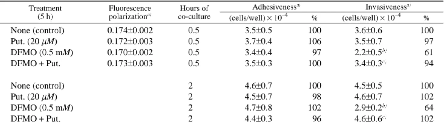
The process of in vitro invasion of LC-AH cells through a CPAE cell layer consists of two steps, adhesion of the tu-mor cells to the cell layer and their migration through it. Neither DFMO nor exogenous putrescine alone affected the membrane fluidity or the adhesiveness of LC-AH cells (Ta-ble I). Under the experimental conditions employed, about 35% of the inoculated cells adhered to a CPAE cell layer within 30 min after their inoculation, and this value in-creased to a maximum of 46% within 2 h after cell inocula-tion. No further increase was observed by 6 h after cell inoculation (data not shown). But this maximal ratio (46%) of adherent cells increased when a smaller number of LC-AH cells was inoculated onto the same-sized CPAE cell monolayer (unpublished data), suggesting a limited number of binding sites in the CPAE cell layer for tumor cells. In control (DFMO-untreated) and putrescine-treated cells which showed high putrescine levels,10) all these adherent
cells migrated through the CPAE cell layer (migratory cells) during the migration period. In DFMO-treated cells,
however, 39% (0.5 h cultured cells) and 36% (2 h co-cultured cells) of the adhered cells did not migrate (nonmi-gratory cells), but remained adhering to the CPAE cell layer after 24 h incubation (Table I). These nonmigratory cells showed no migration even 30 h after inoculation. Addition
of 20 µM putrescine to the medium during the migration
period restored cell invasiveness and all of the nonmigra-tory cells began to penetrate the CPAE cell layer (Table I). Thus, DFMO treatment induced nonmigratory cells, which were transformed to migratory cells in the presence of pu-trescine.
As shown in Fig. 1, there were marked morphological and cytochemical differences between nonmigratory and migratory cells: the former were round with a low [Ca2+] i
(1.5–1.7×10⁻⁷ M), while the latter had a cauliflower-like
shape with a higher [Ca2+]
i (2.2–3.1×10
−7 M). The [Ca2+] i
of all the cells that had penetrated the CPAE cell layer (penetrating cells) was again low (1.6–2.0×10⁻⁷ M).
Simul-taneous observations of fluorescence intensity and mor-phology of single control (DFMO-untreated) LC-AH cells with an ACAS interactive laser cytometer showed that an increase in [Ca2+]
i was apparent 10 min after cell addition
and this increase preceded morphological transformation of round cells to a cauliflower-like shape, which began about 30 min after cell addition (data not shown). Control LC-AH cells also adhered to a glass or plastic surface, but in these cases, showed neither increase in [Ca2+]
i nor
cauli-flower-like transformation (data not shown).
Effects of polyamines on [Ca2+]
i and putrescine levels in
DFMO-treated LC-AH cells The above findings
sug-gested a relationship between invasiveness and the [Ca2+] i
Table I. Effects of DFMO and Putrescine on Fluorescence Polarization, Adhesiveness, and Invasiveness of LC-AH Cells
Treatment (5 h)
Fluorescence
polarizationa) co-cultureHours of
Adhesivenessa) Invasivenessa) (cells/well) × 10–4 % (cells/well) × 10–4 % None (control) 0.174±0.002 0.5 3.5±0.5 100 3.6±0.6 100 Put. (20 µM) 0.172±0.003 0.5 3.7±0.4 106 3.5±0.7 97 DFMO (0.5 mM) 0.170±0.002 0.5 3.4±0.4 97 2.2±0.5b) 61 DFMO + Put. 0.173±0.003 0.5 3.5±0.3 100 3.4±0.3c) 94 None (control) 2 4.6±0.7 100 4.5±0.5 100 Put. (20 µM) 2 4.5±0.7 98 4.6±0.7 102 DFMO (0.5 mM) 2 4.7±0.8 102 2.9±0.2b) 64 DFMO + Put. 2 4.4±0.3 96 4.6±0.6c) 102
LC-AH cells (1×10⁶ cells/ml of 10% FCS-DMEM) were treated with 0.5 mM DFMO and 20 µM putrescine (Put.) for 5 h, and then washed with PBS or 10% FCS-DMEM to remove the drugs. Fluorescence polarization was calculated from the fluorescence intensity of PBS-washed cells as described in “Materials and Methods.” The cells (1×10⁵ cells) washed with 10% FCS-DMEM were suspended in 4 ml of the same medium and co-cultured for 0.5 or 2 h with CPAE cell layers grown on 60 mm dishes. Then cell adhesiveness and invasiveness were determined as described in “Materials and Methods.”
a) Values are means±SD’s for 4 separatem experiments.
b) Significantly different from the control value. c) Putrescine was added during the migration period.
of the cells. Therefore, we examined changes in the [Ca2+] i
of DFMO-induced nonmigratory LC-AH cells after addi-tions of polyamines. Fig. 2 shows that on addition of pu-trescine, the [Ca2+]
i of nonmigratory cells was increased to
the same level as in migratory cells within 10 min, with complete recovery of their invasiveness (Table I). How-ever, addition of spermidine or spermine did not increase the [Ca2+]
i or the invasiveness of the cells.
10) Putrescine did
not cause a further increase in the high [Ca2+]
i of migratory
cells that had not been treated with DFMO (data not shown). Moreover, 1,3-diaminopropane and 1,5-diamino-pentane (cadaverine) did not cause significant recovery of either the Ca2+ level or invasiveness of DFMO-treated
non-migratory cells (data not shown).
We could not measure the putrescine levels of DFMO-treated migratory cells after addition of putrescine because the adhered migratory cells were not easily detached from the CPAE cell layer. When DFMO-treated LC-AH cells were cultured at 37°C without the CPAE cell layer and polyamine levels were followed after addition of putrescine
by the method described in “Materials and Methods,” the putrescine levels (nmol/µg DNA, average values of two ex-periments) were 21.3 (0 min), 43.8 (5 min), 64.3 (10 min), and 80.1 (20 min), indicating that the level 10 min after ad-dition of putrescine was almost the same as that of DFMO-untreated cells (70.5 nmol/µg DNA). No changes in sper-midine and spermine levels were observed. These results suggested that uptake of exogenous putrescine was associ-ated with increase of [Ca2+]
i in DFMO + putrescine-treated
migratory cells.
Effects of Ca2+ channel modulators on the invasiveness
and [Ca2+]
i of LC-AH cells Next we tested the effects of various drugs that modulate cytosolic Ca2+ homeostasis on
the invasiveness of the tumor cells. The drugs tested were
Ca2+ ionophores (ionomycin and A23187), antagonists
(verapamil, nifedipine and diltiazem) of the voltage-gated Ca2+ channel, a blocker (ryanodine) of cyclic
ADP-ribose-triggered Ca2+ release from the endoplasmic reticulum,23)
and an inhibitor (thapsigargin) of endoplasmic reticulum Ca2+-ATPase.24) These drugs were added to the culture
me-dium during the migration period. Fig. 3 shows that none of the calcium ionophores (10 µM) or antagonists (20 µM) of
the voltage-gated Ca2+ channel affected the invasiveness of
control cells, the DFMO-dependent suppression of invasion or the putrescine-dependent restoration of DFMO-induced suppression of the invasion. These calcium channel antago-nists were reported to be effective in various tumor cells at concentrations of less than 20 µM.25–29) On the other hand,
ryanodine and thapsigargin, which modulate Ca2+ release in
the endoplasmic reticulum, had definite influences on the invasiveness. Ryanodine concentration-dependently
low-Fig. 1. Morphology (A) and [Ca2+]
i (B) of LC-AH cells that ad-hered to and penetrated a CPAE cell layer. For A, DFMO-treated (a) and control DFMO-untreated (b and c) LC-AH cells were cul-tured on CPAE cell layers for 2 (b) or 24 (a and c) h. The mor-phologies of cells that adhered to (a and b) and penetrated (c) a CPAE cell layer were observed by scanning electron (a and b) and light (c) microscopes as described in “Materials and Methods.” For B, indo 1-AM was loaded at 0 h (b) or at 24 h (a and c). Col-umns and bars show means and SD’s, respectively, of results in 3 separate experiments. a, a DFMO-treated nonmigratory cell at 24 h; b, a control migratory cell at 2 h; c, a control penetrated cell at 24 h. Scale bar, 10 µm. ∗ Significant difference from (a).
Fig. 2. Effects of polyamines on [Ca2+]
i in DFMO-treated nonmi-gratory cells. DFMO-treated cells were loaded with indo 1-AM as described in “Materials and Methods” and co-cultured on CPAE cell layers for 2 h. The [Ca2+]
i of nonmigratory cells that adhered to the CPAE cell layer was followed after no addition (a), or addition (at the arrow) of 20 µM putrescine (b), spermidine (c) or spermine (d).
ered the invasiveness of control cells and cells treated with DFMO with or without putrescine (Figs. 3 and 4A). Ryan-odine almost completely counteracted the effects of pu-trescine on [Ca2+]
i (Fig. 4B) and the invasiveness of the
cells (Fig. 4A), and the ryanodine-treated nonmigratory cells also had a low [Ca2+]
i (data not shown). In other
words, the [Ca2+]
i remained low in all the nonmigratory
cells induced by either DFMO or ryanodine. In contrast, thapsigargin alone dose-dependently enhanced the inva-siveness of DFMO-treated cells (Fig. 4C) with elevation of their [Ca2+]
i in the absence of putrescine (Fig. 4D). But
thapsigargin alone had no effect on the invasiveness of con-trol cells (Fig. 3A). These effects of thapsigargin were sim-ilar to those of putrescine. Ionomycin had no effect on the invasiveness (Fig. 3), but increased the [Ca2+]
i. Fig. 5
shows the surge of increase in [Ca2+]
i in a DFMO-induced
nonmigratory cell after addition of ionomycin to the me-dium during the migration period. This pattern of increase in [Ca2+]
i was markedly different from those induced by
pu-trescine and thapsigargin (Fig. 4D). The maximal [Ca2+] i
1 min after addition of ionomycin was 3 times that 10 min after addition of putrescine or thapsigargin, but the iono-mycin-induced increase lasted for only 2 min whereas the putrescine- and thapsigargin-induced increase persisted for at least the 10 min measurement period (Figs. 2, 4 and 5). These results suggest that a prolonged increase in [Ca2+]
i is
necessary for restoration of invasiveness after DFMO treat-ment. Nifedipine did not suppress the putrescine-evoked increase in [Ca2+]
i in nonmigratory cells (Fig. 5). DISCUSSION
The present studies by ACAS laser cytometry and mi-croscopy showed that all control LC-AH cells that adhered
to a CPAE cell layer showed an increase in [Ca2+]
i,
fol-lowed by change from a round to a cauliflower-like shape (Fig. 1), and then migration through the CPAE cell layer. After penetration through the CPAE cell layer, the cells again had a low [Ca2+]
i (Fig. 1). We found no transformed
cells (migratory cells) with a low [Ca2+]
i and no
nonmigra-tory cells or cells that had penetrated the cell layer with a high [Ca2+]
i at any time after cell inoculation. These
find-ings suggest that migratory cells maintain a high [Ca2+] i
during their migration period and that this level returns to the original low level after their invasion of the CPAE cell
layer. In DFMO-induced nonmigratory cells, the [Ca2+]
i
was rapidly elevated for a prolonged time to the level in migratory cells by addition of putrescine, but not spermi-dine or spermine (Fig. 2). This implies that a high intracel-lular level of putrescine is necessary for the increase in [Ca2+]
i evoked by adhesion of the tumor cells to the CPAE
cell layer and is prerequisite for their invasion of the layer.
Fig. 3. Effects of Ca2+ channel modulators on the invasiveness of LC-AH cells. LC-AH cells were incubated for 5 h in the absence (A) or presence (B) of 0.5 mM DFMO or 0.5 mM DFMO plus 20 µM putrescine (C). Then they were washed and their invasiveness was
determined in the absence or presence of 10 µM Ca2+
ionophores, 20 µM antagonists of the voltage-gated Ca2+
channel, 10 µM ryanodine
or 5 µM thapsigargin. Columns and bars show means and SD’s, respectively, of results in 3 separate experiments. ∗ Significantly differ-ent from the control value (none).
This implication was supported by the findings that [Ca2+] i
and the invasiveness of the cells were concomitantly de-creased and inde-creased by ryanodine and thapsigargin, re-spectively (Figs. 3 and 4).
The putrescine-dependent increase in [Ca2+]
i in
DFMO-treated nonmigratory cells was probably due to release of Ca2+ from the endoplasmic reticulum because this increase
was not affected by nifedipine (Fig. 5) which inhibits influx of extracellular Ca2+ into the cells, but was completely
inhibited by ryanodine (Fig. 4B), a blocker of the Ca2+
-induced Ca2+-release system through the endoplasmic
ryan-odine receptor.23) The possibility of participation of
putre-scine-dependent Ca2+ release from the endoplasmic
reticu-lum in induction of invasiveness was supported by the putrescine-like effects of thapsigargin (Figs. 3 and 4), which is thought to restore the invasiveness of nonmigra-tory cells due to increase in their [Ca2+]
i by decrease in
influx of cytoplasmic Ca2+ into the endoplasmic reticulum
through the inhibition of endoplasmic Ca2+-ATPase.24)
There are reports that cyclic ADP-ribose is a natural ago-nist of the ryanodine channel in a variety of animal cells30–35)
and that this channel is independently regulated by other endoplasmic reticulum-located Ca2+-releasing systems that
are triggered by inositol 1,4,5-trisphosphate,33–35) and
nico-tinate adenine dinucleotide phosphate.36–38) The complete
inhibitions by ryanodine of putrescine-dependent recover-ies of the [Ca2+]
i and invasiveness of nonmigratory cells
sug-gest that putrescine is involved in either the increase in the cellular level of cyclic ADP-ribose which is initiated by the adhesion of LC-AH cells to a CPAE cell layer or the func-tional expression of this cyclic nucleotide. Bourguignon et
al.39) reported that binding of the ryanodine receptor to
ankyrin, a cytoskeleton protein, evoked cyclic ADP-ribose-induced Ca2+ release from internal vesicles of T-lymphoma Fig. 4. Effects of ryanodine (A and B) and thapsigargin (C and D) on the invasiveness and [Ca2+]
i of LC-AH cells. The invasiveness (A and C) of control (open circles) and DFMO-treated cells was determined in the absence (closed circles) or presence (closed triangles) of 20 µM putrescine and the indicated concentrations of ryanodine (A) or thapsigargin (C). Points indicate mean values in 3–5 separate
ex-periments. SD’s were all less than 11% of the means. ∗ Significantly different from the control (no addition). In experiments on [Ca2+] i (B and D), DFMO-treated and indo 1-AM-loaded LC-AH cells were co-cultured with CPAE cell layers for 2 h. The [Ca2+
]i of nonmigra-tory cells was monitored after additions (at the arrow) of 20 µM putrescine (B and D), 10 µM ryanodine (B) or 5 µM thapsigargin (D).
cells. In connection with the functional expression, Chini
et al.40) reported that cyclic ADP-ribose-induced Ca2+
release in a homogenate of sea urchin eggs was strongly inhibited by spermine, but not putrescine. There are also reports that among natural polyamines, spermine is the most effective in modulating voltage-activated,41–43)
N-methyl-D-aspartate-mediated44) and inositol
trisphosphate-stimulated45) Ca2+ channels, while putrescine has little or no
effect. But these in vitro effects of spermine and spermi-dine observed at high concentrations (mM range) without depletion by specific inhibitors may be explained by their polybasic natures, resulting in their high affinities to nega-tively charged cellular structures such as biomembranes and nucleic acids and low-molecular-weight metabolites. With regard to the latter, Mernissi-Arifi et al.46) reported the
formation of stable complexes of spermine and spermidine with inositol trisphosphate. Moreover, there are reports that the cellular spermine level does not change appreciably under a variety of physiological conditions1, 16, 17) in which
the putrescine level is markedly elevated. Therefore, the relationship between the observed effects of high concen-trations of spermine in vitro and their physiological regula-tory functions in vivo must be considered with caution.1)
Recently, Khan et al.47) reported that KCl
depolarization-induced serotonin release from fish brain synaptosomes is mediated by KCl-stimulated influx of Ca2+ into the
synap-tosomes from the extracellular medium and that DFMO-pretreatment resulted in decreases of both Ca2+ influx and
serotonin release which were restored by putrescine, the tissue content of which is higher than those of spermidine
and spermine in various parts of fish brain. Shinki et al.48)
reported that in vitamin D-deficient chick small intestine, treatment with calcitriol resulted in marked elevation of the putrescine level, mainly due to back-conversion of spermi-dine,17) and stimulation of Ca2+ absorption that is thought to
be accompanied by enhanced Ca2+ passage across both
mu-cous and serous membranes of the epithelial cells. Lower-ing of the putrescine level by inhibitors of polyamine
oxidase and ornithine decarboxylase decreased Ca2+
ab-sorption. These and the present results support the idea that putrescine is a cofactor for the Ca2+ current through
second messenger-stimulated or voltage-gated Ca2+
chan-nels present in either the endoplasmic reticulum or plasma membrane in putrescine-rich cells.
The function of the putrescine-dependent increase in [Ca2+]
i is unknown. Possibly this multifunctional messenger
participates in at least cell migration and morphological transformation of tumor cells and their secretion or exocy-tosis of factors that stimulate their invasiveness, because these cellular events are thought to be accompanied by re-arrangement of the actin-based cytoskeleton regulated by actin-binding proteins that are modulated by changes in [Ca2+]
i.
49–51) For example, with regard to the relationship
be-tween polyamines and cell migration, McCormack et
al.52, 53) found that DFMO treatment reduced the migration
of small intestinal crypt cells concomitantly with decreases in their actin stress fibers and lamellipodia due to redistri-bution of F-actin and tropomyosin from stress fibers to the actin cortex, and that their reduced migration was com-pletely restored by exogenous polyamines, putrescine being the essential polyamine for their migration. Because the endothelial cell layer, basement membrane and extracellu-lar matrix are barriers against tumor cell invasion, tumor cells produce and secrete factors that weaken or remove these barriers. These factors include an endothelial cell re-traction factor54) and a matrix metalloproteinase-2 whose
transcription in human cancer cells is enhanced by recep-tor-operated Ca2+ influx.55)
In summary, we have demonstrated a novel function of putrescine in Ca2+ release from the ryanodine channel. The
precise mechanism of this action of putrescine is unknown, but the findings that 1,3-diaminopropane and 1,5-diamino-pentane had scarcely any effect and the demonstration of an early response (within 2 min, Figs. 2, 4 and 5) of [Ca2+]
i to
putrescine imply that putrescine interacts with a specific site on the cyclic ADP-ribose binding receptor.
ACKNOWLEDGMENTS
The authors thank Drs. K. Yamashita and S. Kitamura of our School and Drs. K. Shinkai and H. Akedo of The Center for Adult Diseases, Osaka, for technical assistance.
(Received August 1, 1997 / Revised October 8, 1997 / Accepted October 16, 1997)
Fig. 5. Effects of ionomycin and nifedipine on [Ca2+]
i of LC-AH cells. DFMO-treated and indo 1-AM-loaded LC-AH cells were cultured on CPAE cell layers for 2 h. The [Ca2+]
i of nonmigratory cells was followed after no addition (a), or addition (at the arrow) of 10 µM ionomycin (b), 20 µM putrescine (c) or 20 µM
REFERENCES
1) Jänne, J., Pösö, H. and Raina, A. Polyamines in rapid growth and cancer. Biochim. Biophys. Acta, 473, 241–293 (1978). 2) Löwkvist, B., Oredsson, S. M., Holm, I., Emanuelsson, H.
and Heby, O. Inhibition of polyamine synthesis reduces the growth rate and delays the expression of differentiated phe-notypes in primary cultures of embryonic mesoderm from chick. Cell Tissue Res., 249, 151–160 (1987).
3) Pegg, A. E. Polyamine metabolism and its importance in neoplastic growth and as a target for chemotherapy. Cancer
Res., 48, 759–774 (1988).
4) Jänne, J., Alhonen, L. and Leinonem, P. Polyamines: from molecular biology to clinical application. Ann. Med., 23, 241–259 (1991).
5) Pohjanpelto, P., Virtanen, I. and Höttä, E. Polyamine star-vation causes disappearance of actin filaments and microtu-bules in polyamine-auxotrophic CHO cells. Nature, 293, 475–477 (1981).
6) Trout, J. J., Lu, C. Y., Goldstone, A. D., Iqbal, Z. and Koenig, H. Polyamines mediate coronary transcapillary mac-romolecular transport in the calcium paradox. J. Mol. Cell.
Cardiol., 26, 369–377 (1994).
7) Johnson, L. R., Brockway, P. D., Madsen, K., Hardin, J. A. and Gall, D. G. Polyamines alter intestinal glucose trans-port. Am. J. Physiol., 268, G416–G423 (1995).
8) Subramaniam, S. and McGonigle, P. Regional heterogene-ity of polyamine effects on the N-methyl-D-aspartate recep-tor in rat brain. J. Neurochem., 60, 2276–2284 (1993). 9) Marvizon, J.-C. and Baudry, M. Allosteric interactions and
modulator requirement for NMDA receptor function. Eur. J.
Pharmacol., 269, 165–175 (1994).
10) Ashida, Y., Kido, J., Kinoshita, F., Nishino, M., Shinkai, K., Akedo, H. and Inoue, H. Putrescine-dependent invasive ca-pacity of rat ascites hepatoma cells. Cancer Res., 52, 5313– 5316 (1992).
11) Andersson, G. and Heby, O. Polyamine and nucleic acid concentrations in Ehrlich ascites carcinoma cells and liver of tumor-bearing mice at various stages of tumor growth. J.
Natl. Cancer Inst., 48, 165–172 (1972).
12) Kubota, S., Ohsawa, N. and Takaku, F. Effects of D,L-α -di-fluoromethylornithine on the growth and metastasis of B16 melanoma in vivo. Int. J. Cancer, 39, 244–247 (1987). 13) Sunkara, P. S. and Rosenberger, A. L. Antimetastatic
activ-ity of D,L-α-difluoromethylornithine, an inhibitor of poly-amine biosynthesis, in mice. Cancer Res., 47, 933–935 (1987).
14) Nakaike, S., Kashiwagi, K., Terao, K., Iio, K. and Igarashi, K. Combined use of α-difluoromethylornithine and an inhib-itor of S-adenosylmethionine decarboxylase in mice bearing P388 leukemia or Lewis lung carcinoma. Jpn. J. Cancer
Res. (Gann), 79, 501–508 (1988).
15) Casero, R. A., Jr. and Pegg, A. E. Spermidine/spermine N1 -acetyltransferase: the turning point in polyamine metabo-lism. FASEB J., 7, 653–661 (1993).
16) Inoue, H., Asada, A., Kato, Y. and Takeda, Y.
Intercon-version of aliphatic polyamines in isoproterenol-stimulated mouse parotid glands. J. Biochem., 84, 719–725 (1987). 17) Shinki, T., Tanaka, H., Kadofuku, T., Sato, T. and Suda, T.
Major pathway for putrescine synthesis induced by 1α ,25-di-hydroxyvitamin D3 in chick duodenum. Gastroenterology, 96, 1494–1501 (1989).
18) Zoli, M., Pedrazzi, P., Zini, I. and Agnati, L. F. Spermidine/ spermine N1-acetyltransferase mRNA levels show marked and region-specific changes in the early phase after transient forebrain ischemia. Mol. Brain Res., 38, 122–134 (1996). 19) Shappell, N. W., Fogel-Petrovic, M. F. and Porter, C. W.
Regulation of spermidine/spermine N1-acetyltransferase by intracellular polyamine pools: evidence for a functional role in polyamine homeostasis. FEBS Lett., 321, 179–183 (1993). 20) Kido, J., Ashida, Y., Shinkai, K., Akedo, H., Isoai, A., Kumagai, H. and Inoue, H. Effects of methylthiodeoxyade-nosine and its analogs on in vitro invasion of rat ascites hepatoma cells and methylation of their phospholipids. Jpn.
J. Cancer Res., 82, 1104–1111 (1991).
21) Akedo, H., Shinkai, K., Mukai, M., Mori, Y., Tateishi, R., Tanaka, K., Yamamoto, R. and Morishita, T. Interaction of rat ascites hepatoma cells with cultured mesothelial cell lay-ers: a model for tumor invasion. Cancer Res., 46, 2416–2422 (1986).
22) Luckhoff, A. Measuring cytosolic free calcium concentra-tion in endothelial cells with indo-1: the pitfall of using the ratio of two fluorescence intensities recorded at different wavelengths. Cell Calcium, 7, 233–248 (1986).
23) Wier, W. G., Yue, D. T. and Marban, E. Effects of ryanodine on intracellular Ca2+ transients in mammalian cardiac mus-cle. Fed. Proc., 44, 2989–2993 (1985).
24) Christensen, S. B., Andersen, A., Poulsen, J.-C. J. and Treiman, M. Derivatives of thapsigargin as probes of its binding site on endoplasmic reticulum Ca2+ ATPase: stereo-selectivity and important functional groups. FEBS Lett., 335, 345–348 (1993).
25) Horio, M., Lovelace, E., Pastan, I. and Gottesman, M. M. Agents which reverse multidrug-resistance are inhibitors of [3H]vinblastine transport by isolated vesicles. Biochim.
Bio-phys. Acta, 1061, 106–110 (1991).
26) Taylor, J. M. and Simpson, R. U. Inhibition of cancer cell growth by calcium channel antagonists in the athymic mouse. Cancer Res., 52, 2413–2418 (1992).
27) Pancrazio, J. J., Oie, H. K. and Kim, Y. I. Voltage-sensitive calcium channels in a human small-cell lung cancer cell line.
Acta Physiol. Scand., 144, 463–468 (1992).
28) Huet, S., Chapey, C. and Robert, J. Reversal of multidrug resistance by a new lipophilic cationic molecule, S9788: comparison with 11 other MDR-modulating agents in a model of doxorubicin-resistant rat glioblastoma cells. Eur. J.
Cancer, 29A, 1377–1383 (1993).
29) Todd, D. G. and Mikkelsen, R. B. Ionizing radiation induces a transient increase in cytosolic free [Ca2+] in human epithe-lial tumor cells. Cancer Res., 54, 5224–5230 (1994).
30) Lee, H. C., Aarhus, R., Graeff, R., Gurnack, M. E. and Walseth, T. F. Cyclic ADP ribose activation of the ryano-dine receptor is mediated by calmodulin. Nature, 370, 307– 309 (1994).
31) Thorn, P., Gerasimenko, O. and Petersen, O. H. Cyclic ADP-ribose regulation of ryanodine receptors involved in agonist evoked cytosolic Ca2+ oscillations in pancreatic aci-nar cells. EMBO J., 13, 2038–2043 (1994).
32) Sitsapesan, R., McGarry, S. J. and Williams, A. J. Cyclic ADP-ribose, the ryanodine receptor and Ca2+ release. Trends
Pharmacol. Sci., 16, 386–391 (1995).
33) Gromada, J., Jorgensen, T. D. and Dissing, S. Cyclic ADP-ribose and inositol 1,4,5-triphosphate mobilize Ca2+ from distinct intracellular pools in permeabilized lacrimal acinar cells. FEBS Lett., 360, 303–306 (1995).
34) Yue, C., White, K. L., Reed, W. A. and Bunch, T. D. The existence of inositol 1,4,5-trisphosphate and ryanodine re-ceptors in mature bovine oocytes. Development (Camb.), 121, 2645–2654 (1995).
35) Gerasimenko, O. V., Gerasimenko, J. V., Belan, P. V. and Petersen, O. H. Inositol trisphosphate and cyclic ADP-ri-bose-mediated release of Ca2+ from single isolated pancreatic zymogen granules. Cell, 84, 473–480 (1996).
36) Chini, E. N. and Dousa, T. P. Enzymatic synthesis and deg-radation of nicotinate adenine dinucleotide phosphate (NAADP), a Ca2+-releasing agonist, in rat tissues. Biochem.
Biophys. Res. Commun., 209, 167–174 (1995).
37) Genazzani, A. A. and Galione, A. Nicotinic acid-adenine dinucleotide phosphate mobilizes Ca2+ from a thapsigargin-insensitive pool. Biochem. J., 315, 721–725 (1996).
38) Aarhus, R., Dickey, D. M., Graeff, R. M., Gee, K. R., Walseth, T. F. and Lee, H. C. Activation and inactivation of Ca2+ release by NAADP+. J. Biol. Chem., 271, 8513–8516 (1996).
39) Bourguignon, L. Y. W., Chu, A., Jin, H. and Brandt, N. R. Ryanodine receptor-ankyrin interaction regulates internal Ca2+ release in mouse T-lymphoma cells. J. Biol. Chem., 270, 17917–17922 (1995).
40) Chini, E. N., Beers, K. W., Chini, C. C. and Dousa, T. P. Specific modulation of cyclic ADP-ribose-induced Ca2+ re-lease by polyamines. Am. J. Physiol., 269, C1042–C1047 (1995).
41) Swärd, K., Nilsson, B.-O. and Hellstrand, P. Polyamines in-crease Ca2+ sensitivity in permeabilized smooth muscle of guinea pig ileum. Am. J. Physiol., 266, C1754–C1763 (1994).
42) Ventura, C., Ferroni, C., Flamigni, F., Stefanelli, C. and Capogrossi, M. C. Polyamine effects on [Ca2+]
i homeostasis and contractility in isolated rat ventricular cardiomyocytes.
Am. J. Physiol., 267, H587–H592 (1994).
43) Gomez, M. and Hellstrand, P. Effects of polyamines on voltage-activated calcium channels in guinea-pig intestinal smooth muscle. Pflugers Arch. Eur. J. Physiol., 430, 501– 507 (1995).
44) Pritchard, G. A., Fahey, J. M., Minocha, S. C., Conaty, C. and Miller, L. G. Polyamine potentiation and inhibition of NMDA-mediated increases of intracellular free Ca2+ in cul-tured chick cortical neurons. Eur. J. Pharmacol., 266, 107– 115 (1994).
45) Sayers, L. G. and Michelangeli, F. The inhibition of the inositol 1,4,5-trisphosphate receptor from rat cerebellum by spermine and other polyamines. Biochem. Biophys. Res.
Commun., 197, 1203–1208 (1993).
46) Mernissi-Arifi, K., Imbs, I., Schlewer, G. and Spiess, B. Complexation of spermine and spermidine by myo-inositol 1,4,5-tris(phosphate) and related compounds: biological sig-nificance. Biochim. Biophys. Acta, 1289, 404–410 (1996). 47) Khan, N. A., Moulinoux, J. Ph. and Deschaux, P.
Putrescine modulation of depolarization-induced [3 H]seroto-nin release from fish brain synaptosomes. Neurosci. Lett., 212, 45–48 (1996).
48) Shinki, T., Tanaka, H., Takito, J., Yamaguchi, A., Naka-mura, Y., Yoshiki, S. and Suda, T. Putrescine is involved in the vitamin D action in chick intestine. Gastroenterology, 100, 113–122 (1991).
49) Schliwa, M. Proteins associated with cytoplasmic actin.
Cell, 25, 587–590 (1981).
50) Lauffenburger, D. A. and Horwitz, A. F. Cell migration: a physically integrated molecular process. Cell, 84, 359–369 (1996).
51) Mitchison, T. J. and Cramer, L. P. Actin-based cell motility and cell locomotion. Cell, 84, 371–379 (1996).
52) McCormack, S. A., Viar, M. J. and Johnson, L. R. Polyamines are necessary for cell migration by a small intes-tinal crypt cell line. Am. J. Physiol., 264, G367–G374 (1993).
53) McCormack, S. A., Wang, J.-Y. and Johnson, L. R. Polyamine deficiency causes reorganization of F-actin and tropomyosin in IEC-6 cells. Am. J. Physiol., 267, C715– C722 (1994).
54) Kusama, T., Nakamori, S., Ohigashi, H., Mukai, M., Shin-kai, K., Ishikawa, O., Imaoka, S., Matsumoto, Y. and Akedo, H. Enhancement of in vitro tumor-cell transcellular migra-tion by tumor-cell-secreted endothelial-cell-retracmigra-tion factor.
Int. J. Cancer, 63, 112–118 (1995).
55) Kohn, E. C., Jacobs, W., Kim, Y. S., Alessandro, R., Stetler-Stevenson, W. G. and Liotta, L. A. Calcium influx modu-lates expression of matrix metalloproteinase-2 (72-kDa type IV collagenase, gelatinase A). J. Biol. Chem., 269, 21505– 21511 (1994).
![Fig. 2. Effects of polyamines on [Ca 2+ ] i in DFMO-treated nonmi- nonmi-gratory cells](https://thumb-ap.123doks.com/thumbv2/123deta/6800918.1165619/4.1000.515.914.200.485/effects-polyamines-dfmo-treated-nonmi-nonmi-gratory-cells.webp)
![Fig. 5. Effects of ionomycin and nifedipine on [Ca 2+ ] i of LC-AH cells. DFMO-treated and indo 1-AM-loaded LC-AH cells were cultured on CPAE cell layers for 2 h](https://thumb-ap.123doks.com/thumbv2/123deta/6800918.1165619/7.1000.86.481.202.498/effects-ionomycin-nifedipine-cells-treated-loaded-cultured-layers.webp)
