Interventional Evaluation of Monoammonium
Glycyrrhizinate-Glycine/DL-Methionine Combination Tablets in Mild Alopecia Areata
Yoshiaki Kubo1*, Toshitatsu Nogita2, Ikuko Kimura3, Mami Chiba4 and Kanako Sakakibara4
1Department of Dermatology, Institute of Biomedical Sciences, Tokushima University Graduate School, Tokushima 2Department of Dermatology, Shinjuku Minamiguchi Hifuka, Tokyo
3Department of Dermatology, Tsubasa Clinic, Tokyo
4Department of Dermatology, Iderea Skin Clinic Daikanyama, Tokyo, Japan
*Corresponding Author: Yoshiaki Kubo, Department of Dermatology, Institute of Biomedical Sciences, Tokushima University Graduate School, 15-18-3 Kuramoto-cho,
Tokushima City, Tokushima 770-8503, Japan, Tel: +81-886-33-7154; E-mail: [email protected] Received date: December 28, 2015; Accepted date: January 10, 2016; Published date: January 12, 2016
Copyright: © 2015 Kubo Y, et al. This is an open-access article distributed under the terms of the Creative Commons Attribution License, which permits unrestricted use, distribution, and reproduction in any medium, provided the original author and source are credited.
Abstract
Objective: Although monoammonium glycyrrhizinate/glycine/DL-methionine (MG) combination tablets have been
widely used widely for the treatment of alopecia areata (AA), there are few studies on efficacious combinations with MG. This study was conducted to determine the efficacy and safety of MG plus 5% carpronium chloride (CC).
Methods: In the present interventional study, MG tablets plus 5% carpronium chloride (CC) were compared with
CC monotherapy in 31 patients with AA.
Results: There were no significant differences in efficacy between the two groups, and the AA area at 8 and 12
weeks was significantly reduced in both. The results of subanalysis stratified by the presence of allergic factors as determined by IgE level showed that there were also significant decreases in the areas of AA 8 and 12 weeks after the start of the combination therapy in patients with allergic factors (p<0.05). No serious adverse events were observed in either group.
Conclusion: It is suggested that combination therapy with MG and CC has better therapeutic effects than CC
monotherapy, with a significant decrease in the area of AA from 4 weeks of treatment even in mild AA patients with allergic factors.
Keywords: Alopecia areata; Glycyrrhizinate-glycine/DL-methionine combination tablets; Carpronium chloride; Allergic factors; IgE
Introduction
Alopecia areata (AA) is the most frequent form of acquired alopecia. It was reported that it has a 0.1-2% prevalence rate in the USA, with a lifetime prevalence rate of 1.7%, and those figures are estimated to be comparable in Japan [1,2]. This condition can occur in all ethnic groups, genders, and age-groups. Recently, genes involved in the development of AA have been identified. A tendency for AA to occur in severe atopic dermatitis patients with filaggrin gene abnormalities has been reported, a family history is often seen, and therefore AA is considered to be a multifactorial genetic disease [3-5]. In addition, there is a high rate of concomitant atopic disease and autoimmune disease [6-8]. Even in the initial stage of AA, some studies found that the IgE level is elevated [9,10]. It is well known that glycyrrhizin has antiinflammatory effects, and thus the effects of monoammonium glycyrrhizinate/glycine/DL-methionine (MG) on AA may be related to immunological activity. The hair follicle has immunological privilege (IP) and is not susceptible to attack by normal immunocompetent cells. Theoretically, the autoimmune reaction to the hair follicle tissue due to the collapse of the IP is the main etiology of AA, which can be triggered by widespread infection, autoimmune
disease, or psychological stress in those with a genetic predisposition to develop it [11,12].
Various treatments for AA have been proposed, but many cases relapse and/or prove refractory, and thus the establishment of appropriate treatment is necessary. The Japanese Dermatological Association Alopecia Areata Clinical Practice Guidelines were proposed in 2010 and recommend treatment according to the severity and stage of disease [13]. MG tablets have been used for the treatment of AA for more than 50 years, and those guidelines state that they can also be used in combined treatment. However, there are few detailed reports on agents that can be combined with MG tablets for the treatment of this condition. We therefore performed a preliminary investigation of the appropriate use of MG tablets for the treatment of AA and compared the efficacy and safety of combined treatment with MG tablets and carpronium chloride (CC; Furozin) solution and treatment with CC solution alone.
Patients and Methods
Outpatients with AA who met the following criteria were enrolled in this study: five or fewer separate areas of hair loss or a hair loss area of less than 25% of the scalp; and aged 20 years or older who gave written informed consent for study participation. Exclusion criteria were: severe dermatitis or eczema of the scalp; hair loss score of 3 or more in the pull test; received oral, inhalation, or local-injection
steroids or topical treatment of the scalp within 1 month prior to the study; the use of agents to promote hair growth or prevent hair loss within 1 month prior to the study; a history of allergy to glycyrrhizin or carpronium; a diagnosis of malignancy; complications from severe liver, kidney, or heart disease; pregnant, potentially pregnant, hoping to become pregnant, or lactating during the study period; and deemed ineligible by the attending physician for other reasons.
Eleven patients were assigned to the single-treatment group and 20 to the combined-treatment group using the minimization method to ensure a balance between the two factors of age and number of areas of hair loss. In the single-treatment group, the recommended dosage of CC was applied to the affected area twice daily (morning and evening). In the combined-treatment group, the recommended dosage of CC solution was applied to the affected area twice daily (morning and evening) and 3. It contains the components in one tablet.as monoammonium glycyrrhizinate 35 mg (glycyrrhizinate 25 mg), Glycine 25 mg, DL-methionine 25 mg. MG tablets were taken three times daily (after meals). During the study period, the patients did not receive any other drugs for the treatment of AA. In addition, pharmaceutical products likely to induce hirsutism side effects such as minoxidil, steroids, and cyclosporine; antiandrogenic agents such as finasteride; antihistamines, other drugs, and quasi-drugs; and cosmetics promoting hair growth or hair loss prevention were prohibited.
For all patients, date of birth, gender, number of areas of hair loss, size of areas of hair loss, results of the pull test, and second-degree family history of AA or atopic predisposition were recorded in addition to meeting the exclusion criteria and serum IgE levels. When more than one area of hair loss was present, the area of maximum hair loss was defined as the area for observation at the start of the study.
Item Comparison study start with Evaluation Score
Number of areas of hair loss Increased Worsened -1 No change No change 0 Decreased Improved 1 Size of area (area observed) of hair loss Increased Worsened -1 No change No change 0 Decreased Improved 1
Disappeared Markedly improved 2 Degree of hair breakage and callous Increased Worsened -1 No change No change 0 Decreased Improved 1
Disappeared Markedly improved 2
Terminal hair growth
No Changing No change 0
Growing at low density Improved 1 Growing at high
density Markedly improved 2
Table 1: Physicians’ findings.
At the beginning of the study, and at 4, 8, and 12 weeks, the attending physician recorded the number of areas of hair loss, size of the areas, degree of hair breakage, and callous hair inside and outside the observed areas, in addition to the growth of terminal hair. At each evaluation visit, the attending physician compared the patients’ conditions with that at the beginning of treatment and assigned scores (Table 1).
The areas observed were also photographed at each visit. The scores at 4, 8, and 12 weeks were summed, and the treatment effect was assessed as shown in Table 2. In addition, at 4, 8, and 12 weeks, the attending physician asked all patients about their impressions of the treatment effects compared with their conditions at the start of the study, and their responses were recorded in the same six categories (Table 3).
Evaluation Criteria Overview of effect
Worsened Total score –1 Progressive hair loss No change Total score 0 No change Slightly
improved Total score 1–2 Trend toward improvement Moderately
improved Total score 3–4 Developing improvement Markedly
improved Total score 5 Improvement Not
determined Missing data
Table 2: Treatment effect evaluation.
At the eligibility survey, beginning of the study, and at 4, 8, and 12 weeks, the attending physician interviewed the patients on their general health and recorded subjective symptoms and objective findings reported. Pulse rates and blood pressure were measured, and laboratory blood testing was performed at the beginning of the study and at 12 weeks.
Evaluation Impression
Worsened Progressive hair loss
No change No change
Improved Terminal hair growth Markedly improved Nearly recovered Not determined Missing data
Table 3: Patients’ impression of treatment effects.
At 12 weeks, the attending physician made a comprehensive evaluation of the six categories of treatment effects, taking into account the patients’ own impressions, photographic evidence, the occurrence of side effects, and other data including subjective symptoms, objective findings, blood pressure, pulse rate, and blood test results. This comprehensive evaluation was the primary endpoint of the study. The safety evaluation was based on the incidence of adverse events including abnormal changes in blood test results and side effects for which a causal relationship with the study drugs could not be ruled out. As the secondary endpoints, efficacy and safety were evaluated
separately. In addition, the reduction in areas of hair loss was evaluated by measuring each area using Image J 1.47v software and comparing them with the areas at the start of the study.
Statistical analysis
Statistical analyses of the comprehensive evaluation, patients’ impressions, and treatment effects were performed using Fisher’s exact
test on the results aggregated for the frequency of the categorical variables in the contingency table. Changes in the area of hair loss and physicians’ findings were compared in the rank-sum Wilcoxon test. In the safety evaluation, the χ2 test was used to determine the incidence of
adverse events and Student’s t-test for blood test results, blood pressure, and pulse rate changes. The statistical analysis software SAS ver. 9.2 or later (SAS Institute, Cary, NC, USA) was used.
Single treatment Combined treatment p-value Note
Average SD Average SD -5 % % Age 11 43.9 ± 11.2 20 42.7 ± 13.2 0.7909 1 Gender Male 2 18.2 6 30 0.4634 2 Female 9 81.8 14 70
Number of areas of hair loss
Average 11 2.1 ± 1.6 20 1.4 ± 0.6 0.0681 1 1 7 63.6 14 70 0.039 3 2 0 0 5 25 3 1 9.1 1 5 4 2 18.2 0 0 5 1 9.1 0 0
Area of hair loss
<25 11 100 20 100 - 2 ≥ 25 0 0 0 0 Pull test 2 11 100 20 100 - 2 3 0 0 0 0
Family history of alopecia areata
Yes 2 18.2 4 20 0.9021 2 No 9 81.8 16 80 Atopic predisposition Yes 4 36.4 4 20 0.3255 2 No 7 63.6 16 80 Lactating No 9 81.8 14 70 - 4 Pregnant No 9 81.8 14 70 - 4
History of oral steroid treatment
No 11 100 20 100 Topical steroid treatment for alopecia areata
Yes 0 0 1 5
0.3442 2
No 11 100 19 95
Allergy to carpronium or glycyrrhizin
Yes 0 0 0 0
- 2
No 11 100 20 100
Dermatitis and eczema on scalp
Yes 0 0 0 0 - 2 No 11 100 20 100 Diagnosis of malignancy Yes 0 0 0 0 - 2 No 11 100 20 100
Severe liver, kidney, or heart disease
Yes 0 0 0 0 - 2 No 11 100 20 100 Concomitant drug Yes 0 0 4 20 0.0505 2 No 11 100 16 80 IgE RIST 117.4 ± 156.4 218.2 ± 391.6 0.4225 1 Notes
1. Comparison between groups using Student’s t-test. 2. Comparison between groups using the χ2 test.
3. Comparison between groups using Fisher’s exact test. 4. Women only.
5. -, p-value not calculated.
Table 4: Patient characteristics.
Result
Patient characteristics
The characteristics of patients are shown in Table 4. The single-treatment group had more areas of hair loss than the
combined-treatment group at the start of the study (p=0.0390). Four patients in the combined-treatment group had received drugs other than the study drugs (p=0.0505). No significant differences were found in other characteristics between the two groups.
Evaluation
Single treatment Combined treatment p-value
n % n %
Markedly improved 4 36.4 4 21.1
0.7954
Moderately improved 3 27.3 8 42.1
No change 0 0 1 5.3
Worsened 0 0 1 5.3
Not determined 0 0 0 0
Total 11 100 19 100
-Table 5: Comprehensive evaluation. Comparison between groups using Fisher’s exact test.
Efficacy evaluation (primary endpoint)
The comprehensive evaluation results evaluated by the attending physicians (11 patients in the single-treatment group and 19 in the combined-treatment group after 2 dropped out of the study after 8
weeks) are shown in Table 5. No significant difference between the two groups was seen. Seven of 11 patients (63.6%) in the single-treatment group and 12 of 19 (63.2%) in the combined-treatment group showed moderate or greater improvement.
4 weeks 8 weeks 12 weeks
Single Treatment n % n % n % Yes 1 9.1 0 0 0 0 No 10 90.9 11 100 11 100 Combined Treatment Total 11 100 11 100 11 100 Yes 3 15.8 1 5.9 0 0 No 16 84.2 16 94.1 17 100 Total 19 100 17 100 17 100 p-value 0.5939 0.3121
-Table 6: Adverse events during the study period [Comparison between groups using the χ2 test].
Safety evaluation (primary endpoint)
The number and incidence rate of adverse events at 4, 8, and 12 weeks; the number and incidence rate of subjective symptoms and objective findings; and the blood test results at the beginning of the study and at 12 weeks are shown in Tables 6-9, respectively. Seven adverse events (1 in the single-treatment and 6 in the combined-treatment groups) occurred in 5 patients (1 in the single-combined-treatment
group and 4 in the combined-treatment groups), and a causal relationship with the study drugs could not be ruled out in 3 of those adverse events (all in the combined-treatment group). The 3 adverse events that were possibly associated with the study drugs were 2 episodes of headache and 1 of itching at the topical application site. The symptoms were mild, and all patients recovered.
Start 4 weeks 8 weeks 12 weeks
n % n % n % n % Single treatment Yes 0 0 0 0 0 0 0 0 No 11 100 11 100 11 100 11 100 Total 11 100 11 100 11 100 11 100 Combined Treatment Yes 1 5 2 10.5 1 5.9 1 5.9 No 19 95 17 89.5 16 94.1 16 94.1 Total 20 100 19 100 17 100 17 100 p-value 0.3442 0.1671 0.3121 0.3121
Three patients dropped out of the study, 1 after 4 weeks and 2 after 8
weeks. The compliance of other patients was good, with no deviation in dosage and administration. Data on the patients who dropped outwere aggregated and treated as missing values.
Start 4 weeks 8 weeks 12 weeks
Single n % n % n % n % treatment Yes 0 0 0 0 0 0 0 0 No 11 100 11 100 11 100 11 100 Total 11 100 11 100 11 100 11 100 Combined n % n % n % n % treatment Yes 1 5 1 5.3 1 5.9 0 0 No 19 95 18 94.7 16 94.1 17 100 p-value 0.3442 0.334 0.3121
Table 8: Objective findings [Comparison between groups using the χ2 test].
Changes in findings, treatment evaluation, and patient
impressions (secondary endpoints)
Changes in physicians’ findings, evaluation of treatment, and patients’ impressions at 4, 8, and 12 weeks compared with those at the
start of study are shown in Tables 10-12, respectively. No significant differences between the two groups were seen throughout the study period.
Single treatment Item
Start 12 weeks
n Ave. ± SD n Ave. ± SD p-value1
SBP 11 115.5 ± 27.1 11 112.3 ± 18.6 0.508 DBP 11 74.3 ± 16.3 11 72.6 ± 12.5 0.6382 Puls 11 73.3 ± 11 11 69.7 ± 12.9 0.3365 BUN 11 14 ± 4.9 11 13.3 ± 2.8 0.5577 CRE 11 0.64 ± 0.09 11 0.65 ± 0.1 0.4579 AST 11 19 ± 3.4 11 19.2 ± 4.8 0.8591 ALT 11 16 ± 4.4 11 17.2 ± 4.1 0.4802 ALP 11 189 ± 28.8 11 185.5 ± 36.1 0.556 LDH 11 193.3 ± 61.6 11 182.1 ± 29.4 0.4305 γ-GTP 11 22 ± 10 11 19.7 ± 7.8 0.203 CRP 11 0.05 ± 0.03 11 0.08 ± 0.09 0.2745 WBC 11 6554.5 ± 1465.9 11 5990.9 ± 1363.4 0.1034 RBC 11 451.2 ± 41.9 11 450.6 ± 41.1 0.9334 Hgb 11 13.8 ± 1.3 11 13.7 ± 1.2 0.6116 PLT 11 26.9 ± 6 11 25.5 ± 6.2 0.242 11 100 ± 0 11 100 ± 0 -Eos 11 1 ± 1 11 0.8 ± 0.8 0.6761 Baso 11 2.1 ± 1.8 11 3 ± 2.6 0.4171
Neut-Stab 11 2 ± 1 11 1.6 ± 0.8 0.3705
Neut-Seg 11 58.9 ± 11.1 11 50.8 ± 6 0.0569
Ly 11 32 ± 9.1 11 37.9 ± 7 0.1246
Mono 11 4 ± 2.3 11 5.8 ± 3.7 0.1852
Table 9: (a) Showing single treatment blood test results.
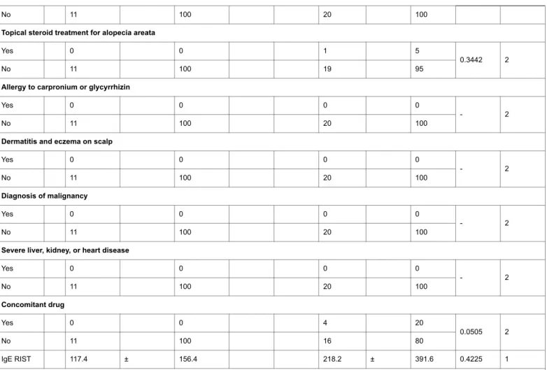
Combined treatment Item
Start 12 weeks Abort
n Ave. ± SD n Ave. ± SD n Ave. ± SD p-value1
SBP 20 118.2 ± 24.1 17 117.2 ± 13.7 2 127 ± 32.5 0.687 DBP 20 77.5 ± 15.1 17 76.6 ± 13.9 2 72.5 ± 3.5 0.777 Puls 20 76.2 ± 14.4 17 75.4 ± 11.5 2 66 ± 5.7 0.8455 BUN 20 14.5 ± 3.1 17 13.6 ± 3.1 2 10.5 ± 3.5 0.1762 CRE 20 0.67 ± 0.1 17 0.72 ± 0.14 2 0.6 ± 0.1 0.003 AST 20 22.8 ± 9.2 17 21.9 ± 11.8 2 19 ± 1.4 0.1441 ALT 20 23.2 ± 15 17 23.9 ± 20.1 2 13 ± 1.4 0.6551 ALP 20 188.6 ± 38.6 17 195.2 ± 37 2 191 ± 67.9 0.3037 LDH 20 168.7 ± 29.2 17 174.7 ± 27.5 2 173.5 ± 33.2 0.6236 γ-GTP 20 29 ± 25.3 17 29.5 ± 24.8 2 14.5 ± 3.5 0.3906 CRP 20 0.07 ± 0.08 17 0.08 ± 0.09 2 0.04 ± 0.01 0.4484 WBC 20 5315 ± 1458.3 17 5705.9 ± 2011.7 2 4050 ± 1343.5 0.4471 RBC 20 450.2 ± 51 17 454.9 ± 57.5 2 428.5 ± 26.2 0.8707 Hgb 20 13.5 ± 1.5 17 13.6 ± 1.6 2 13.4 ± 1.8 0.7747 PLT 20 24.2 ± 4.2 17 24.2 ± 5 2 25.4 ± 3 0.8832 20 100 ± 0 17 100 ± 0 2 100 ± 0 -Eos 20 0.9 ± 0.9 17 0.8 ± 0.9 2 2 ± 1.4 0.8899 Baso 20 2.7 ± 2.2 17 4.9 ± 4.3 2 5 ± 7.1 0.0765 Neut 1 55.1 - - - -Neut-Stab 19 1.5 ± 0.6 16 1.6 ± 0.7 2 2 ± 0 0.5805 Neut-Seg 19 56.5 ± 7.8 16 53.8 ± 8.1 2 55.5 ± 12 0.3085 Ly 20 33.9 ± 7.7 17 33.6 ± 6.9 2 30.5 ± 4.9 0.897 Mono 20 4.8 ± 2.3 17 5 ± 1.6 2 5 ± 1.4 0.6925
1. Comparison between start and 12 weeks within each group using the paired t-test. 2. Comparison between groups using the paired t-test (start).
3. Comparison between groups using the paired t-test (12 weeks).
Table 9: (b) Showing combined treatment blood test results.
0.7848 0.4283 0.5895 0.4446 0.5636 0.2387 0.753 0.7507 0.4678 0.1632 0.2019 0.4712 0.1326 0.2837 0.9763 0.497 0.1407 0.5051 0.3886 0.2207 0.5107 0.9164 0.0315 0.6841 0.9548 0.8339 0.5729 0.7551 0.1522 0.5415 - -0.7063 0.9869 0.4681 0.2024 - -0.0832 0.8063 0.4943 0.306 0.5516 0.119 0.378 0.4285
Table 9: Blood test results.
Changes in areas of hair loss (secondary endpoint)
Areas of hair loss at 4, 8, and 12 weeks in 11 patients in the single-treatment group and 17 in the combined-single-treatment group, excluding the patients who dropped out, were compared with the areas at the start of the study. No significant difference was seen between the two groups. Although no reduction in the size of areas of hair loss was observed in the single-treatment group at any time point, a reduction was observed in the combined-treatment group at 4 weeks and thereafter (Table 13 and Figure 1).
In patients with allergic factors, as indicated by “IgE ≥ 171” in Table 14, comprising 6 in the single-treatment group and 7 in the combined-treatment group, the areas of hair loss at each observation were compared with those at the start of the study. In the combined-treatment group, the areas of hair loss at 8 and 12 weeks were reduced significantly (both p<0.05, Table 14, Figure 1).
Figure 1: Change in size of areas of hair loss in patients with atopic predispositions in the combined-treatment and single-treatment groups at the start of the study and at 12 weeks.
Discussion
The Alopecia Areata Clinical Practice Guidelines were formulated based on the results of extensive clinical practice and other
recommendations [11]. However, additional treatment options are required because the condition has a major effect on patients’ quality of life, refractory and recurrent cases are common, and regeneration of
the hair may take a long time. MG tablets have been used widely for many years in treating AA and are recommended as combined therapy in the clinical practice guidelines. This study investigated combined treatment with MG tablets and CC solution. It was reported that this
combination showed efficacy in 60% of AA patients with moderate or more severe disease [14]. However, no randomized, controlled study evaluating the efficacy of MG tablets has been conducted, as pointed out in the guidelines.
Start to 4 weeks
Single treatment Combined treatment p-value
Items n Ave. SD Median n Ave. SD Median
Number of areas of hair loss 11 0 ± 0 0 19 -0.1 ± 0.2 0 0.4891
Size of area of hair loss 11 0.4 ± 0.8 1 19 0.1 ± 0.6 0 0.2413
Hair breakage and callous 11 0.5 ± 0.7 0 19 0.2 ± 0.4 0 0.3283
Terminal hair growth 11 0.8 ± 0.6 1 19 0.7 ± 0.7 1 0.7149
Total score 11 1.6 ± 1.6 2 19 1 ± 1.2 1 0.3149
Start to 8 weeks
Number of areas of hair loss 11 0 ± 0 0 17 0.1 ± 0.4 0 0.6613
Size of area of hair loss 11 0.9 ± 0.3 1 17 0.6 ± 0.8 1 0.236
Hair breakage and callous 11 0.5 ± 0.7 0 17 0.6 ± 0.6 1 0.3565
Terminal hair growth 11 1.4 ± 0.7 1 17 1.1 ± 0.7 1 0.3655
Total score 11 2.7 ± 1.1 3 17 2.4 ± 1.9 2 0.5755
Start to 12 weeks
Number of areas of hair loss 11 0.4 ± 0.5 0 17 0.3 ± 0.7 0 0.9155
Size of area of hair loss 11 1.3 ± 0.6 1 17 0.9 ± 1 1 0.4417
Hair breakage and callous 11 1.2 ± 0.9 1 17 1.1 ± 0.9 1 0.8603
Terminal hair growth 11 1.3 ± 0.8 1 17 1.2 ± 0.7 1 0.8371
Total score 11 4.1 ± 2.6 3 17 3.6 ± 2.6 3 0.7558
Table 10: Physicians’ findings [Comparison between groups using the Wilcoxon rank-sum test]. Therefore, we compared the effectiveness of MG tablets alone with
the combination of MG tablets and CC solution. No significant differences in efficacy, clinical findings, and patients’ impressions of efficacy were observed between the two groups. A significant reduction in areas of hair loss was not observed in the single-treatment group but was observed in the combined treatment group at 4 weeks and thereafter. This result may have been due to the difference in the number of patients (11 versus 17) in the two groups. No serious adverse events occurred during the study, and the incidence of adverse events in the two groups did not differ significantly.
It has been reported that the IgE level as an allergic factor is elevated in the early stage of AA [15] .Therefore we conducted an analysis by allergic factors. The area of AA was significantly decreased in patients with allergic factors 8 and 12 weeks after treatment as compared with baseline, while a reduction in the AA area was observed in patients without allergic factors but was not significant.
4 weeks
Single treatment Combined treatment p-value
Evaluation n % n %
Worsened 1 9.1 2 10.5
0.8064
Slightly improved 6 54.5 9 47.4 Moderately improved 1 9.1 2 10.5 Markedly improved 1 9.1 0 0 Total 11 100 19 100 -8 weeks Worsened 0 0 1 5.9 0.8539 No change 0 0 2 11.8 Slightly improved 5 45.5 6 35.3 Moderately improved 5 45.5 6 35.3 Markedly improved 1 9.1 2 11.8 Total 11 100 17 100 -12 weeks Worsened 0 0 2 11.8 0.6644 No change 1 9.1 0 0 Slightly improved 2 18.2 3 17.6 Moderately improved 3 27.3 6 35.3 Markedly improved 5 45.5 6 35.3 Total 11 100 17 100
-Table 11: Treatment evaluation [Comparison between groups using Fisher’s exact test]. It was shown in vitro that glycyrrhizin acid, which is the active
ingredient in MG tablets, is hydrolyzed by β-D-glucuronidase and metabolized to glycyrrhetinic acid [16,17]. Since glycyrrhetinic acid has an inhibitory effect on 11β-HSD2, the enzyme that metabolizes
inactive cortisone to cortisol, it is possible that the antiinflammatory effects of cortisol in the body are indirectly affected by glycyrrhetinic acid, resulting in improved AA [18].
Single treatment Combined treatment p-value
Evaluation n % n % 4 weeks Worsened 0 0 2 10.5 0.5851 No change 6 54.5 6 31.6 Improved 5 45.5 10 52.6 Markedly improved 0 0 1 5.3 Not determined 0 0 0 0 Total 11 100 19 100 -8 weeks Worsened 0 0 1 5.9 0.6832 No change 1 9.1 4 23.5 Improved 9 81.8 10 58.8
Markedly improved 1 9.1 2 11.8 Not determined 0 0 0 0 Total 11 100 17 100 -12 weeks Worsened 0 0 0 0 0.7005 No change 3 27.3 4 23.5 Improved 4 36.4 4 23.5 Markedly improved 4 36.4 9 52.9 Not determined 0 0 0 0 Total 11 100 17 100
-Table 12: Patients’ impression of treatment effect [Comparison between groups using Fisher’s exact test]. When steroids are administered by local injection, Samrao et al.
recommended monitoring the bone mineral density (BMD) of AA patients to avoid the risk of steroidal osteoporosis because they found abnormal BMD in 50% of patients after 20 weeks or longer treatment with triamcinolone acetonide in 4-8 week cycles and because the cumulative dose of triamcinolone acetonide is a risk factor for steroidal osteoporosis [18]. On the other hand, it was reported that glycyrrhizic acid prevents steroid-induced osteoporosis in rats [19]. Glycyrrhetinic acid was reported to inhibit 11β-HSD1 of the enzyme metabolizing
cortisone to cortisol [20], and thus it was inferred that glycyrrhetinic acid prevents osteoporosis by promoting the metabolism of cortisol produced in excess due to steroid treatment. In other words, glycyrrhetinic acid acts on both the enzyme that activates and the enzyme that inactivates cortisol and plays a role in the treatment of AA by promoting cortisol production when the response to corticotropin-releasing hormone is decreased. Glycyrrhetinic acid also prevents side effects by increasing the metabolism of excess cortisol produced in response to steroid administration.
Average area (cm2)
n Start 4 weeks 8 weeks 12 weeks
Single treatment 11 1.85 ± 1.22 1.44 ± 1.15 1.08 ± 0.95 0.89 ± 1.24
p-value (within group)* 0.1581 0.126 0.0966
Combined treatment 17 2.63 ± 2.78 2.06 ± 2.54 1.56 ± 2.25 0.95 ± 1.54
p-value (within group)* 0.0387 0.0078 0.0009
p-value (between groups)** 0.7419 0.7598 0.9064 1
Reduction rate (%)
Single treatment -20.84 ± 29.56 -35.88 ± 40.64 -46.64 ± 55.84
Combined treatment -15.83 ± 41.91 -45.79 ± 37.25 -61.12 ± 48.57
p-value (between groups)** 0.8323 0.4659 0.6592
*Comparison wihin each group using the Bonferroni multiple procedure. **Comparison between groups using Wilcoxon’s exact test.
Table 13: Change in area (area observed) of hair loss.
Since it has been recognized that the serum oxidative stress marker level is higher and the antioxidative stress marker is lower in AA patients in than in healthy people, it was suggested that oxidative stress is involved in the development of the condition [21,22]. Glycyrrhizic acid is known to exert antioxidative effects [23-25], suggesting that it could improve symptoms by decreasing oxidative stress in AA.
CC hydrate, the active ingredient in CC solution, has local vasodilator activity, increasing blood flow in capillaries and promoting local metabolism by stimulating acetylcholine receptors of vascular smooth muscle. CC hydrate promotes hair growth by acting on degraded hair follicles [26,27].
Among the various treatments for AA, steroids have been administered most frequently because their mechanism of action is
clear. However, many patients have reservations about steroid treatment even under the supervision of a physician. MG tablets are viewed as safe in terms of both lack of side effects and treatment
efficacy and they contribute to the maintenance of homeostasis of corticosteroids in hair follicle tissue.
Average area (cm2)
n Start 4 weeks 8 weeks 12 weeks
Single treatment 6 2.11 ± 1.52 1.63 ± 1.53 1.3 ± 1.26 1.17 ± 1.62
p-value (within group)* 0.6564 0.4689 0.6564
Combined treatment 7 2.95 ± 3.84 2.34 ± 3.45 1.27 ± 2 0.46 ± 0.69
p-value (within group)* 0.3282 0.0468 0.0468
p-value (between
groups)** 1 0.6682 0.5677 0.7072
Reduction rate (%)
Single treatment -22.62 ± 34.48 -41.53 ± 35.08 -45.32 ± 66.18
Combined treatment -26.84 ± 30.3 -65.80 ± 20.02 -86.76 ± 13
p-value (between groups)** 1 0.0865 0.4989
*Comparison within each group using the Bonferroni multiple procedure. **Comparison between groups using Wilcoxon’s exact test.
Table 14: Change in area (area observed) of hair loss in patients with atopic predisposition. The limitations of this study were, because it was an interventional
comparison, the number of patients differed between the two groups, and the overall sample size was small. Therefore, no statistically significant difference was seen between groups. Since the present combination therapy obviously showed the decrease in the area of AA and the effects in patients with allergic factors, we proceed to further larger randomized trials to confirm the results in this study.
In conclusion, in the present interventional study, MG tablets combined with CC solution therapy showed decreases in areas of AA without serious adverse events, especially in patients with allergic factors.
Acknowledgment
This study was carried out under a multicenter academic research grant from the Waxman Foundation, which is gratefully acknowledged.
References
1. Safavi K (1992) Prevalence of alopecia areata in the First National Health and Nutrition Examination Survey. Arch Dermatol 128: 702.
2. Safavi KH, Muller SA, Suman VJ, Moshell AN, Melton LJ 3rd (1995) Incidence of alopecia areata in Olmsted County, Minnesota, 1975 through 1989. Mayo Clin Proc 70: 628-633.
3. Martinez-Mir A, Zlotogorski A, Gordon D, Petukhova L, Mo J, et al. (2007) Genomewide scan for linkage reveals evidence of several susceptibility loci for alopecia areata. Am J Hum Genet 80: 316-328.
4. Petukhova L, Duvic M, Hordinsky M, Norris D, Price V, et al. (2010) Genome-wide association study in alopecia areata implicates both innate and adaptive immunity. Nature 466: 113-117.
5. Betz RC, Pforr J, Flaquer A, Redler S, Hanneken S, et al. (2007) Loss-of-function mutations in the filaggrin gene and alopecia areata: strong risk
factor for a severe course of disease in patients comorbid for atopic disease. J Invest Dermatol 127: 2539-2543.
6. Katagiri K, Arakawa S, Hatano Y (2007) In vivo levels of IL-4, IL-10, TGF-beta1 and IFN-gamma mRNA of the peripheral blood mononuclear cells in patients with alopecia areata in comparison to those in patients with atopic dermatitis. Arch Dermatol Res 298: 397-401.
7. Kasumagić-Halilović E (2008) Thyroid autoimmunity in patients with alopecia areata. Acta Dermatovenerol Croat 16: 123-125.
8. Friedmann PS (1981) Alopecia areata and auto-immunity. Br J Dermatol 105: 153-157.
9. Zhang B, Zhao Y, Cai Z, Caulloo S, McElwee KJ, et al. (2013) Early stage alopecia areata is associated with inflammation in the upper dermis and damage to the hair follicle infundibulum. Australas J Dermatol 54: 184-191.
10. Yang DQ, You LP, Song PH, Zhang LX, Bai YP (2012) A randomized controlled trial comparing total glucosides of paeony capsule and compound glycyrrhizin tablet for alopecia areata. Chin J Integr Med 18: 621-625.
11. Gilhar A, Ullmann Y, Berkutzki T, Assy B, Kalish RS (1998) Autoimmune hair loss (alopecia areata) transferred by T lymphocytes to human scalp explants on SCID mice. J Clin Invest 101: 62-67.
12. Ito T (2013) Recent advances in the pathogenesis of autoimmune hair loss disease alopecia areata. Clin Dev Immunol 2013: 348546.
13. Arase S, Tsuboi R, Yamazaki M, Inui S, Itami S, et al. (2010) The Japanese Dermatological Association Alopecia Areata Clinical Practice Guidelines 2010. Jpn J Dermatol 120: 1841-1859.
14. Maruo K, Kayashima K, Ono T, Ikeda I (2004) Study of Grychiron tablets monotherapy on alopecia areata. Rinsyo to Kenkyu 81: 179-183.
15. Zhao Y, Zhang B, Caulloo S, Chen X, Li Y, et al. (2012) Diffuse alopecia areata is associated with intense inflammatory infiltration and CD8+ T cells in hair loss regions and an increase in serum IgE level. Indian J Dermatol Venereol Leprol 78: 709-714.
16. Stewart PM, Wallace AM, Valentino R, Burt D, Shackleton CH, et al. (1987) Mineralocorticoid activity of liquorice: 11-beta-hydroxysteroid dehydrogenase deficiency comes of age. Lancet 2: 821-824.
17. Nakayama H (2001) Therapeutic results of long-term administration of Grychiron tablets concomitant with Chinese medicine on intractable alopecia areata. Nishinihon Dermatol 63: 191-196.
18. Samrao A, Fu JM, Harris ST, Price VH (2013) Bone mineral density in patients with alopecia areata treated with long-term intralesional corticosteroids. J Drugs Dermatol 12: e36-40.
19. Ramli ES, Suhaimi F, Asri SF, Ahmad F, Soelaiman IN (2013) Glycyrrhizic acid (GCA) as 11ß-hydroxysteroid dehydrogenase inhibitor exerts protective effect against glucocorticoid-induced osteoporosis. J Bone Miner Metab 31: 262-273.
20. Shamsa F, Ohtsuki K, Hasanzadeh E, Rezazadeh Sh (2010) The Anti-inflammatory and Anti-viral Effects of an Ethnic Medicine: Glycyrrhizin. J Med Plants 9: 1-28.
21. Al-Wasiti EA, A-Tammimy SM,Wasan Taha Al-Rubayee(2010) The Role of Oxidative Stress in Vitiligo and Alopecia Areata. Iraqi J Commun Med 4: 287-291.
22. Bilgili SG, Ozkol H, Karadag AS, Ozkol HU, Seker A, et al. (2013) Serum paraoxonase activity and oxidative status in subjects with alopecia areata. Cutan Ocul Toxicol 32: 290-293.
23. Yildirim AO, Ince M, Eyi YE, Tuncer SK, Kaldirim U, et al. (2013) The effects of glycyrrhizin on experimental acute pancreatitis in rats. Eur Rev Med Pharmacol Sci 17: 2981-2987.
24. Lee CH, Park SW, Kim YS, Kang SS, Kim JA, et al. (2007) Protective mechanism of glycyrrhizin on acute liver injury induced by carbon tetrachloride in mice. Biol Pharm Bull 30: 1898-1904.
25. Sil R, Ray D, Chakraborti AS (2013) Glycyrrhizin ameliorates insulin resistance, hyperglycemia, dyslipidemia and oxidative stress in fructose-induced metabolic syndrome-X in rat model. Indian J Exp Biol 51: 129-138.
26. Takeda K, Matsumoto A, Shimada M(1966) Dermatological application and experimental study of MTB (methyl N-trimethyl-gamma-aminobutyrate chloride). Hifu to Hinyo 28: 719-732.
27. Minamiyama M, Minato T, Yamamoto A, Kaihatsu T, Tsunoda K (2006) Effects of carpronium chloride on the microvascular blood flow in rat mesentery using intravital videomicroscopy. Clin Hemorheol Microcirc 34: 125-129.


![Table 8: Objective findings [Comparison between groups using the χ 2 test].](https://thumb-ap.123doks.com/thumbv2/123deta/6817650.1167221/6.892.60.841.210.448/table-objective-findings-comparison-groups-using-χ-test.webp)

![Table 11: Treatment evaluation [Comparison between groups using Fisher’s exact test].](https://thumb-ap.123doks.com/thumbv2/123deta/6817650.1167221/10.892.53.840.168.651/table-treatment-evaluation-comparison-groups-using-fisher-exact.webp)


