博 士 学 位 論 文
廃 用 性 筋 萎 縮 の 回 復 に お い て 老 化 が
ラ ッ ト ヒ ラ メ 筋 の 基 底 板 に 与 え る 影 響
金
澤
佑
治
廃 用 性 筋 萎 縮 の 回 復 に お い て 老 化 が ラ ッ ト ヒ ラ メ 筋 の 基 底 板 に 与 え る 影 響 金 澤 佑 治近 畿 大 学 大 学 院
医 学 研 究 科 医 学 系 専 攻
Doctoral Dissertation
Effects of aging on basement membrane of the soleus
muscle during recovery following disuse atrophy in rats
Yuji Kanazawa
October 2017
Department of Anatomy and Neurobiology, Major in Medical Sciences Kindai University Graduate School of Medical Sciences
Effects of aging on basement membrane of the soleus
muscle during recovery following disuse atrophy in
rats
Yuji Kanazawa1, 2, Keisuke Ikegami3, Mitsugu Sujino3, Satoshi Koinuma3, Mamoru
Nagano2, 3, Yuki Oi4, Tomoya Onishi4, Shinichi Sugiyo1, Isao Takeda1, Hiroshi Kaji5,
Yasufumi Shigeyoshi2, 3§
1
Department of Physical Therapy, Osaka University of Human Sciences, Shojyaku, Settsu, 566-8501, Japan
2
Department of Anatomy and Neurobiology, Graduate school of Medical Sciences, Kindai University, Ohnohigashi, Osakasayama, 589-8511, Japan
3
Department of Anatomy and Neurobiology, Kindai University Faculty of Medicine, Ohnohigashi, Osakasayama, 589-8511, Japan
4
Faculty of Health Care Sciences, Takarazuka University of Medical and Health care, Hanayashiki Midorigaoka, Takarazuka, 666-0162, Japan
5
Department of Physiology and Regenerative Medicine, Kindai University Faculty of Medicine, Ohnohigashi, Osakasayama, 589-8511, Japan
§
Corresponding author:
Yasufumi Shigeyoshi, Ph.D. Professor, Department of Anatomy and Neurobiology, Kindai University Faculty of Medicine, 377-2, Ohnohigashi, Osakasayama, 589-8511, Japan.
Abstract
1
Aging is known to lead to the impaired recovery of muscle after disuse as well as the 2
increased susceptibility of the muscle to damage. Here, we show that, in the older rats, 3
reloading after disuse atrophy, causes the damage of the muscle fibers and the basement 4
membrane (BM) that structurally support the muscle fibers. Male Wistar rats of 3- 5
(young) and 20- (older) months of age were subjected to hindlimb-unloading for 2 6
weeks followed by reloading for a week. In the older rats, the soleus muscles showed 7
necrosis and central nuclei fiber indicating the regeneration of muscle fibers. 8
Furthermore, ectopic immunoreactivity of collagen IV, a major component of the BM, 9
remained mostly associated with the necrotic appearance, suggesting that the older rats 10
were impaired with the ability of repairing the damaged BM. Further, after unloading 11
and reloading, the older rats did not show a significant alteration, although the young 12
rats showed clear response of Col4a1 and Col4a2 genes, both coding for collagen IV. In 13
addition, during the recovery phase, the young rats showed increase in the amount of 14
Hsp47 and Sparc mRNA, which are protein folding-related factor genes, while the older
15
rats did not show any significant variation. Taken together, our findings suggest that the 16
atrophic muscle fibers of the older rats induced by unloading were vulnerable to the 17
weight loading, and that attenuated reactivity of the BM-synthesizing fibroblast to 18
gravity contributes to the fragility of muscle fibers in the older animals. 19
20
Keywords: aging; skeletal muscle; basement membrane; hindlimb unloading; reloading
1. Introduction
Skeletal muscle mass and functionality can be changed in response to physical activity. Mechanical overload like resistance exercise improves the muscle mass and the muscular strength (Schiaffino et al., 2013) and, on the other hand, disuse causes muscle atrophy (Thomason et al., 1990). After reloading, compared with the young animals, the older animals show delayed recovery from disuse muscle atrophy (Hwee et al., 2009; White et al., 2015). The reason for the sluggish recovery in the older animals, however, is poorly understood. Previous studies have revealed age-related dysfunction in the recovery process from disuse atrophy. Decreased function of satellite cells with aging was suggested to contribute to the impaired response of muscle to reloading (Gallegly et al., 2004). During the muscle recovery, some molecules necessary for protein synthesis shows attenuated responses such as delay in the activation of Akt (Hwee et al., 2009), or
a decreased phosphorylation of p70S6K in the muscle fibers of the aged animals (Morris,
2004).
Basement membrane (BM) is a layer of extracellular matrix material, which coats muscle fiber as a static structure that provides mechanical support (Sanes, 2003). Previous studies have shown that BM plays a critical role in maintaining muscle structure and function. Deficiency of the molecular components of BM results in muscular dystrophy associated with fragility of muscle fibers (Girgenrath et al., 2009; Labelle-Dumais et al., 2011). Therefore, the slow recovery of muscles in the older animals can possibly be attributed to the malfunction of BM.
slow-twitch postural muscle and is used for the study of disuse atrophy induced by
hindlimb unloading. The soleus muscles of rats showed atrophy after unloading of
hindlimb and recovery of mass after reloading such as weight bearing (Oishi et al., 2008). Flück et al., (2003) have shown that reloading of atrophied rat soleus muscle induces tenascin-C, a marker of damaged muscle fibers, suggesting that reloading of body weight would be a mechanical stress that damages atrophied muscle fibers. Here, we hypothesized that, after unloading, muscle fibers of the older animals are at high risk of damage by reloading. In the present study, we have investigated the effects of aging on the recovery process during the periods of hindlimb unloading followed by reloading. We have compared the soleus muscles of the young rats with the older rats to observe the morphological and molecular changes of muscle fiber and BM in the recovery process from disuse atrophy.
2. Materials and Methods
2.1. Animals
Male Wistar rats (n = 32) were obtained from Japan SLC (Shizuoka, Japan). Three-month-older rats were used as young rats and 20-month-older rats were used as older rats. All animals were housed in individual cages and allowed free access to food and water constantly. The environmental conditions were maintained at 22 ± 2 °C and a 12:12 h light:dark cycle.
This study was approved by the Committee of Animal Care and Use of the Kindai University Faculty of Medicine (KAME-26-049). All experimental procedures were
conducted in accordance with institutional guidelines for the use of experimental animals.
2.2. Experimental protocol
The young and older rat groups (n = 16 each) were divided into 4 groups each (n = 4 per group): non-unloaded ambulatory controls (Cont), hindlimb unloaded for 14 days (HU14d), where the hindlimbs of rats were unloaded for 14 days and, were then reloaded. After the unloaded period, the rats were allowed to move freely in the cage for one (RE1d) or seven (RE7d) days.
2.3. Hindlimb unloading and reloading
Hindlimb unloading was performed as previously described with minor modifications (Morey-Holton et al. 2002). Briefly, hindlimbs of rats in the HU14d, RE1d, and RE7d groups were prevented from bearing weight by tail suspension and the rats were allowed free access to food and water by their forelimbs during the hindlimb unloading. The unloading continued for 14 days, after which the rats in the RE1d and RE7d groups were released from the tail suspension. Hindlimbs of the rats were reloaded in order to recover their muscle atrophy for both RE1d and RE7d groups.
2.4. Sampling
At the end of the experimental period, rats were anesthetized using sodium pentobarbital, after which the soleus muscles were removed and weighed. A part of the
soleus muscle belly of each rat was excised and preserved in RNAlater (Thermo Fisher Scientific, Hanover Park, IL) and other parts of the soleus muscles were frozen immediately in isopentane, cooled in dry ice, and stored at -80 °C for further analyses.
2.5. Histochemical analyses
Summation of extracellular matrix, muscle fibers and tissue fluid is evaluated by the muscle wet- weight. Therefore, in order to evaluate the muscle fibers alone, fiber cross sectional area (FCSA) that measures the cross-sectional area of the muscle fiber was adopted. Transverse tissue sections (10-µm thickness) were cut from the middle part of the soleus muscle belly using a cryostat (CM1950; Leica, Wetzlar, Germany) at -25 °C and were mounted onto amino silane-coated glass slides. Then, to know the ratio of type I and type II fibers and fiber type specific FCSA, an ATPase staining was carried out using a calcium method (Brook et al., 1970). In short, the sections were treated with myofibrillar adenosine triphosphate (ATPase) staining after alkaline pre-incubation (pH 10.7). The sections were then washed in 1 % CaCl2, reacted with 2 % CoCl2 and finally washed in 0.005 M sodium barbital. After washing with distilled water, the sections were visualized by staining with 2 % ammonium sulfide. After staining, the sections were dehydrated with a graded series of ethanol rinsing, immersed in xylene and embedded in Entellan (Merck, Darmstadt, Germany). For measurement of FCSA and observation of histopathologic finding to investigate the effect of hindlimb unloading and reloading, the sections were stained using Hematoxylin and Eosin (HE) and observed under microscope. The FCSA were measured using Image J software (NIH,
Bethesda, Maryland).
2.6. Electron microscopy
Longitudinal tissue sections (1-mm thickness) were cut from the middle part of the soleus muscle belly at -25 °C. The sections were fixed with 4 % paraformaldehyde / 2 % glutaraldehyde and then treated using osmium tetraoxide, after which they were dehydrated using a series of ethanol gradients. The sections were finally embedded in Epon. Ultrathin sections (90-nm thickness) were cut with an ultra-microtome and stained with 4 % uranyl acetate and 1 % lead citrate. The sections were analyzed by transmission electron microscopy (HT7700; Hitachi, Tokyo, Japan).
2.7. Immunohistochemical analyses
Transverse sections were fixed in 4 % paraformaldehyde and were rinsed with PBS
(pH 7.4). The sections were then bleached with 3 % H2O2, rinsed with PBS, and
incubated for 1 h at 4 °C in PBS containing 1 % normal goat serum and 0.3 % Triton X-100. The sections were then incubated for 24 hours at 4 °C in rabbit polyclonal anti-collagen IV antibody (ab6586; Abcam, Cambridge, MA) diluted 1:500 in PBS containing 0.3 % Triton X-100. The sections were subsequently incubated for 1 h at room temperature in biotinylated anti-rabbit IgG (Vectastain ABC kit; Vector Laboratories, Berlingame, CA) diluted 1:1000 in PBS, after which they were incubated in avidin-biotin complex (Vectastain ABC kit) for 1 h at 4 °C. After rinsing with PBS, the sections were washed with Tris-HCl buffer (pH 7.4) and incubated with
diaminobenzidine (0.035 % DAB) in Tris-HCl buffer (0.003 % H2O2) for 15 min at
room temperature. After the DAB reaction, the sections were also stained with Hematoxylin and dehydrated with a graded series of ethanol rinses, immersed in xylene, and embedded in Entellan (Merck, Darmstadt, Germany).
2.8. Quantitative PCR
Total RNA extracts of the soleus muscle belly saved in RNAlater were prepared using TRIZOL reagent (Invitrogen, Carlsbad, CA). RNA (1 µg) was reverse-transcribed using ReverTra Ace (Toyobo, Osaka, Japan) with random primers. Quantitative PCR was performed in a StepOnePlus Real-Time PCR System (Applied Biosystems, Carlsbad, CA) using SYBR Premix Ex Taq II (Takara Bio, Shiga, Japan). The quantity of each target gene PCR product was normalized to Gapdh levels and up- or downregulation was calculated as fold change relative to the Cont group in the young rats. The oligonucleotide primer sets used in these PCR analyses were as follows:
Col4a1, 5 ′ -ATGCCAGGAAGAGCAGGAAC-3 ′ (Forward) and 5 ′ -
CGACTACCAGGAAAGCCAACTC-3′ (Reverse);
Col4a2, 5 ′ -ACAGGGACCCTCTGGAACTG-3 ′ (Forward) and 5 ′ -
CGATATTTGTCACGGTCCTCTTT-3′ (Reverse);
Hsp47, 5 ′ -CGCAGCAGTAAGCAACACTACA-3 ′ (Forward) and 5 ′ -
TCCACATCCTTGGTGACCTCT-3′ (Reverse);
Sparc, 5 ′ -GACTACATCGGACCATGCAAATAC-3 ′ (Forward) and 5 ′ -
Mmp14, 5 ′ -GGATACCCACTTTGATTCTGCTG-3 ′ (Forward) and 5 ′ -
GGAGGGGTCGTTGGAATGT-3′ (Reverse);
Gapdh, 5 ′ -TGCACCACCAACTGCTTA-3 ′ (Forward) and 5 ′ -
GGATGCAGGGATGATGTTC-3′ (Reverse).
2.9. Statistical analyses
All data are represented as mean ± SEM. The t-test was performed on differences between means of both the young and older rats and the Tukey HSD test was performed on differences between means of each group for both the young and older rats (Kaleidagraph statistical analysis software version 4.5.1; Synergy Software, Reading, PA). Differences with p <0.05 were considered statistically significant.
3. Results
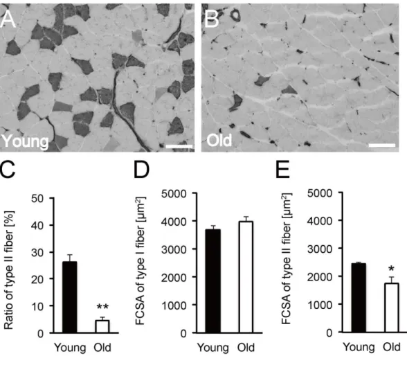
3.1. Ratio of type II fiber and fiber type-specific FCSA
Aging induces a decrease in type II fiber (fast type fiber) and causes fast type fiber-specific atrophy (Lexell et al., 1988; Deschenes et al., 2001; Deschenes et al., 2004; Nilwik et al., 2013). To confirm the aging of the soleus muscles in the older rats, we calculated the ratio of type I and type II fibers and fiber type-specific FCSA by ATPase staining images (Fig. 1A and B). The FCSA of type I fiber showed no significant difference between the young and older rats (Fig. 1D), however, the ratio of type II fiber in the older rats was significantly lower than that in the young rats (Fig. 1C) and the FCSA of type II fiber in the older rats was smaller than that in the young
rats (Fig. 1E). The findings on the soleus muscles of the older rats showed typical morphological characteristics of muscles in the aged animals. Hence, the aged rats were considered appropriate for further investigation.
Fig. 1 Ratio of type II fiber and fiber type-specific FCSA. Cross sections of soleus
muscle were stained by ATPase staining (pre-incubation at pH 10.7) in the young (A) and older (B) rats. Dark stained fibers are type II fibers. Scale bar represents 100 µm. Ratio of type II fiber (C) in the young (counting of 608 fibers in 4 soleus muscles in 4 rats) and older rats (counting of 510 fibers in 4 soleus muscles in 4 rats). FCSA of type I
(D) and type II (E) fiber in the soleus muscle from the young and older rats. *<0.05 vs. Young; **<0.001 vs. Young. Data are presented as mean ± SEM, n = 4 per group.
3.2. Soleus muscle weight
Several lines of evidence suggest that aging suppresses the decrease in muscle weight by hindlimb unloading and the recovery of muscle weight after reloading (Hwee et al., 2009; White et al., 2015). To confirm this, we measured the wet weight of muscle soleus of the young and the older rats of the Cont, HU14d, RE1d, and RE7d groups (Fig. 2). Soleus muscle wet weight showed a significant decrease, both in the young rats and older rats by hindlimb unloading compared to the respective control level (Fig. 2). The ratio of the decrease in the muscle wet weight was lesser in the older rats (17 %) than in the young rats (45 %). After reloading, the young rats showed a significant increase by 33 % in RE7d group compared to HU14d group while the older RE7d rat muscle did not show significant fluctuation (Fig. 2). Moreover, the response to unloading and reloading of soleus muscle weight in the older rats was blunted compared with the young rats, consistent with previous reports (Hwee et al., 2009; White et al., 2015). This result suggested that the recovery of muscle weight after disuse atrophy was delayed or impaired in the older rats.
Fig. 2 Soleus muscle weight. Changes in soleus muscle weight in response to hindlimb
unloading and reloading in the young and older rats. *<0.05 vs. Young; **<0.0001 vs. Young; †<0.05 vs. Cont; ††<0.001 vs. Cont; †††<0.0001 vs. Cont; ‡<0.05 vs. HU14d. Data are presented as mean ± SEM, n = 4 per group.
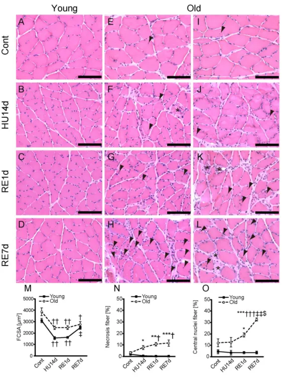
3.3. FCSA and histopathologic findings
A previous study also suggests that aging suppresses the decrease in FCSA by hindlimb unloading and the recovery of FCSA after reloading (White et al., 2015). To confirm this, we measured FCSA of the control, HU14d, RE1d, and RE7d group of the young and older rats by HE staining image (Fig. 3A-L). HE staining revealed that FCSA
of the soleus muscle showed a significant decrease both in the young rats and older rats by hindlimb unloading compared with the control (Fig. 3M). The decrease in FCSA was lesser in the older rats (36 %) than in the young rats (49 %). FCSA showed a significant increase of 57 % in RE7d group compared to HU14d group in the young rats, while a 13% recovery was observed in the old RE7d rats, which was not statistically significant (Fig. 3M). The blunted responses in FCSA to unloading and reloading in the older rats compared with the young rats, were consistent with a previous report (White et al., 2015). This result therefore suggested that, in the older rats, after reloading, the
recovery of the muscle fiber after the disuse atrophy is either delayed or impaired. The findings correspond well with the changes of the muscle wet weight and suggested that the delayed recovery of the wet weight of the muscle after reloading is due to the delayed or impaired response of the muscle fibers themselves.
Histopathlogic observations of cross sections of the muscles showed that the number of necrosis fibers in the older rats was increased after reloading at RE1d and RE7d (Fig. 3 asterisks in K and L). The number of necrosis fibers in the older rats was significantly higher than those in the young rats (Fig. 3N). According to previous studies (Dobek et al., 2013; Stauber et al., 2004), necrosis of muscle fibers is characterized by the existence of pale or hyper-contracted region in the muscle fibers,
an altered fiber shape and phagocytosis in HE staining.
During the regeneration of muscle fibers after necrosis, centrally located nuclei fibers, or central nuclei fibers are observed as immature muscle fibers (Ambrosio et al., 2010; Banks et al., 2008; Sciorati et al., 2016). Therefore, the existence of central nuclei
fibers suggests the sequential degeneration and regeneration events in muscle fibers. Also in the present study, several central nuclei fiber were detected in the older rats after reloading (Fig. 3 arrowheads in G, H, K, and L), whereas only few central nuclei fibers were observed in the young rats (Fig. 3C and D). In addition to these high amounts, the number of central nuclei fiber in the older rats further is increased after reloading (Fig. 3O). Taken together, all these results suggested that muscle fiber could be injured in the recovery phase following disuse atrophy in the older rats.
Fig. 3 FCSA and histopathologic findings. Cross sections of the soleus muscle were
stained by HE in the young (A–D) and older (E–L) rats. Staining images of Cont (A, E, and I), HU14d (B, F, and J), RE1d (C, G, and K), and RE7d (D, H, and L) are shown. Asterisks indicate necrosis fiber, and arrowheads indicate central nuclei fiber. Scale bar
represents 100 µm. Changes in FCSA (M), number of necrosis fibers (N), and number of central nuclei fibers (O) in response to hindlimb unloading and reloading in the young and older rats. FCSA (M) was measured by using HE staining images. Changes in number of necrosis fibers (N) in response to hindlimb unloading and reloading in the young (counting 4926 fibers in 4 soleus muscles from 4 rats) and older (counting 3745 fibers in 4 soleus muscles from 4 rats) rats. Changes in numbers of central nuclei fibers (O) in response to hindlimb unloading and reloading in the young (counting 5760 fibers in 8 soleus muscles from 8 rats) and older (counting 3992 fibers in 8 soleus muscles from 8 rats) rats. *<0.05 vs. Young; **<0.001 vs. Young; ***<0.0001 vs. Young;
†<0.05 vs. Cont; ††<0.001 vs. Cont; †††<0.0001 vs. Cont; ‡<0.05 vs. HU14d;
‡‡<0.0001 vs. HU14d; $<0.01 vs. RE1d. Data are presented as mean ± SEM, n = 4 per group.
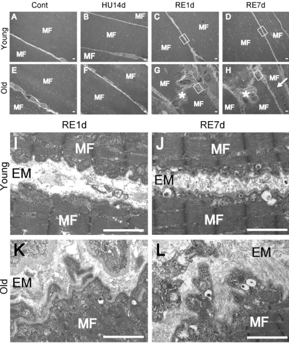
3.4. Electron microscopy analysis
To further elucidate the detailed structures of the injured muscles, we carried out electron microscopic analysis (Fig. 4). In the young rats, morphological abnormality such as injured muscle fiber and abnormal structure of BM, was not observed (Fig. 4A-D, I, and J). However, in the older rats, after reloading, we observed not only necrosis and central nuclei fiber (Fig. 4G and H), but also tearing and tortuous BM (Fig. 4K and L). This finding clearly suggested that BMs of the older rats were more easily damaged by reloading of the weight after disuse atrophy than the young animals in which the fragility of the muscle fiber were increased due to the vulnerability of BM.
Fig. 4 Electron microscopy analysis. Longitudinal sections of the soleus muscle were
observed by transmission electron microscopy in the young (A–D) and older (E–H) rats. Muscle fiber (MF) images of Cont (A and E), HU14d (B and F), RE1d (C and G), and RE7d (D and H) are shown. Necrosis fibers (asterisks in G and H) and central nuclei
fiber (allow in H) were observed in the older RE1d and older RE7d. BM image of young RE1d (I), the young RE7d (J), older RE1d (K), and older RE7d (L) rats. The images (I, J, K, and L) are extended images of white rectangles in C, D, G, and H. EM means extracellular matrix. Tearing and tortuous BMs were observed in the older RE1d (K) and older RE7d (L) rats. Scale bar represents 2 µm.
3.5. Localization of Collagen IV
Collagen IV is one of the major components of BM (Sanes, 2003). Col4a1 gene codes for collagen IV, and the Col4a1 mutant mice shows fragility of BM (Labelle-Dumais et al., 2011). Therefore, we postulated that the abnormal distribution of BM could be detected by immunohistochemistry analysis for collagen IV detection (Fig. 5A-H). Collagen IV-immunoreactivity (ir) in normal muscle fiber was confined to the region of BM in the young rats, making a single thin layer surrounding the muscle fibers (Fig. 5A-D). However, in the older rats, ectopic collagen IV-ir that appears to invade into the cytoplasm of muscle fibers was observed (Fig. 5 asterisks in G and H). In addition, time series quantification revealed that the number of the ectopic collagen IV-ir showed marked increase after reloading in the older rats but not in the young rats (Fig. 5I). The above results also suggested the fragility of BM in the older rats.
Fig. 5 Localization of Collagen IV. Cross sections of the soleus muscle were stained
by anti-collagen IV antibody in young (A–D) and older (E–H) rats. Staining images of Cont (A and E), HU14d (B and F), RE1d (C and G), and RE7d (D and H) are shown. Scale bar represents 100 µm. In addition, ectopic localizations of collagen IV-ir (asterisks in G and H) were observed. Changes in the number of muscles containing ectopic collagen IV-ir (I) in response to hindlimb unloading and reloading in the young (counting 4603 fibers in 8 soleus muscles from 8 rats) and older (counting 3671 fibers in 8 soleus muscles from 8 rats) rats. *<0.05 vs. Young; **<0.001 vs. Young; †<0.05 vs. Cont. Data are presented as mean ± SEM, n = 4 per group.
3.6. Histopathologic finding of ectopic collagen IV-ir
To confirm whether fibers containing ectopic collagen IV-ir fibers show other histopathologic findings, we performed immunohistochemistry analysis (Fig. 6A) and HE staining (Fig. 6B) to analyze the changes of collagen IV expressions in the alternating sections of soleus muscle fibers. On analysis, it was observed that the ectopic collagen IV-ir was mostly co-localized with necrotic appearance at cellular level (Fig. 6 asterisks in A and B). We observed that 98 % of ectopic collagen IV-ir (n=321) was co-localized with necrotic appearance (Fig. 6C). Conversely, we also observed that 95 % muscle fibers showing necrotic appearance (n = 333) showed ectopic collagen IV-ir (Fig. 6D). These results, therefore suggest that ectopic collagen IV-ir in the muscle fibers is mostly found associated with necrotic appearance and vice versa.
Fig. 6 Histopathologic finding of ectopic collagen IV-ir. Serial cross sections stained
anti-collagen IV antibody (A) and by HE (B) in the older RE7d rats. Scale bar represents 100 µm. Ectopic collagen IV-ir (asterisks in A) was shown in necrosis fibers (asterisks in B). Concordance rate (Ectopic collagen IV-ir/necrosis fiber) was 98 % (C). Concordance rate (Necrosis fiber/Ectopic collagen IV-ir) was 95 % (D).
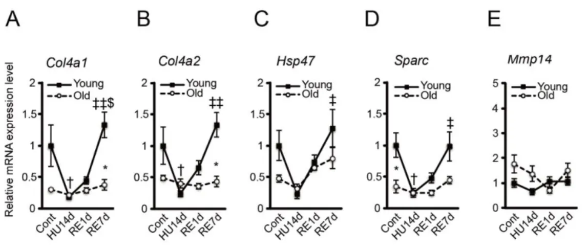
3.7. Synthetic and degradative factors of collagen IV
Collagen IV, encoded by Col4a1 and Col4a2 genes (Kuo et al., 2012) has a triple spiral structure and its synthesis is regulated by two proteins - heat shock protein 47 (Hsp47) and secreted protein acidic and cysteine rich (Sparc) (Kuo et al., 2012). On the other hand, the degradation of collagen IV is regulated by matrix metalloproteinase-14 (Mmp14) (Snyman et al., 2015). Here, we investigated whether aging affects collagen IV synthesis and degradation process during unloading and reloading. On analysis, we observed that the amount of Col4a1, Col4a2, Hsp47 and Sparc mRNA expression in the soleus muscle of the young rats was significantly decreased during unloading, which later increased up to similar levels as that of control (Fig. 7A-D). On the contrary, in the older rats, these gene expressions were significantly lower than those in the young rats and displayed attenuated fluctuations compared with young rats (Fig. 7A-D). When we quantified Mmp14 mRNA expression, we did not observe any significant differences between the young and older rats or between each treatment group of rats (Fig. 7E). Thus, from the above results, it is plausible that collagen IV synthetic (Col4a1, Col4a2,
following disuse atrophy.
Fig. 7 Synthetic and degradative factors of collagen IV. Changes in relative mRNA
expression levels of Col4a1 (A), Col4a2 (B), Hsp47 (C), Sparc (D), and Mmp14 (E) in response to hindlimb unloading and reloading in the young and older soleus muscle. *<0.05 vs. Young; †<0.05 vs. Cont; ‡<0.01 vs. HU14d; ‡‡<0.001 vs. HU14d; $<0.01 vs. RE1d. Data are presented as mean ± SEM, n = 3 or 4 per group.
4. Discussion
In the present study, we investigated the effect of aging on the soleus muscle during recovery following disuse atrophy. The key findings of the present study were as follows: (1) necrosis fiber and central nuclei fiber were increased in the older rats in the recovery phase after disuse atrophy, but not in the young rats; (2) tearing and tortuous BMs were observed in the recovery phase in the older rats, but not in the young rats; (3) collagen IV in the soleus muscle fiber of the older rats exhibited ectopic localization in
necrosis fiber; and (4) Col4a1 and Col4a2 mRNA expression levels were significantly decreased by unloading and then increased by reloading, respectively, in the young rats, but did not change in the older rats. In addition, collagen IV synthetic factors (Hsp47 and Sparc) were not increased in the recovery phase in the older rats. The findings suggest that not only muscle fiber but also dysfunction of BM were involved in contributing to the vulnerability of muscle fiber in the older rats.
The older rats showed necrosis fibers and central nuclei fibers, especially after reloading (Fig. 3). In contrast, the young rats did not show similar features of muscle damage. The finding suggests that, even with weight bearing, the older rats show muscle damage after the muscle atrophy induced by unloading. Similar histopathologic findings have also been previously observed in mice that lack both dystrophin and integrin in a normal gravity environment (Rooney et al., 2006). Also, muscle contraction-induced membrane injury and neuromuscular junction instability have been observed in the older rats, but not in the young rats (Hughes et al., 2016). In accordance with the previous reports, our study suggests that the older rats possess fragility in muscle fibers, and that the mechanical stimulus of reloading to atrophied muscle could be a potential damage to the muscle fibers.
Unlike the young rats, the older rats showed abnormal structures of BM such as tearing and tortuosity in soleus muscle fibers after reloading. The finding suggests that reloading following disuse atrophy not only affects muscle fiber but also the BM structure in the older rats. The muscle damage by mechanical stimuli is accompanied by the destruction of BM (Stauber et al., 2004; Winkler et al., 2011). Skeletal muscles in
mice with muscular dystrophy showed disrupted BM under normal breeding (Han, 2009). Col4a1 mutant mice demonstrated muscle fiber damage after exercise (Labelle-Dumais et al., 2011). Drosophila BM col4a1 mutations showed an irregular structure of the basal lamina in myofibers (Kelemen-Valkony et al., 2012), suggesting that collagen IV is necessary for maintaining the normal structure of BM. In line with previous studies, our present observation suggests that the fragility of BM in the older rats probably contributes to the disruption of muscle fibers by reloading after muscle atrophy induced by unloading.
In the present study, ectopic immunoreactivity of collagen IV, a major component of the BM, was mostly associated with the necrotic appearance of muscle fibers (Fig. 6). This finding suggests that the necrosis of muscle fibers is caused by the ectopic distribution of collagen IV, however, it is also possible that necrosis of muscle fibers always causes ectopic distribution of collagen IV. Several previous studies have demonstrated that the destruction of membrane leads to necrosis. For example, the accumulation of muscle membrane damage ultimately leads to necrosis of muscle fibers in muscular dystrophies, although muscle fibers originally possess an ability to repair damaged muscular membrane (Doherty et al., 2003; Han et al., 2007; Han et al., 2009; Lennon et al., 2003). Therefore, it is probable that the decreased ability of repairing the damaged BM causes necrosis in the soleus muscle of the older rats.
The older rats showed attenuated responses in collagen IV synthetic factors (Col4a1,
Col4a2, Hsp47, and Sparc) after hindlimb unloading and reloading compared with the
after reloading. Previous studies have demonstrated that the component of extracellular matrix reacts in response to gravity information (Chaillou et al., 2015; Riso et al., 2010). For synthesis of collagen I in fibroblasts, integrin transmits mechanical loading from the outside to the inside of the cell (Kjaer et al., 2004). Intracellularly, transcription of collagen gene in nuclei occurs followed by the translation of pre-procollagen mRNA (Kjaer et al., 2004). In collagen IV, procollagen subunits undergo extensive post-translational modifications and assemble as heterotrimers for incorporation into extracellular matrix where they polymerize into a network and interact with other extracellular and membrane bound molecules (Kuo et al., 2012). In this process, HSP47 works as a collagen-specific molecular chaperone and SPARC interacts with collagen in the endoplasmic reticulum, thereby influencing collagen IV folding (Kuo et al., 2012). Additionally, previous studies revealed that aging reduces synthesis of collagen I and collagen I mRNA in fibroblasts (Furth et al., 1991; Varani et al., 2006). Together with the previous reports, the present finding of attenuation of gene transcription for collagen IV synthesis in the older rats suggests that the response to gravity and the production of enough amount of mature collagen IV for BM construction are impaired in fibroblasts of the older rats.
5. Conclusions
To summarize, from the findings presented here, it is clear that the BM of the soleus muscle in the older rats was damaged during the recovery phase following disuse atrophy. The collagen IV of the older rats, furthermore, exhibited the ectopic
localization in necrosis fibers. Collagen IV synthetic factors (Col4a1, Col4a2, Hsp47, and Sparc) did not display an increase in the recovery phase in the older rats. These results suggest that aging affects the synthesis of collagen IV and causes muscle fiber fragility in the recovery phase after disuse atrophy.
Acknowledgments
This work was supported by JSPS KAKENHI (Grant Number 15K21540). The authors have no conflicts of interest to disclose. We would like to thank Life Science Research Institute at Kindai University for their technical support.
References
Ambrosio, F., Ferrari, R.J., Distefano, G., Plassmeyer, J.M., Carvell, G.E., Deasy, B.M., Boninger, M.L., Fitzgerald, G.K., Huard, J., 2010. The synergistic effect of treadmill running on stem-cell transplantation to heal injured skeletal muscle. Tissue Eng. Part A 16 (3), 839–849.
Banks, G.B., Combs, A.C., Chamberlain, J.R., Chamberlain, J.S., 2008. Molecular and cellular adaptations to chronic myotendinous strain injury in mdx mice expressing a truncated dystrophin. Hum. Mol. Genet. 17 (24), 3975–3986.
Brooke, M.H., Kaiser, K.K., 1970. Muscle fiber types: how many and what kind? Arch. Neurol. 23 (4), 369–379.
Chaillou, T., Jackson, JR., England, J.H., Kirby, T.J., Richards-White, J., Esser, K.A., Dupont-Versteegden, E.E., McCarthy, J.J., 2015. Identification of a conserved set of
upregulated genes in mouse skeletal muscle hypertrophy and regrowth. J. Appl. Physiol. 118 (1), 86–97.
Deschenes, M.R., 2004. Effects of aging on muscle fibre type and size. Sports Med. 34 (12), 809–824.
Deschenes, M.R., Britt, A.A., Chandler, W.C., 2001. A comparison of the effects of unloading in young adult and aged skeletal muscle. Med. Sci. Sports Exerc. 33 (9), 1477–1483.
Dobek, G.L. Fulkerson, N.D., Nicholas, J., Schneider, B.S.P., 2013. Mouse model of muscle crush injury of the legs. Comp. Med. 63 (3), 227–232.
Doherty, K.R., McNally, E.M., 2003. Repairing the tears: dysferlin in muscle membrane repair. Trends Mol. Med. 9 (8), 327–330.
Flück, M., Chiquet, M., Schmutz, S., Mayet-Sornay, M.H., Desplanches, D., 2003. Reloading of atrophied rat soleus muscle induces tenascin-C expression around damaged muscle fibers. Am. J. Physiol. Regul. Integr. Comp. Physiol. 284 (3), R792– R801.
Furth, J.J., 1991. The steady-state levels of type I collagen mRNA are reduced in senescent fibroblasts. J. Gerontol. 46 (3), B122–124.
Gallegly, J.C., Turesky, N.A., Strotman, B.A., Gurley, C.M., Peterson, C.A., Dupont-Versteegden, E.E., 2004. Satellite cell regulation of muscle mass is altered at old age. J. Appl. Physiol. 97 (3), 1082–1090.
Girgenrath, M., Beermann, M.L., Vishnudas, V.K., Homma, S. Miller, J.B., 2009. Pathology is alleviated by doxycycline in a laminin-α2-null model of congenital muscular dystrophy. Ann. Neurol. 65 (1), 47–56.
Han, R., Campbell, K.P., 2007. Dysferlin and muscle membrane repair. Curr. Opin. Cell Biol. 19 (4), 409–416.
Han, R., Kanagawa, M., Yoshida-Moriguchi, T., Rader, E.P., Ng, R.A., Michele, D.E., Muirhead, D.E., Kunz, S., Moore, S.A., Iannaccone, S.T., Miyake, K., McNeil, P.L., Mayer, U., Oldstone, M.B.A., Faulkner, J.A., Campbell, K.P., 2009. Basal lamina strengthens cell membrane integrity via the laminin G domain-binding motif of α -dystroglycan. Proc. Natl. Acad. Sci. U. S. A. 106 (31), 12573–12579.
Hughes, D.C., Marcotte, G.R., Marshall, A.G., West, D.W.D., Baehr, L.M., Wallace, M.A., Saleh, P.M., Bodine, S.C. Baar, K., 2016. Age-related differences in dystrophin: impact on force transfer proteins, membrane integrity, and neuromuscular junction stability. J. Gerontol. 72 (6), 640-648
Hwee, D.T., Bodine, S.C., 2009. Age-related deficit in load-induced skeletal muscle growth. J. Gerontol. A Biol. Sci. Med. Sci. 64 (6), 618–628.
Kelemen-Valkony, I., Kiss, M., Csiha, J., Kiss, A., Bircher, U., Szidonya, J., Maroy, P., Juhasz, G., Komonyi, O., Csiszar, K., Mink, M., 2012. Drosophila basement membrane collagen col4a1 mutations cause severe myopathy. Matrix Biol. 31 (1), 29–37.
Kjaer, M., 2004. Role of extracellular matrix in adaptation of tendon and skeletal muscle to mechanical loading. Physiol. Rev. 84 (2), 649–698.
Kuo, D.S., Labelle-Dumais, C., Gould, D.B., 2012. Col4a1 and col4a2 mutations and disease: insights into pathogenic mechanisms and potential therapeutic targets. Hum.
Mol. Genet. 21 (R1) R97–R110.
Labelle-Dumais, C., Dilworth, D.J., Harrington, E.P., de Leau, M., Lyons, D., Kabaeva, Z., Manzini, M.C., Dobyns, W.B., Walsh, C.A., Michele, D.D., Gould, D.B., 2011. COL4A1 mutations cause ocular dysgenesis, neuronal localization defects, and
myopathy in mice and walker-warburg syndrome in humans. PLoS Genet. 7 (5) 1–14.
Lennon, N.J., Kho, A., Bacskai, B.J., Perlmutter, S.L., Hyman, B.T., Brown, R.H.Jr., 2003. Dysferlin interacts with annexins A1 and A2 and mediates sarcolemmal wound-healing. J. Biol. Chem. 278 (50), 50466–50473.
Lexell, J., C.C. Taylor, M. Sjöström, 1988. What is the cause of the ageing atrophy? Total number, size and proportion of different fiber types studied in whole vastus lateralis muscle from 15- to 83-year-old men. J. Neurol. Sci. 84 (2-3), 275–294.
Morey-Holton, E.R., Globus, R.K., 2002. Hindlimb unloading rodent model: technical aspects. J. Appl. Physiol. 92 (4), 1367–1377.
Morris, R.T., Spangenburg, E.E., Booth, F.W., 2004. Responsiveness of cell signaling pathways during the failed 15-day regrowth of aged skeletal muscle. J. Appl. Physiol. 96 (1), 398–404.
Nilwik, R., Snijders, T., Leenders, M., Groen, B.B.L., van Kranenburg, J., Verdijk, L.B., van Loon, L.J.C., 2013. The decline in skeletal muscle mass with aging is mainly attributed to a reduction in type II muscle fiber size. Exp. Gerontol. 48 (5), 492–498.
Oishi, Y., Ogata, T., Yamamoto, K.I., Terada, M., Ohira, T., Ohira, Y., Taniguchi, K., Roy, R.R., 2008. Cellular adaptations in soleus muscle during recovery after hindlimb unloading. Acta Physiol. (Oxf) 192 (3), 381–395.
Riso, E.M., Ahtikoski, A.M., Takala, T.E.S., Seene, T., 2010. The effect of unloading and reloading on the extracellular matrix in skeletal muscle: changes in muscle strength and motor activity. Biol Sport 27 (2), 89–94.
Rooney, J.E., Welser, J.V., Dechert, M.A., Flintoff-Dye, N.L., Kaufman, S.J., Burkin, D.J., 2006. Severe muscular dystrophy in mice that lack dystrophin and alpha 7 integrin. J. Cell Sci. 119 (11), 2185–2195.
Sanes, J.R., 2003. The basement membrane/basal lamina of skeletal muscle. J. loiB. mehC.278 (15), 12601–12604.
Schiaffino, S., Dyar, K.A., Ciciliot, S., Blaauw, B., Sandri, M., 2013. Mechanisms regulating skeletal muscle growth and atrophy. FEBS J. 280 (17), 4294–4314.
Sciorati, C., Rigamonti, E., Manfredi, A.A., Rovere-Querini, P., 2016. Cell death, clearance and immunity in the skeletal muscle. Cell Death Differ. 23 (6), 927–937. Snyman, C., Niesler, C.U., 2015. MMP-14 in skeletal muscle repair. J. Muscle Res. Cell Motil. 36 (3), 215–225.
Stauber, W.T., 2004. Factors involved in strain-induced injury in skeletal muscles and outcomes of prolonged exposure. J. Electromyogr. Kinesiol. 14 (1), 61–70.
Thomason, D.B., Booth, F.W., 1990. Atrophy of the soleus muscle by hindlimb unweighting. J. Appl. Physiol. 68 (1), 1–12.
Varani, J., Dame, M.K., Rittie, L., Fligiel, S.E., Kang, S., Fisher, G.J., Voorhees, J.J., 2006. Decreased collagen production in chronologically aged skin: roles of age-dependent alteration in fibroblast function and defective mechanical stimulation. Am. J. Pathol. 168 (6), 1861–1868.
White, J.R., Confides, A.L., Moore-Reed, S., Hoch, J.M., Dupont-Versteegden, E.E., 2015. Regrowth after skeletal muscle atrophy is impaired in aged rats, despite similar responses in signaling pathways. Exp. Gerontol. 64, 17–32.
Winkler, T., von Roth, P., Matziolis, G., Schumann, M.R., Hahn, S., Strube, P., Stoltenburg-Didinger G., Perka, C., Duda, G.N., Tohtz, S.V., 2011. Time course of