1 2 3 4 5 6 7 8 9 10 11 12 13 14 15 16 17 18 Original Article
Pulmonary aspergillosis as a late complication after surgery for locally advanced non-small cell lung cancer treated with induction chemoradiotherapy
Seiichiro Sugimoto 1, Junichi Soh 1, Ken Suzawa 1, Kentaroh Miyoshi 1, Shinji Otani 1,
Hiromasa Yamamoto 1, Mikio Okazaki 1, Masaomi Yamane 1, Takahiro Oto 1, Susumu
Kanazawa 2, Katsuyuki Kiura 3, Shinichi Toyooka 1
1 Department of General Thoracic Surgery, Okayama University Hospital, Japan 2 Department of Radiology, Okayama University Hospital, Japan
3 Department of Respiratory Medicine, Okayama University Hospital, Japan
Correspondence:
Seiichiro Sugimoto, MD, PhD
Department of General Thoracic Surgery, Okayama University Hospital, 2-5-1 Shikata-cho, Kita-ku, Okayama 700-8558, Japan
E-mail: [email protected]
19 20 21
Abstract 1
Purpose: Some long-term survivors after surgery for locally advanced non-small cell lung 2
cancer (NSCLC) treated with induction chemoradiotherapy (trimodality treatment) develop
3
chronic pulmonary aspergillosis (CPA). The aim of our study was to assess the
4
characteristics and outcomes of CPA that develops after trimodality treatment.
5
Methods: We retrospectively reviewed the data of 187 NSCLC patients who underwent 6
trimodality treatment between 1999 and 2018.
7
Results: Six male ever-smoker patients developed CPA. All 6 patients had undergone 8
extended resection for NSCLC and had a history of either adjuvant chemotherapy (n=3)
9
or radiation pneumonitis (n=4). Among the 4 patients with CPA localized in a single lung,
10
3 patients were treated surgically (completion pneumonectomy or cavernostomy) and 1
11
patient was treated with antifungal therapy alone. Both treatments led to the improved
12
control of CPA. In contrast, patients with CPA in both lungs were not candidates for surgery,
13
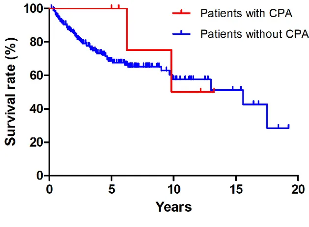
and died of CPA. The survival rates after trimodality treatment in the CPA group and the
14
group without CPA were comparable (10-year survival rate, 50.0% vs. 57.6%, P=0.59).
15
Conclusion: The early diagnosis of CPA localized in a single lung after NSCLC surgery 16
is critical to improving control and survival in patients with CPA.
17 18
Keywords: lung cancer; aspergillosis; surgery; radiation; chemotherapy 19
Introduction 1
Chronic pulmonary aspergillosis (CPA), including simple aspergilloma and chronic
2
cavitary pulmonary aspergillosis, sometimes develops in non-immunocompromised
3
patients with prior or current lung disease [1]. Risk factors for CPA include chemotherapy
4
[2], radiotherapy, thoracic surgery, and lung cancer [3-6], which are inevitable in patients
5
who have undergone induction chemoradiotherapy followed by surgery for non-small cell
6
lung cancer (NSCLC). Induction chemoradiotherapy followed by surgery has been shown
7
to be a feasible therapeutic option for patients with locally advanced NSCLC [7], and this
8
trimodality therapy for NSCLC has been shown to provide favorable long-term results [8],
9
which have led to an increase in survivors of NSCLC. Thus, long-term survivors may
10
develop CPA due to risk factors for CPA that are involved in trimodality therapy, despite
11
the irradiated lung containing the NSCLC being resected at surgery. Furthermore, the risk
12
of CPA may be increased by chronic obstructive lung disease, which is a common
13
comorbidity of ever-smoker NSCLC patients as well as by prolonged corticosteroid
14
therapy for radiation pneumonitis, which sometimes develops as a complication after
15
chemoradiotherapy [1, 6, 9]. Although evidence has accumulated on the treatment of
16
NSCLC by induction chemoradiotherapy followed by surgery [10], little information is
17
available in relation to CPA after the trimodality therapy. The aim of our study was to
18
assess the characteristics and outcomes of CPA after surgery for locally advanced NSCLC
19
treated by induction chemoradiotherapy.
20 21 Methods 22 Patients 23
Trimodality therapy has been performed to treat NSCLC patients with mediastinal nodal
metastasis. It has also been selectively applied to the treatment of localized N3 or
T3-1
4N0-1M0 and, at the physician’s discretion, to patients with large or invasive tumors, such
2
as bulky N1 tumors with chest wall invasion, or T4 involvement, as a means of achieving
3
complete resection with a pathologic safety margin [11, 12]. This study was a retrospective
4
review of cases of locally advanced NSCLC treated by induction chemoradiotherapy and
5
surgery at Okayama University Hospital between January 1999 and December 2018. A
6
total of 187 patients were included in this study. The inclusion criteria were an Eastern
7
Cooperative Oncology Group performance status of 0 to 1 and adequate functional
8
reserves of the major organs, as previously described [13]. Staging was performed
9
according to the International Association for the Study of Lung Cancer TNM Staging
10
System for NSCLC, eighth edition [14]. The study protocol (No. 1055) was approved by
11
the Institutional Review Board of Okayama University Hospital. The requirement for
12
patient consent was waived due to the retrospective nature of the study and the patients
13
were informed of their right to opt out.
14 15
Induction chemoradiotherapy followed by surgery 16
The details of trimodality treatment as initial therapy targeting a primary NSCLC tumor
17
have been described previously [8, 11, 15]. Briefly, most patients received cisplatin and
18
docetaxel as induction chemotherapy, and some patients received alternative
19
chemotherapy regimens. On the first day of chemotherapy, radiotherapy was initiated with
20
a planned total radiation dose of 40–46 Gy. Dose escalation up to 60 Gy was allowed
21
when tumors responded poorly. The surgical procedure after induction
22
chemoradiotherapy was decided on the basis of the disease extent before the start of
23
induction therapy. Although the preferred procedure was pulmonary lobectomy with
complete ipsilateral mediastinal and subcarinal nodal dissection, bilobectomy or
1
pneumonectomy was performed to achieve complete resection when necessary. The
2
patients received postoperative adjuvant therapy at the physician’s discretion. After the
3
completion of trimodality treatment, the patients were followed up in accordance with our
4
follow-up regimen [8].
5 6
Management of pulmonary aspergillosis 7
The diagnosis of CPA during the follow-up period was confirmed on the basis of the clinical,
8
laboratory, and radiographic findings, including testing for 1,3-beta-D-glucan and
9
galactomannan antigen, cultures, bronchoscopy, chest X-ray, and chest computed
10
tomography. After confirming the diagnosis of CPA, the patients were initially treated with
11
antifungal agents. In accordance with the guidelines for the treatment of CPA published
12
by the European Respiratory Society [1], after a careful risk assessment, surgery was
13
considered for improved disease control in patients whose CPA was refractory to medical
14 management. 15 16 Statistical analysis 17
All statistical analyses were performed using the GraphPad Prism 7.03 software program
18
(San Diego, CA, USA). Overall survival was defined as the interval between the start of
19
induction therapy and the date of death or the last follow-up examination. The survival
20
rates were analyzed by the Kaplan–Meier method, and the log-rank test was used to
21
compare the differences between groups. P values of <0.05 were considered to indicate
22
statistical significance.
23 24
Results 1
Patient characteristics 2
As shown in Table 1, six patients developed CPA after trimodality treatment for NSCLC.
3
All 6 patients were male ever-smokers and had been histologically diagnosed with
4
adenocarcinoma. All 6 patients underwent extended resection: combined resection in 4
5
patients, N3 nodal dissection in one patient (Case 4), and bilobectomy in one patient
6
(Case 6). The total number of resected lung segments was ≥4 in 5 patients. After surgery,
7
3 patients received adjuvant chemotherapy, and 4 patients developed radiation
8
pneumonitis.
9
Table 2 summarizes the details with regard to CPA. Five patients had cough and 10
fever, and 3 patients had hemosputum. The intervals between the initial therapy for
11
NSCLC and the diagnosis of CPA ranged from 1.3 years to 9.9 years. Testing for
1,3-beta-12
D-glucan and galactomannan antigen was positive in two patients each. Aspergillus
13
fumigatus was detected in 4 patients, and Pseudomonas aeruginosa was detected in the
14
sputum culture of one patient (Case 2). It is noteworthy that in each patient computed
15
tomography showed consolidation in the single remaining lung or both lungs as well as a
16
cavity at the resection site. A fungus ball was demonstrated in 4 patients, and a
17
bronchopleural fistula was diagnosed in the other 2 patients, who were diagnosed with
18
empyema (Case 1 and 2). Cavernostomy with fenestration was initially performed in the
19
2 empyema cases, and one patient (Case 1) subsequently underwent completion
20
pneumonectomy. Completion pneumonectomy was performed as the initial surgery in the
21
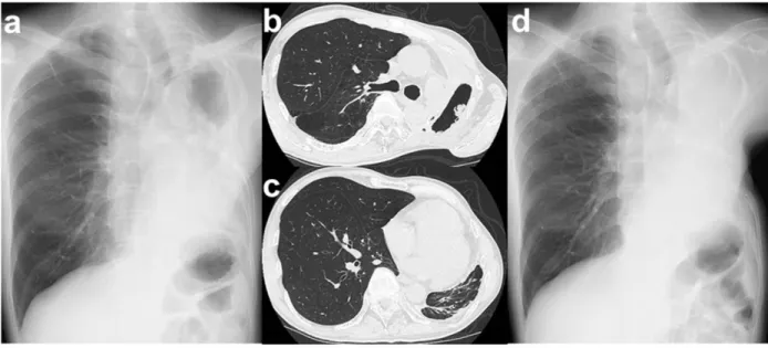
patient (Case 3) who had no comorbidities (Fig. 1). A prompt diagnosis of CPA was
22
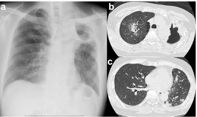
achieved in Case 4 during close follow-up of glucocorticoid tapering for radiation
23
pneumonitis, and CPA was subsequently successful controlled with antifungal agents (Fig.
2). The 2 patients with bilateral CPA lesions (Fig. 3) were not considered to be candidates
1
for surgery and died of CPA. The 4 patients with a unilateral CPA lesion are still alive. The
2
overall survival rate of the CPA group was similar to that of the group without CPA after
3
trimodality treatment for NSCLC (P=0.59). It is noteworthy that no cancer recurrence or
4
death was observed among the NSCLC patients who developed CPA, which is an
5
indication of the importance of CPA control in improving the outcomes of trimodality
6 treatment. 7 8 Discussion 9
In this study we elucidated the characteristics of CPA that developed in patients who had
10
undergone surgery for locally advanced NSCLC after induction chemoradiotherapy and
11
the outcomes of patients with CPA after trimodality treatment. All patients who
12
subsequently developed CPA had undergone extended resection for NSCLC and had a
13
history of either adjuvant chemotherapy or radiation pneumonitis. CPA developed at the
14
resection site of all 6 patients, and the 4 patients whose CPA was localized in a remaining
15
lobe in a single lung at the time of the diagnosis were considered to be candidates for
16
surgery, which resulted in improved CPA control and long-term survival. To the best of our
17
knowledge, this is the first report describing CPA after trimodality treatment.
18
Trimodality treatment for NSCLC, which consists of chemotherapy, radiotherapy,
19
and surgery, may itself be a risk factor for CPA [2-6]. Adjuvant chemotherapy may further
20
increase the risk of developing CPA [2], and radiation pneumonitis may contribute to the
21
development of a destroyed lung, which is susceptible to aspergillus infection. In addition,
22
extended lung resection may lead to the compensatory overexpansion of the remaining
23
lobes, especially in the emphysematous lungs of ever-smokers, as was observed in this
study, and contribute to cavity formation in the pulmonary parenchyma. Furthermore,
1
treatment of lung infections and second primary cancer may prolong patient survival after
2
trimodality treatment for NSCLC [16]. In view of these factors, physicians should be aware
3
of CPA as a possible late complication after trimodality treatment for NSCLC.
4
The diagnosis of CPA requires a consistent appearance on computed tomography,
5
such as a cavity and fungus ball, which are direct evidence of Aspergillus infection, or
6
evidence of an immunological response to Aspergillus species and the exclusion of other
7
diagnoses [1]. No Aspergillus species were detected in 2 of our patients (Cases 2 and 4).
8
Because an antagonistic relationship has been shown to exist between Aspergillus
9
fumigatus and Pseudomonas aeruginosa [17], in Case 2, in which a fungal infection was
10
histologically diagnosed based on the examination of the surgical specimen, the
11
pseudomonal infection may have resulted in a false-negative Aspergillus culture. In Case
12
4, the presence of a fungus ball and a positive galactomannan antigen test contributed to
13
the early diagnosis of CPA. An early diagnosis of CPA is required for effective treatment
14
to prevent the spread of CPA from one lung to the contralateral lung.
15
The localization of CPA in a single lung after NSCLC surgery at the time of the
16
diagnosis may be a key to the improved control of CPA after trimodality treatment for
17
NSCLC. Three patients with CPA in a single lung after NSCLC surgery underwent
18
successful surgery, including completion pneumonectomy. Another patient, whose CPA
19
remains well controlled with antifungal agents, would still be a candidate for surgery, even
20
if their condition deteriorated. Because pneumonectomy has been shown to provide
21
favorable results as a treatment for complex aspergilloma [18], completion
22
pneumonectomy may be a therapeutic option for operable CPA patients after trimodality
23
treatment for NSCLC. Survivors of NSCLC after trimodality treatment who have risk
factors for CPA might benefit from prophylactic antifungal therapy, which is routinely
1
administered to lung transplant recipients [19]. The further accumulation of cases of CPA
2
after trimodality treatment will be necessary for a more detailed evaluation.
3
In conclusion, careful follow-up is necessary to detect CPA as a late complication
4
after surgery for locally advanced NSCLC treated with induction chemoradiotherapy,
5
especially in ever-smoker patients who have undergone extended resection and have a
6
history of either adjuvant chemotherapy or radiation pneumonitis. The early detection and
7
diagnosis of CPA localized in a single lung after NSCLC surgery is critical to improving the
8
control of CPA with antifungal agents and surgery as well as long-term survival after
9
trimodality treatment.
10 11
Compliance with ethical standards 12
Conflict of interest: Shinichi Toyooka received research grants from Astellas Pharma 13
Inc., Chugai Pharmaceutical Co., Ltd., and Taiho Pharmaceutical Co., Ltd. Katsuyuki Kiura
14
received research grants from Bristol-Myers Squibb K. K., Chugai Pharmaceutical Co.,
15
Ltd., Nippon Boehringer Ingelheim Co., Ltd., and Ono Pharmaceutical Co., Ltd. The other
16
authors declare no conflicts of interest in association with the present study.
17 18
References 1
1. Denning DW, Cadranel J, Beigelman-Aubry C, Ader F, Chakrabarti A, Blot S, et al.
2
Chronic pulmonary aspergillosis: rationale and clinical guidelines for diagnosis and
3
management. Eur Respir J 2016;47:45-68.
4
2. Vento S, Cainelli F, Temesgen Z. Lung infections after cancer chemotherapy. Lancet
5
Oncol 2008;9:982-92.
6
3. Saraceno JL, Phelps DT, Ferro TJ, Futerfas R, Schwartz DB. Chronic necrotizing
7
pulmonary aspergillosis: approach to management. Chest 1997;112:541-8.
8
4. Denning DW, Riniotis K, Dobrashian R, Sambatakou H. Chronic cavitary and fibrosing
9
pulmonary and pleural aspergillosis: case series, proposed nomenclature change,
10
and review. Clin Infect Dis 2003;37 Suppl 3:S265-80.
11
5. Camuset J, Nunes H, Dombret MC, Bergeron A, Henno P, Philippe B, et al. Treatment
12
of chronic pulmonary aspergillosis by voriconazole in nonimmunocompromised
13
patients. Chest 2007;131:1435-41.
14
6. Smith NL, Denning DW. Underlying conditions in chronic pulmonary aspergillosis
15
including simple aspergilloma. Eur Respir J 2011;37:865-72.
16
7. Toyooka S, Kiura K, Shien K, Katsui K, Hotta K, Kanazawa S, et al. Induction
17
chemoradiotherapy is superior to induction chemotherapy for the survival of
non-18
small-cell lung cancer patients with pathological mediastinal lymph node metastasis.
19
Interact Cardiovasc Thorac Surg 2012;15:954-60.
20
8. Toyooka S, Kiura K, Takemoto M, Oto T, Takigawa N, Fujiwara T, et al. Long-term
21
outcome of induction chemoradiotherapy with docetaxel and cisplatin followed by
22
surgery for non-small-cell lung cancer with mediastinal lymph node metastasis.
23
Interact Cardiovasc Thorac Surg 2012;14:565-9.
24
9. Kosmidis C, Denning DW. The clinical spectrum of pulmonary aspergillosis. Thorax
25
2015;70:270-7.
26
10. Bryan DS, Donington JS. The Role of Surgery in Management of Locally Advanced
27
Non-Small Cell Lung Cancer. Current treatment options in oncology 2019;20:27.
28
11. Shien K, Toyooka S, Kiura K, Matsuo K, Soh J, Yamane M, et al. Induction
29
chemoradiotherapy followed by surgical resection for clinical T3 or T4 locally
30
advanced non-small cell lung cancer. Ann Surg Oncol 2012;19:2685-92.
31
12. Sato H, Toyooka S, Soh J, Hotta K, Katsui K, Yamamoto H, et al. The Feasibility of
32
Median Sternotomy With or Without Thoracotomy for Locally Advanced Non-Small
33
Cell Lung Cancer Treated With Induction Chemoradiotherapy. Ann Thorac Surg
34
2016;102:985-92.
35
13. Oken MM, Creech RH, Tormey DC, Horton J, Davis TE, McFadden ET, et al. Toxicity
36
and response criteria of the Eastern Cooperative Oncology Group. Am J Clin Oncol
37
1982;5:649-55.
38
14. Goldstraw P, Chansky K, Crowley J, Rami-Porta R, Asamura H, Eberhardt WE, et al.
39
The IASLC Lung Cancer Staging Project: Proposals for Revision of the TNM Stage
40
Groupings in the Forthcoming (Eighth) Edition of the TNM Classification for Lung
41
Cancer. J Thorac Oncol 2016;11:39-51.
42
15. Katayama H, Ueoka H, Kiura K, Tabata M, Kozuki T, Tanimoto M, et al. Preoperative
43
concurrent chemoradiotherapy with cisplatin and docetaxel in patients with locally
44
advanced non-small-cell lung cancer. Br J Cancer 2004;90:979-84.
45
16. Makimoto G, Kubo T, Oze I, Ohashi K, Hotta K, Tabata M, et al. Second primary cancer
in survivors of locally advanced non-small cell lung cancer treated with concurrent
1
chemoradiation followed by surgery. Jpn J Clin Oncol 2018;48:287-90.
2
17. Mowat E, Rajendran R, Williams C, McCulloch E, Jones B, Lang S, et al.
3
Pseudomonas aeruginosa and their small diffusible extracellular molecules inhibit
4
Aspergillus fumigatus biofilm formation. FEMS Microbiol Lett 2010;313:96-102.
5
18. Shiraishi Y, Katsuragi N, Nakajima Y, Hashizume M, Takahashi N, Miyasaka Y.
6
Pneumonectomy for complex aspergilloma: is it still dangerous? Eur J Cardiothorac
7
Surg 2006;29:9-13.
8
19. Sugimoto S, Yamane M, Otani S, Kurosaki T, Okahara S, Hikasa Y, et al. Airway
9
complications have a greater impact on the outcomes of living-donor lobar lung
10
transplantation recipients than cadaveric lung transplantation recipients. Surg Today
11 2018;48:848-55. 12 13 14 15 16 17 18 19 20 21 22 23 24 25 26 27 28 29 30
1 Ta b le 1 . P a ti e n t ch a ra ct e ri st ics N o . A g e a t th e d ia g n o si s o f C P A (ye a rs) S e x S m o ki n g h ist o ry H ist o lo g y T u m o r lo ca ti o n c-S ta g e C h e m o th e ra p y R a d ia ti o n (G y) T h o ra co to m y P u lm o n a ry re se ct io n C o m b in e d re se ct io n L ym p h n o d e d isse ct io n T o ta l n u m b e r o f re se ct e d lu n g se g m e n ts P a th o lo g ica l e va lu a ti o n A d ju va n t ch e m o th e ra p y C o m o rb id it ie s 1 47 M a le E ve r AD R t. S 1 IV A , T 4 N 2 M 1 b (r ib ) V N R 60 M S , T RUL R t. cl a vi cl e , S V C , p h e re n ic n e rve , w e d g e o f rt . S 6 N 2 le ve l 3 M in o r Y e s R a d ia ti o n p n e u m o n it is 2 55 M a le E ve r AD L t. S 1 + 2 II IB , T 4 N 2 M 0 C D D P + D O C 46 MS L U L W e d g e o f lt. S 6 N 2 le ve l 5 o r m o re M a jo r Y e s A lco h o lic ch ir rh o si s, D ia b e te s m e lli tu s, R ig h t p n e u m o th o ra x 3 52 M a le E ve r AD L t. S 6 II B , T 3 N 0 M 0 C D D P + D O C 46 PL LLL W e d g e o f lt. S 1 + 2 , ch e st w a ll N 2 le ve l 4 o r m o re C o m p le te No R a d ia ti o n p n e u m o n it is 4 73 M a le E ve r AD L t. S 1 + 2 II IA , T 1 cN2 M 0 C D D P + D O C 46 M S , T L U L N o n e N 3 le ve l 5 C o m p le te No R a d ia ti o n p n e u m o n it is tr e a te d w it h g lu co co rt ico id , P u lm o n a ry ve n o u s th ro m b u s 5 57 M a le E ve r AD L t. S 1 + 2 II B , T 2 a N 1 M 0 C D D P + D O C 46 PL L U L P u lm o n a ry a rt e ry N 2 le ve l 5 M a jo r Y e s L e ft a d re n a l m e ta st a si s tr e a te d w it h su rg e ry a n d ch e m o th e ra p y 9 m o n th s a ft e r in it ia l t h e ra p y 6 56 M a le E ve r AD R t. S 4 II IB , T 3 N 2 M 0 C D D P + D O C 46 PL R M L , R L L N o n e N 2 le ve l 7 M a jo r No R a d ia ti o n p n e u m o n it is AD a d e n o ca rci n o m a , CDDP ci sp la ti n , LLL le ft lo w e r lo b e , L t le ft , L U L le ft u p p e r lo b e , MS m e d ia n st e rn o to m y, PL p o st e ro la te ra l t h o ra co to m y, R L L r ig h t lo w e lo b e , R M L r ig h t m id d le lo b e , R t ri g h t, R U L r ig h t u p p e r lo b e , S se g m e n t o f th e lu n g , SVC su p e ri o r ve n a ca va , T t ra n sve rse t h o ra co to m y, V N R vi n o re lb in e
1 Ta b le 2 . S u m m a ry o f ch ro n ic p u lm o n a ry a sp e rg ill o si s F e ve r C o u g h S p u tu m 1 ,3 -b e ta -D -g lu ca n (p g /m L ) GM CRP (m g /d L ) C o n so lid a ti o n C a vi ty in t h e re se ct io n si te F u n g u s b a ll E m p ye m a w it h b ro n ch o -p le u ra l fi st u la 1 Y e s Y e s N o n e 9 .9 2 4 .8 N e g a ti ve 0 .2 1 5 .7 6 A sp e rg ill u s fu m ig a tu s P le u ra l e ff u si o n U n ila te ra l Y e s No Y e s IT C Z , V R C Z , M C F G C a ve rn o st o m y w it h fe n e st ra ti o n f o llo w e d b y co m p le ti o n p n e u m o n e ct o m y F u n g u s in fe ct io n A live (1 3 .3 ) 2 Y e s Y e s P u ru le n t 9 .4 < 6 .0 P o si ti ve 4 .5 1 .6 7 N e g a ti ve S p u tu m , BAL U n ila te ra l Y e s No Y e s IT C Z , V R C Z , C P F G C a ve rn o st o m y w it h fe n e st ra ti o n f o llo w e d b y m u scl e p lo m b a g e F u n g u s in fe ct io n A live (1 2 .2 ) 3 Y e s Y e s N o n e 4 .2 7 .9 N /A 6 .7 4 A sp e rg ill u s fu m ig a tu s S p u tu m U n ila te ra l Y e s Y e s No IT C Z , V R C Z C o m p le ti o n p n e u m o n e ct o m y w it h t h o ra co p la st y F u n g u s in fe ct io n A live (5 .0 ) 4 No No B lo o d y 1 .3 < 6 .0 P o si ti ve 1 .1 0 .7 5 N e g a ti ve S p u tu m U n ila te ra l Y e s Y e s No IT C Z , V R C Z , M C F G N o n e N o n e A live (5 .6 ) 5 Y e s Y e s B lo o d y 6 .3 < 6 .0 N e g a ti ve 0 .1 1 6 .7 6 A sp e rg ill u s fu m ig a tu s BAL B ila te ra l Y e s Y e s No V R C Z , M C F G N o n e N o n e D e a d (6 .3 ) 6 Y e s Y e s B lo o d y 3 .6 < 6 .0 N e g a ti ve 0 .3 2 .9 4 A sp e rg ill u s fu m ig a tu s S p u tu m B ila te ra l Y e s Y e s No V R C Z , L -A M B N o n e N o n e D e a d (9 .8 ) H ist o lo g y B A L b ro n ch o a lve o la r la va g e , C P A ch ro n ic p u lm o n a ry a sp e rg ill o si s, C P F G ca sp o fu n g in , CRP C -r e a ct ive p ro te in , CT co m p u te d t o m o g ra p h y, GM g a la ct o m a n n a n , IT C Z i tr a co n a zo le , L -A M B li p o so m a l a m p h o te ri ci n B , M C F G m ica fu n g in , N S C L C n o n -sm a ll ce ll lu n g ca n ce r, V R C Z vo ri co n a zo le N o . In te rva l b e tw e e n in it ia l t h e ra p y fo r N S C L C a n d t h e d ia g n o si s o f C P A (ye a rs ) A n ti fu n g a l a g e n ts O u tco m e (ye a rs a ft e r in it ia l t h e ra p y fo r N S C L C ) O p e ra ti ve p ro ce d u re S ym p to m s L a b o ra to ry fi n d in g s A sp e rg ill u s sp e ci e s C T f in d in g s C u ltu re sp e ci m e n
Figure legends 1
Fig. 1 Representative diagnostic images of surgical cases of chronic pulmonary 2
aspergillosis in a single lung after trimodality treatment (Case 3). A preoperative chest
X-3
ray film (a) and computed tomography scan (b) showed a cavity and fungus ball in the
4
remaining left upper lobe. A postoperative chest X-ray film showed fluid accumulation in
5
the left post-pneumonectomy space (c).
6 7 8 9 10 11 12 13 14 15 16
Fig. 2 A case of successful medical management of chronic pulmonary aspergillosis in a 1
single lung after trimodality treatment (Case 4). A chest X-ray film (a) and computed
2
tomography scan (b) revealed a cavity and fungus ball (arrow) in the left lower lobe
3
remnant. A chest X-ray film (c) and computed tomography scan (d) after antifungal therapy
4
showed that the fungus ball had shrunk.
5 6 7 8 9 10
Fig. 3 Representative diagnostic images of chronic pulmonary aspergillosis in both lungs 1
(Case 5). A chest X-ray film (a) and computed tomography scans showed a cavity with a
2
thickened wall in the remaining lower lobe of the left lung (b) and consolidation in both
3 lungs (b, c). 4 5 6 7 8 9 10 11 12 13
Fig. 4 Survival of patients who developed chronic pulmonary aspergillosis (CPA) and 1
those who did not develop CPA after trimodality treatment for locally advanced non-small
2
cell lung cancer. The survival rates of the CPA and non-CPA groups were comparable
(10-3
year survival rate, 50.0% vs. 57.6%, respectively, P=0.59).
4