INTRODUCTION
Stem cell therapies have great potential for bone tissue engineering applications. The ideal cell type for regenerative therapy would be one that is easily accessible and abundant, does not induce immunological rejection, and has the capacity for extensive self-renewal and expression of the phenotype of the desired tissue1,2). A recent breakthrough was the reprogramming of somatic cells from various tissues by the forced expression of defined exogenous factors into induced pluripotent stem cells (iPSCs)3,4). iPSCs offer the potential of producing a high output of custom-made stem cells. These cells have a similar potential to ESCs, but without the ethical or immunogenic concerns associated with ESCs5,6). These patient-specific iPSCs can be derived from various tissues, including fibroblasts7), keratinocytes8), BMSCs9) and blood cells10). They can serve as an inexhaustible cell source with the intriguing potential for use in various clinical applications, including bone regeneration11).
TGF-β is abundant in bone and has a biphasic effect
on bone turnover and homeostasis which can promote or inhibit osteogenesis12-14). TGF-β inhibits osteoblast differentiation through Smad3-mediated suppression of runt-related transcription factor 2 (Runx2) function and its expression15,16). The small molecule SB431542 (C22H16N4O3, Sigma-Aldrich, St. Louis, MO, USA) is a synthetic inorganic molecule that can inhibit activin receptor-like kinase4 and activin receptor-like kinase 5 (ALK5, the TGF-β type I receptor) (Fig. 1). Compared to other conventional drugs (natural or chemically-synthesized), gene-related methods or biological factors in bone tissue engineering, SB431542 has a simple structure and single function; thus, it is more effective and easier to control, and has fewer side effects17). It can provide a cost-effective way to stimulate iPSC differentiation into MSCs and subsequently activate bone morphogenetic protein (BMP) signalling of the TGF superfamily on the osteoblasts18-20). This is supported by the finding that SB431542 could induce iPSCs into MSCs in a simple one-step and cost-effective method18) and could promote high BMP2 expression expressin in preosteoblasts19,20). Thus, investigating the effects of SB431542 on stem cell differentiation may lead to
Bioactive small molecules in calcium phosphate scaffold enhanced osteogenic
differentiation of human induced pluripotent stem cells
Bing SONG1,2*, Haijun FU3*, Jianwei LIU3, Ke REN4, Michael D. WEIR2, Abraham SCHNEIDER5, Ping WANG2, Yang SONG2,6, Liang ZHAO1,2 and Huakun XU2,7,8
1 Department of Orthopedic Surgery, Shunde Hospital of Southern Medical University, Shunde, Guangdong 528300, China 2 Department of Advanced Oral Sciences and Therapeutics, University of Maryland School of Dentistry, Baltimore, MD 21201, USA
3 Department of Operative Dentistry and Endodontics, Guanghua School of Stomatology, Sun Yat-sen University; Guangdong Provincial Key Laboratory of Stomatology, Guangzhou, Guangdong 510055, China
4 Department of Neural and Pain Sciences, University of Maryland School of Dentistry, Baltimore, MD 21201, USA 5 Department of Oncology and Diagnostic Sciences, University of Maryland School of Dentistry, Baltimore, MD 21201, USA
6 Department of Prosthodontics, Guanghua School of Stomatology, Sun Yat-sen University, Guangdong Provincial Key Laboratory of Stomatology, Guangzhou, Guangdong 510055, China
7 Center for Stem Cell Biology and Regenerative Medicine, University of Maryland School of Medicine, Baltimore, MD 21201, USA
8 University of Maryland Marlene and Stewart Greenebaum Cancer Center, University of Maryland School of Medicine, Baltimore, MD 21201, USA Corresponding author, Liang ZHAO; E-mail: [email protected]
Human induced pluripotent stem cells (hiPSCs) are exciting for regenerative medicine due to their multi-potent differentiation. SB431542 bioactive molecule can activate bone morphogenetic protein-signalling in osteoblasts. The objectives were to: (1) develop a novel injectable calcium phosphate cement (CPC)-SB431542 scaffold for dental/craniofacial bone engineering; and (2) investigate cell proliferation and osteo-differentiation of hiPSC-derived mesenchymal stem cells (hiPSC-MSCs) on CPC-SB431542 scaffold. Three groups were tested: CPC control; CPC with SB431542 inside CPC (CPCSM); CPC with SB431542 in osteogenic medium (CPC+SMM). SB431542 in CPC promoted stem cell proliferation and viability. hiPSC-MSCs differentiated into osteogenic lineage and synthesized bone minerals. CPC with SB431542 showed much greater osteo-expressions and more bone minerals than those without SB431542. In conclusion, hiPSC-MSCs on CPC scaffold containing SB431542 showed excellent osteo-differentiation and bone mineral synthesis for the first time. CPC was a suitable scaffold for delivering stem cells and SB431542 to promote bone regeneration in dental/ craniofacial applications.
Keywords: Bone tissue engineering, Calcium phosphate cement, Induced-pluripotent stem cells, Small molecule SB431542, Osteogenic differentiation
*These authors contributed equally to this work.
Color figures can be viewed in the online issue, which is avail-able at J-STAGE.
Received Aug 23, 2019: Accepted Jul 7, 2020
Fig. 1 The chemical structure of SB431542.
enhanced bone tissue engineering efficacy21).
Calcium phosphate cement (CPC) was first developed by Brown and Chow in the 1980s and has been increasingly used as bone substitutes and drug delivery carrier for orthopedic and dental applications due to its excellent biocompatibility, osteoconductivity, injectability, self-setting ability, and ability to be replaced by new bone22,23). It was approved for the repair of craniofacial defects in 1996 by the Food and Drug Administration (FDA). However, the brittleness, poor solubility, insufficient mechanical properties and low degradation rate limit further clinical applications24). Our previous studies have shown much better load-bearing, physical and biological properties of CPC by using methods involving chitosan25), alginate26), fibers27), mannitol28), gas-forming porosity29), growth factors30) and biofunctionalization31). However, there has been no report on the incorporation of small molecule SB431542 into CPC to form a biofunctionalized scaffold and the effects on iPSC-MSCs for bone tissue engineering.
The present study investigated whether SB431542 could enhance the osteogenic differentiation and bone mineral synthesis of hiPSC-MSCs seeded on CPC scaffold for the first time. We hypothesized that the small molecule SB431542 could upregulate the expression of osteogenic genes and induce osteogenic differentiation for hiPSC-MSCs, thus promoting bone regeneration. The objectives of this study were: (1) to develop a novel self-setting CPC scaffold containing SB431542 for bone engineering; and (2) to investigate the effects of SB431542 in CPC paste and in culture medium on hiPSC-MSC viability, proliferation, osteogenic differentiation and bone mineral synthesis.
MATERIALS AND METHODS
Fabrication of SB431542-chitosan-CPC scaffold
CPC powder consisted of tetracalcium phosphate [TTCP: Ca4(PO4)2O] and dicalcium phosphate anhydrous (DCPA: CaHPO4)32). The TTCP particles were produced using a solid-state reaction of CaCO3 with DCPA (both from
J.T. Baker, Philipsburg, NJ, USA). The blended powder was heat-treated for 6 h in a high-temperature furnace (Model 51333, Lindberg, Watertown, WI, USA) at 1,500°C. Then the reacted material was milled in a ball mill (Retsch PM4, Brinkman, NY, USA), which produced particles with a median diameter of approximately 5 μM. The as-received DCPA powder was milled in a ball mill using 95% ethanol, which refined the DCPA particles to have a median diameter of approximately 1 μm. The TTCP and DCPA powders, using a 1:3 molar ratio, were blended together, forming the CPC powder.
A nature polymer chitosan was used due to its low toxicity, ability to be degraded by enzymes, and good biocompatibility33). Chitosan can provide more rapid setting and mechanical strength to CP34). Due to its hydrophilicity, the SB431542 release process can be controlled by controlling the water content. SB431542 was dissolved in dimethyl sulfoxide (DMSO) with a maximum absorption peak at 308 nm. Chitosan lactate (referred to as chitosan; VANSON, Redmond, WA, USA) was mixed with water containing 100 μM of SB431542 and dissolved in 0.2% DMSO at a chitosan/ (chitosan+SB431542+water) mass fraction of 15% to form the SB431542-chitosan liquid, as described in our previous study35). This concentration was chosen because a previous study showed that a 10 μM SB431542 solution could achieve the differentiation of ESCs and iPSCs36-38). We therefore selected 100 μM SB431542 in the CPC because in our preliminary study, it could achieve the release process of 10 μM SB431542 every 2 days to meet the needed drug amounts for the differentiation processes. A chitosan/(chitosan+water) mass fraction 15% was used to form the chitosan liquid for CPC control without SB431542. A CPC powder: liquid mass ratio of 2:1 was used to form a flowable CPC paste. The following stem cell-seeded groups were tested: CPC control, CPC with SB431542 inside CPC (CPCSM), CPC with SB431542 in osteogenic medium (CPC+SMM) to evaluate the stimulatory effects of SB431542 in the culture medium. The mixed cement was put into a mould, which had a 10 mm diameter and 1 mm thickness. The samples were maintained in a 37°C humidor for 1 day. Epoxyethane gas sterilization was performed for CPC which was degassed for 7 days. The CPC disks were then incubated in 24-well plates containing one disk in each well.
Release profile of SB431542-loaded CPC scaffold
Carbon14[14C]-labelled materials are used to evaluate the drug release profiles. The release of SB431542 was assessed following a previous study39). Briefly, CPCSM disks were cultured in 24-well plates with 1 mL of cell culture medium in an incubator, and 100 μL cell culture medium was withdrawn at each time interval. CPCSM (SM containing 1/10 [14C]-adenosine) scaffold were cultured in 24-well plates with 1 mL of cell culture medium in an incubator. The released solution was analyzed at various time points (1, 3, 7, 14, and 21 days). At every pre-determined time point, centrifugation was performed for the microsphere suspension. Then the PBS without microspheres was used for measuring
the [14C]-SB431542 concentration. Then, PBS (100 μL) was withdrawn at each time interval and transferred to a scintillation tube containing 3 mL Biodegradable Counting Cocktail buffer (Fisher Scientific, Pittsburgh, PA, USA). The amount of adenosine released from the CPC scaffold was quantified via counting the radioactivity using a scintillation method (Beckman LS6500 Counter, Brea, CA, USA).
Cell culture
The hiPSCs cultures was performed as described previously40). The study was approved by the University of Maryland Baltimore Institutional Review Board, and the manuscript did not have any information that would lead to the identification of the participants. All the methods were used following the guidelines and policies. The informed consent was obtained from every participant. Briefly, the hiPSC BC1 line was obtained from bone marrow cells (CD34+) in adults. The hiPSC-MSCs harvested from bone marrow stem cells exhibited high ALP activity, osteocalcin gene expression and bone minerals, compared to other cells such as hiPSC-MSCs from foreskin and other tissues used in bone tissue engineering41). Human primary mononuclear cells from a healthy adult marrow donor (code: BM2426) were isolated with a standard gradient protocol by Ficoll-Paque Plus (p=1.077). The mononuclear cells were magnetically labelled with CD34 microbead kit (MACS No.130100453), and then magnetically separated using an appropriate MACS column and MACS separator according to the number of total cells and the number of CD34+ cells, following the manufacture’s protocol. CD34+ cells were purified with a magnetically activated cell sorting system and cultured for 4 days using haematopoietic cytokines. Then the cells were reprogrammed using a single episomal vector, pEB-C542). BM-iPSCs were transferred to murine embryonic fibroblasts (MEF) feeding layer cells in hiPSC medium for purification and amplification. When 80% confluence was reached, these were detached and disintegrated to become clumps by using 1 mg/mL collagenase type IV for 6 min in DMEM/F12 at 37°C. Then mechanical scraping was performed. The disintegrated clumps of BM-hiPSC were obtained via a sedimentation method. They were then re-placedin an embryoid body (EB) differentiation medium, which had the same composition to the medium for hiPSCs but without β-FGF. The samples were placed into 25 cm2 ultra-low attachment cell culture flasks (Corning, Corning, NY, USA). The culture medium was changed every other day. After culturing for 10 days, the EBs were then placed onto 0.1% gelatine-coated culture dishes. The cells that grew out of the EBs were then cultured. When 70% confluence was reached, those outgrowing cells (referred to as P0) were then selectively harvested via the use of cell scrapers. The samples were then sub-cultured in the MSC medium. The differentiated cells were passaged until a homogeneous fibroblastic morphology appeared. These cells were referred to as the hiPSC-derived MSCs (hiPSC-MSCs)43). The use of the hiPSC-MSCs was
approved by the University of Maryland Baltimore. The obtained hiPSC-MSCs were maintained in a tissue culture disk containing low glucose Dulbecco’s modified Eagle’s medium (DMEM) supplenmented with 10% foetal bovine serum (FBS), 5 mg/mL penicillin and 5 mg/mL streptomycin (Invitrogen, Carlsbad, CA, USA) and incubated at 37°C in a humidified CO2 incubator. The medium was changed every two days until the cells reached 80–90% confluence. Then the hiPSC-MSCs were detached by trypsin (Invitrogen) and passaged at 6,000 cells/cm2. hiPSC-MSCs at passage 7 were used for the subsequent experiments.
Flow cytometry analysis of iPSC-MSCs
A previous report confirmed that this method produced hiPSC-MSCs that indeed had surface markers typical for MSCs, including CD29, CD44, CD166, CD7344,45). Furthermore, they did not express for haematopoietic (CD34), endothelial (CD31) or pluripotent markers such as TRA-1-81 and OCT 3/444,45). The cell surface antigen expression profiles of these iPSC-MSCs were characterized using flow cytometry as described previously41). The iPSC-MSCs were obtained using trypsin-ethylenediaminetetraacetic acid (EDTA). They were then washed using cold phosphate buffered saline (PBS) which had 1% bovine serum albumin (BSA). The samples were then placed with a cell density of 1×10⁶ cells in 50 mL of the cold PBS that had 1% BSA. The cells were labelled on ice using the optimal dilution of fluorescein isothiocyanate conjugated (FITC) monoclonal antibodies (mAbs; all obtained from Invitrogen, unless otherwise noted) against CD29, CD31, CD34, CD44, CD166, CD73, and HLA-DA, phycoerythrin(PE)-conjugated mAbs against Oct3/4 (BD, San Diego, CA, USA) and CD166 (BD), and an Alexa Fluor 488-conjugated mAb against CD105 in the dark. After a 30-min incubation, cells were washed with cold PBS containing 1% BSA. Nonspecific fluorescence was determined by incubating cells with isotype-matched conjugated mAbs. At least 10,000 events were collected from each flow cytometry run. Data were analyzed using Cell Quest software (Becton Dickinson). The fluorescence histogram for each mAb was displayed alongside the control antibody. The percentages of positive cells were subtracted from the isotype control antibody for each conjugate.
Cell attachment and viability assay
Consistent with a previous study, 3×10⁵ cells were re-suspended in 1 mL of cell culture medium, seeded on a 10×1 mm disk in a 24-well plate, and cultured in an incubator at 37°C46). After the seeding cells had attached to CPC and CPC-SB431542 disks at day 2, the medium was changed with osteogenic medium that containing 100 nM dexamethasone, 10 mM β-glycerophosphate, 0.05 mM ascorbic acid, and 10 nM 1a, 25-dihydroxyvitamin (Sigma-Aldrich). The osteogenic medium supplemented with 10 μM SB431542 were referred to as CPC+SMM47). The culture was incubated for 1, 4, 7 and 14 days and the culture medium was changed every other day.
(Invitrogen) with 1 μL ethidium homodimer-1 and 1 μL calcein-AM dissolved in 1 mL PBS at 1, 7 and 14 days. An epifluorescence microscope (Eclipse TE2000-S, Nikon, Melville, NY, USA) was used. The percentage of live cells was referred to as P. The live cell density was termed D. P and D were measured from two randomly-chosen fields for each disk. The images were evaluated vis the Image-Pro Plus software48). P=live cell number/ (live cell number+dead cell number). D=live cell number in the image/the area of the image. This evaluation was repeated by three times at different time periods.
The viability of the stem cell was measured using cell counting kit-8 assay (Enzo Life Sciences, Farmingdale, NY, USA). The kit solution was added to each well at a ratio of to the 1:10 with cell culture mixture and maintained for 2 h in a humidified 5% CO2 atmosphere. The absorbance, which was proportional to the cell viability, was evaluated using the absorbance at 450 nm
via a microplate reader.
ALP assay
Cells were seeded with 3×10⁵ cells/well on each CPC scaffold in 24-well plates. The ALP activity was evaluated at 1, 4, 7 and 14 days with the ALP kit (Wako, Japan). The specimens were washed in PBS before adding 0.5 mL cell lysis buffer that had 0.2% Triton X-100 (Sigma) with 10 mM Tris (pH 7.0) and 1 mM EDTA (Sigma) onto CPC scaffolds. The samples were then incubated for 20 min, and the placed into a −80°C freezer for 30 min. Then the samples were thawed for 30 min at room temperature. This freeze-thawing method was repeated two times to lyse cells and collect all the ALP samples. Then the specimens were placed into a 96 well plate. Finally, the activity was evaluated at 405 nm concurrently alongside with the standard samples. The ALP activity was measured relative to the total cellular protein concentrations: [pNpp (μM/min)]/[Protein (mg)]. The protein amount in every specimen was evaluated using a BCA protein kit (Thermo Scientific, Rockford, IL, USA). For the ALP activity assays, every specimen was measured in triplicate and the results were repeated by performing at least three independent tests.
Osteogenic differentiation of seeding cells
The osteogenic gene expression analyses were performed using BioRad real-time quantitative reverse transcription-polymerase chain reaction (RT-qPCR) system (Applied Biosystems, Foster City, CA, USA). The following primers were used: ALP forward: CTATCCTGGCTCCGTGCTC,
reverse: GCTGGCAGTGGTCAGATGTT; Runx2
forward: GACTGTGGTTACCGTCATGGC,
reverse: ACTTGGTTTTTCATAACAGCGGA;
COL1 forward: CAGGCTGGTGTGATGGGATT,
reverse: AGCTCCAGCCTCTCCATCTT; OC
forward: CTCACACTCCTCGCCCTATT, reverse:
TTGGACACAAAGGCTGCAC; and GAPDH
forward: TCAACGACCCCTTCATTGAC, reverse:
ATGCAGGGATGATGTTCTGG. At 1, 7 and 14 days, the total RNA was extracted with Trizol reagent and PureLink RNA Mini Kit (Invitrogen) from the cell-seeded
CPC disks. 4,000 ng of RNA was reverse-transcribed into cDNA using a high-capacity cDNA Reverse Transcription kit (Applied Biosystems). Real-time RT-PCR analysis was performed using SYBR Premix Ex gene expression kits to quantify the targeted genes. The average fold of changes from three separate tests were evaluated by using the 2-ΔΔCT technique and normalized to the CT values of the housekeeping gene GAPDH. The CT value of the hiPSC-MSCs grown for 1 day on the CPC scaffolds in the growth medium was employed as the calibrating group.
Mineral synthesis by the seeded cells
Alizarin red staining (ARS) was performed to evaluate the mineral synthesis by hiPSC-MSCs. At 1, 7 14 and 21 days, the disks were fixed using 10% formaldehyde for 30 min, washed by PBS and stained with 2% Alizarin Red S (Sigma) for 45 min. The disks were rinsed with PBS two times to visualize the calcified deposits. The minerals were dissolved with 10% cetylpyridinium chloridemonohydrate (Sigma) and the ARS concentration was measured at 550 nm using a microplate reader. CPC without cells was also analyzed as a control to exclude the effects of the CPC scaffold at the same time periods. The CPC control’s absorbance was subtracted from that of the cell-seeded scaffolds to obtain the net mineral concentration synthesized by the cells. This analysis was independently repeated three times.
Statistical analysis
Statistical analysis was done using one-way analyses of variance (ANOVA), as well as post hoc least significant difference (LSD) analyzes via a statistical software (SPSS 20.0). All the data were shown as the mean value±the standard deviation (SD). Kolmogorov–Smirnrov test and Levene test were first done to verify the equal variance and the normality in the data. The p<0.05 value was considered to be significant statistically.
RESULTS
Drug relase profile of SB431542-loaded CPC scaffold
The drug release of SB431542 loaded into CPC scaffold was analyzed at pH 7 and 37°C as shown in Fig. 2. The
in vitro diffusion of SB431542 by chitosan-based systems
in cell culture medium was sustained throughout the tested cell culture time period. The SB431542 release concentration increased with time, reaching a peak of 91.2% at 21 days. The release pattern of SB431542 in chitosan-CPC scaffold showed a linear profile with an initial release of approximately 16% every 2 days and a subsequent sustained release of approximately 10% every 2 days. The results indicated that the SB431542 release from CPC+chitosan scaffold was maintained for a relatively long time period, which was able to satisfy the requirement of a drug carrier to provide releases for at least 7–12 days in order to enhance the hiPSC-MSCs for osteogenic differentiation.
Fig. 2 The controlled release kinetics of SB431542 from CPC scaffold.
The results represent the mean±SD of three independent experiments.
Fig. 3 Immunophenotyping of human iPSC-MSCs. Flow cytometry analysis showed that hiPSC-MSCs expressed typical cell surface markers characteristic of MSCs, and were negative for pluripotent and endothelial cell markers. The names of the antigens are listed inside each plot. Each plot represents the percentage of positive cells.
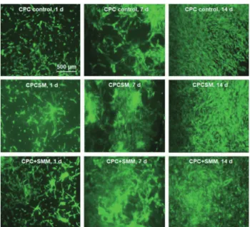
Fig. 4 Live/dead staining images of hiPSC-MSCs seeded on CPC control, CPCSM and CPC+SMM, for 1, 7, and 14 days.
Live cells, stained in green, were prevalent in all groups. Dead cells, stained in red, were few for all groups.
Flow cytometry analysis of iPSC-MSCs
Flow cytometry evaluation of hiPSC-MSCs showed that the MSC surface markers were highly and consistently expressed (Fig. 3). The MSC surface markers CD29, CD44, CD166, CD73 and CD105 were expressed at >95% levels in these iPSC-MSCs. In comparison, the haematopoietic markers CD31 and CD34 expressions levels were <0.5% in the iPSC-MSCs. The expression was <0.5% for hiPSC pluripotency marker HLA-DR (Class II histocompatibility Antigen). These results indicate that the representative MSC markers were greatly expressed in the iPSC-MSCs.
Viability of the attached cells on the CPC scaffold
The viability of hiPSC-MSCs was not negatively influenced by SB431542 as shown by the live/dead pictures (Fig. 4). Cells attached and proliferated well in all scaffold groups. Numerous live cells (green staining) and few dead cells (red staining) were present. The live cell density on the scaffold surfaces was enhanced by increasing the culture time because of cell proliferation (Fig. 5A). Live cells percentages on CPC scaffold control and CPCSM, CPC+SMM groups were approximately 90% at day 7 (Fig. 5B), and their cell percentage and live cell density were significantly greater than those of the CPC control (p<0.05). However, no noticeable difference was shown between CPCSM and CPC+SMM groups, indicating that SB431542 in both the CPC scaffold and in culture medium could promote the stem cell proliferation and viability.
Fig. 5 Cell proliferation and viability of hiPSC-MSCs attached to CPC scaffold.
(A) Live cell density, (B) Percentage of live cells, and (C) CCK-8 cell proliferation. Each value in the bar graph represents the mean±SD of three independent experiments. Significant differences are indicated by an asterisk (*p<0.05).
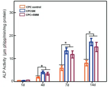
Fig. 6 hiPSC-MSC ALP activity using the PNPP method. The ALP activity values for CPCSM and CPC+SMM were significantly greater than that of CPC control (*p<0.05). Each value in the bar graph represents the mean±SD of three independent experiments.
Cell proliferation
CCK-8 analysis demonstrated that hiPSC-MSCs cells were strongly attached to the scaffold and their viability was high on day 1, 4, 7 and 14 as shown in Fig. 5C. The OD value suggests that there were no significant differences in the cell viability for the three groups
(p>0.05), indicating that SB431542 in the CPC scaffold was not cytotoxic to cells.
ALP activity
The ALP activity of hiPSC-MSCs was quantified and normalized by the amount of protein per sample [(mM pNPP/min)/(mg protein)] (Fig. 6). For each group, the values using different letters in each plot indicate that they are significantly different (p<0.05). In all groups, ALP activity was minimal at day 1 and there was no significant difference among the groups. The increase in ALP activity was time-dependent and reached a maximum at day 21. The highest ALP activity occurred in the CPCSM group, increasing from (8.28±0.95) for CPC control to (17.32±2.06) at day 14, which was significantly greater than the control groups (p<0.05). This indicates that CPC combined with SB431542 was optimal for the osteogenic differentiation of hiPSC-MSCs. These findings indicate a high promise of the hiPSC-MSCs+CPC+SB431542 method as an optimal “stem cell-small molecule-scaffold” construct to enhance bone tissue engineering.
Real time PCR analysis of osteogenic differentiation
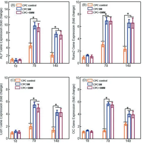
The hiPSC-MSC osteogenic differentiation in the three groups was evaluated, which showed time-dependent increasing expressions: including ALP (Fig. 7A), Runx2 (Fig. 7B), collagen type I (COL1) (Fig. 7C), and osteocalcin (OC) (Fig. 7D). The expression trend was sharply increased using SB431542. At day 1, there was no difference between the groups. The expression trend was sharply increased in the presence of SB431542. ALP, Runx2, COL1 and OC mRNA expression levels peaked at day 7 and then slightly decreased at day 14 in
Fig. 7 hiPSC-MSC osteogenic differentiation on CPC control, CPCSM, and CPC+SMM groups.
RT-PCR analysis of ALP (A), Runx2 (B), COL1 (C), and OC (D). expression levels, hiPSC-MSC CT value in growth media at day 1 were used as a calibrator. Each value in the bar graph represents the mean±SD of three independent experiments. Significant differences are indicated by an asterisk (*p<0.05).
CPCSM and CPC+SMM which were significantly higher than CPC control (p<0.05). These data confirmed that the use of SB431542 with CPC scaffold could stimulate hiPSC-MSC differentiation into osteogenic cells.
Mineralization
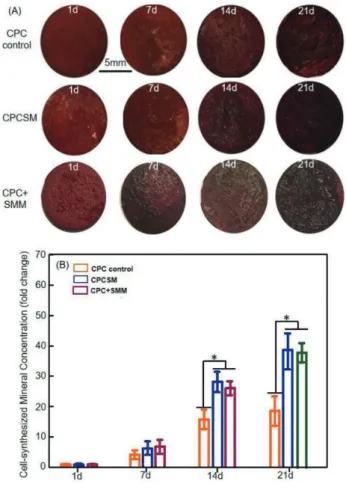
Representative staining images of mineralized extracellular bone matrix deposits produced by hiPSC-MSCs on scaffold surfaces are shown in Fig. 8A. There was a layer of new mineral matrix synthesized by the cells covering the scaffold in CPC control, CPCSM and CPC+SMM groups. The thick bone matrix mineralization covered not only the scaffold’s top surface, but also the scaffold sides. The staining of the synthesized bone mineral matrix indicated that it had an increase in the staining density and darkness. This showed a substantial increase in the bone mineral synthesis by the cells from day 1 to 21. The results from the cetylpyridinium chloride monohydrate are plotted in Fig. 8B. For each group, the amount of mineral synthesized by the cells grew substantially from day 1 to 21 (p<0.05). At each
time point, the mineral amount was greater in CPCSM and CPC+SMM group compared with the CPC control (p<0.05). For instance, at day 21, the iPSC-MSCs in CPCSM group synthesized 2–3 folds more bone mineral than CPC control group.
DISCUSSION
In the present study, we developed a novel biofunctional CPC scaffold that greatly improved hiPSC-MSC attachment, proliferation, osteogenic differentiation and bone mineralization. We generated hiPSC-MSCs from hiPSCs and directly compared, for the first time, the performance of hiPSC-MSCs seeded on CPC, CPCSM and CPC+SMM scaffolds for bone tissue engineering.
In vitro experiments demonstrated that CPC, CPCSM
and CPC+SMM had good affinities for cell attachment, without negative influence on the cell viability. Stem cells in each scaffold group underwent osteogenic differentiation. CPCSM and CPC+SMM scaffolds substantially enhanced the hiPSC-MSC functions.
Fig. 8 Bone matrix mineral synthesis by hiPSC-MSCs. (A) The bone mineral exhibited a denser and darker red staining that increased with increasing cell culture time. (B) The cell-synthesized bone mineral concentration was measured using cetylpyridinium chloride monohydrate. Each value in the bar graph represents the mean±SD of three independent experiments. Significant differences are indicated by an asterisk (*p<0.05).
Live cell density was substantially enhanced due to the SB431542 addition into the CPC scaffold. The mineralization induced by hiPSC-MSCs was highly promoted by the SB431542 in the CPC. Therefore, incorporating the SB431542 into the CPC paste is a promising method to generate biofunctional CPC scaffolds to promote cell function and enhance bone regeneration.
Several studies have identified and described various protocols for iPSC-based bone regeration to promote the osteogenic differentiation of the iPSCs, including directly seeding the cells in the MSC growth media and adding extra chemical inducers46,49), small molecules21), growth factors50), and gene modifications44,51), or seeding cells on three-dimensional scaffolds combined with osteogenic agents52,53), and even mechanical stimulation54). Each method has certain limitations and advantages. All these methods have succeeded in differentiating cells into the osteoblastic lineage, showing ALP activity, osteogenic
gene expression and mineral synthesis by iPSCs. Here we incorporated SB431542 into CPC. Various reports have indicated that SB431542 can alter TGF-β signalling and elevate the expression of BMP219,20,55). In addition, SB431542 can also induce hiPSCs differentiation into MSCs in a simple one-step and cost-effective manner56,57). Shi et al. reported that SB431542 alone could robust osteogenesis of autologous human gingival mesenchymal stem cells to successfully repair minipig severe maxillofacial bone defects58-61). Here we selected hiPSC-MSCs as the initial seeding cells. Instead of using hiPSCs directly, hiPSC-MSCs were used because hiPSCs could not attach strongly to the CPC scaffold or cell culture plate. SB431542 could induce the transformation of hiPSCs into hiPSC-MSCs to promote bone regeneration. To our knowledge, this represents the first report showing that the osteogenic effect of hiPSC-MSCs on CPC scaffold was enhanced in vitro via SB431542. This occurred via the inhibition of the TGF-β signalling and the activation of the BMP2 signalling pathway. The CPC scaffold containing SB431542 could promote cell attachment and proliferation via enhancement of BMP2 expression on the hiPSC-MSCs. In addition, SB431542 did not exert any cytotoxic effect on the cells. A significant difference in osteogenic effect
in vitro was observed between SB431542-treated and
untreated CPC scaffolds seeded with hiPSC-MSCs. These results indicate the potential of SB431542 to replace the use of the extremely expensive BMP2 as a cost-effective and simple method for bone tissue engineering.
Despite of these intriguing finding, the present work has limitations. The accurate and quantitative control of the SB431542 amount is technically difficult. There are controversies surrounding even well-investigated small molecules, and previous studies based on different models often yielded different results33,62). Thus, further in-depth investigation should be performed to identify the mechanism of SB431542-mediated osteogenesis stimulation at the molecular level. Overall, our results showed a potentially important application of SB431542 as a much less expensive method than BMP2 administration to significantly enhance bone regeneration. Further in vivo experiments should be performed using an animal model to demonstrate the feasibility for clinical applications.
CONCLUSION
The present study showed for the first time that a CPC scaffold seeded with hiPSC-MSCs in combination with SB431542 resulted in significantly greater osteogenic effects than scaffolds without SB431542. The mechanism was postulated to involve SB431542 stimulation of the MSCs and their subsequent induction into osteogenic cells by inhibiting endogenous TGF-β signalling. The percentage of live cells in the CPCSM and CPC+SMM groups were higher than those in CPC control. ALP activity of the CPCSM and CPC+SMM groups were higher than those of CPC control; CPCSM group had the most increase among all groups. Real time PCR analysis
revealed an enhanced effect of SB431542 on osteogenic differentiation. Bone mineral synthesis by hiPSC-MSCs in CPCSM and CPC+SMM groups was greater than CPC control. SB431542 with CPC constructs had the strongest osteogenic differentiation and the greatest bone mineral synthesis. Therefore, CPC with SB431542 represents a promising approach to substantially enhance osteogenesis for dental, craniofacial and orthopedic applications.
ACKNOWLEDGMENTS
Authors would like to thank Dr. Linzhao Cheng for providing the hiPSC BC1 cells. This study was supported by the National Natural Science Foundation of China No. 3171051, Guangzhou Science and Technology Planning Project (201803010114), University of Maryland School of Dentistry bridge fund, and University of Maryland Baltimore Seed Grant.
REFERENCES
1) Watt FM, Hogan BL. Out of Eden: stem cells and their niches. Science 2000; 287: 1427-1430.
2) Bianco P, Robey PG. Stem cells in tissue engineering. Nature 2001; 414: 118-121.
3) Takahashi K, Yamanaka S. Induction of pluripotent stem cells from mouse embryonic and adult fibroblast cultures by defined factors. Cell 2006: 126: 663-676.
4) Yu J, Vodyanik MA, Smuga-Otto K, Antosiewicz-Bourget J, Frane JL, Tian S, et al. Induced pluripotent stem cell lines derived from human somatic cells. Science 2007; 318: 1917-1920.
5) Takahashi K, Yamanaka S. Induced pluripotent stem cells in medicine and biology. Development 2013; 140: 2457-2461. 6) Luzzani CD, Miriuka SG. Pluripotent stem cells as a robust
source of mesenchymal stem cells. Stem Cell Rev 2017; 13: 68-78
7) Park I, Zhao R, West JA, Yabuuchi A, Huo H, Ince TA, et al. Reprogramming of human somatic cells to pluripotency with defined factors. Nature 2007; 451: 141-146.
8) Li W, Zhou H, Abujarour R, Zhu S, Young Joo J, Lin T, et al. Generation of human induced pluripotent stem cells in the absence of exogenousSox2. Stem Cells 2009; 27: 2992-300. 9) Yamanaka S. Strategies and new developments in the
generation of patient-specific pluripotent stem cells. Cell Stem Cell 2007; 1: 39-49.
10) Loh YH, Agarwal S, Park IH, Urbach A, Huo H, Heffner GC, et al. Generation of induced pluripotent stem cells from human blood. Blood 2009; 113: 5476-5479.
11) Ono N, Kronenberg HM. Bone repair and stem cells. Curr Opin Genet Dev 2016; 40: 103-107.
12) Bonewald LF, Dallas SL. Role of active and latent transforming growth factor beta in bone formation. J Cell Biochem 1994; 55: 350-357.
13) Ignotz RA, Massague J. Cell adhesion protein receptors as targets for transforming growth factor-beta action. Cell 1987; 51: 189-197.
14) Robey PG, Young MF, Flanders KC, Roche NS, Kondaiah P, Reddi AH, et al. Osteoblasts synthesize and respond to transforming growth factor-type beta (TGF-beta) in vitro. J Cell Biol 1987; 105: 457-463.
15) Alliston T, Choy L, Ducy P, Karsenty G, Derynck R. TGF-beta-induced repression of CBFA1 by Smad3 decreases cbfa1 and osteocalcin expression and inhibits osteoblast differentiation. EMBO J 2001; 20: 2254-2272.
16) Kang JS, Alliston T, Delston R, Derynck R. Repression of Runx2 function by TGF-beta through recruitment of class II histone deacetylases by Smad3. EMBO J 2005; 24: 2543-2555.
17) Han Q, Du Y, Yang P. The role of small molecules in bone regeneration. Future Med Chem 2013; 5: 1671-1684. 18) Chen YS, Pelekanos RA, Ellis RL, Horne R, Wolvetang EJ,
Fisk NM. Small molecule mesengenic induction of human induced pluripotent stem cells to generate mesenchymal stem/stromal cells. Stem Cells Transl Med 2012; 1: 83-95. 19) Kawahara T, Yamashita M, Ikegami K, Nakamura T,
Yanagita M, Yamada S, et al. TGF-beta negatively regulates the BMP2-dependent early commitment of periodontal ligament cells into hard tissue forming cells. PLoS One 2015; 10: e125590.
20) Schindeler A, Morse A, Peacock L, Mikulec K, Yu NY, Liu R, et al. Rapid cell culture and pre-clinical screening of a transforming growth factor-beta (TGF-beta) inhibitor for orthopaedics. BMC Musculoskelet Disord 2010; 11: 105. 21) Kanke K, Masaki H, Saito T, Komiyama Y, Hojo H, Nakauchi
H, et al. Stepwise differentiation of pluripotent stem cells into osteoblasts using four small molecules under serum-free and feeder-free conditions. Stem Cell Reports 2014; 2: 751-760. 22) Xu HH, Wang P, Wang L, Bao C, Chen Q, Weir MD, et al.
Calcium phosphate cements for bone engineering and their biological properties. Bone Res 2017; 5: 17056
23) Frankenburg EP, Goldstein SA, Bauer TW, Harris SA, Poser RD. Biomechanical and histological evaluation of a calcium phosphate cement. J Bone Joint Surg Am 1998; 80: 1112-1124.
24) Ambard AJ, Mueninghoff L. Calcium phosphate cement: Review of mechanical and biological properties. J Prosthodont 2006; 15: 321-328.
25) Xu H. Synergistic reinforcement of in situ hardening calcium phosphate composite scaffold for bone tissue engineering. Biomaterials 2004; 25: 1029-1037.
26) Zhao L, Weir MD, Xu HH. An injectable calcium phosphate-alginate hydrogel-umbilical cord mesenchymal stem cell paste for bone tissue engineering. Biomaterials 2010; 31: 6502-6510.
27) Xu HH, Quinn JB. Calcium phosphate cement containing resorbable fibers for short-term reinforcement and macroporosity. Biomaterials 2002; 23: 193-202.
28) Tang M, Weir MD, Xu HHK. Mannitol-containing macroporous calcium phosphate cement encapsulating human umbilical cord stem cells. J Tissue Eng Regen Med 2012; 6: 214-224. 29) Chen W, Zhou H, Weir MD, Bao C, Xu HH. Umbilical cord
stem cells released from alginate–fibrin microbeads inside macroporous and biofunctionalized calcium phosphate cement for bone regeneration. Acta Biomater 2012; 8: 2297-2306. 30) Xu HHK, Weir MD, Simon CG. Injectable and strong
nano-apatite scaffolds for cell/growth factor delivery and bone regeneration. Dent Mater 2008; 24: 1212-1222.
31) Chen W, Thein-Han W, Weir MD, Chen Q, Xu HH. Prevascularization of biofunctional calcium phosphate cement for dental and craniofacial repairs. Dent Mater 2014; 30: 535-544.
32) Weir MD, Xu HHK. Human bone marrow stem cell-encapsulating calcium phosphate scaffolds for bone repair. Acta Biomater 2010; 6: 4118-4126.
33) Hu Q, Luo Y. Polyphenol-chitosan conjugates: Synthesis, characterization, and applications. Carbohydr Polym 2016; 151: 624-639.
34) Costa-Pinto AR, Reis RL, Neves NM. Scaffolds based bone tissue engineering: the role of chitosan. Tissue Eng Part B Rev 2011; 17: 331-347.
35) Weir MD, Xu HH. Culture human mesenchymal stem cells with calcium phosphate cement scaffolds for bone repair. J Biomed Mater Res B Appl Biomater 2010; 93: 93-105.
36) Chen G, Park C, Xie R, Ji RR. Intrathecal bone marrow stromal cells inhibit neuropathic pain via TGF-β secretion. J Clin Investig 2015; 125: 3226-3240.
37) Sagi I, Chia G, Golan-Lev T, Peretz M, Weissbein U, Sui L, et al. Derivation and differentiation of haploid human embryonic stem cells. Nature 2016; 532: 107-111.
38) Wang L, Meece K, Williams DJ, Lo KA, Zimmer M, Heinrich G, et al. Differentiation of hypothalamic-like neurons from human pluripotent stem cells. J Clin Investig 2015; 125: 796-808.
39) Wang P, Ma T, Guo D, Hu K, Shu Y, Xu HHK, et al. Metformin induces osteoblastic differentiation of human induced pluripotent stem cell-derived mesenchymal stem cells. J Tissue Eng Regen Med 2018; 12: 437-446
40) Tang M, Chen W, Weir MD, Thein-Han W, Xu HH. Human embryonic stem cell encapsulation in alginate microbeads in macroporous calcium phosphate cement for bone tissue engineering. Acta Biomater 2012; 8: 3436-3445.
41) Wang L, Zhang C, Li C, Weir MD, Wang P, Reynolds MA, et al. Injectable calcium phosphate with hydrogel fibers encapsulating induced pluripotent, dental pulp and bone marrow stem cells for bone repair. Mater Sci Eng C 2016; 69: 1125-1136.
42) Mali P, Ye Z, Hommond HH, Yu X, Lin J, Chen G, et al. Improved efficiency and pace of generating induced pluripotent stem cells from human adult and fetal fibroblasts. Stem Cells 2008; 26: 1998-2005.
43) Cheng L, Hansen NF, Zhao L, Du Y, Zou C, Donovan FX, et al. Low Incidence of DNA sequence variation in human induced pluripotent stem cells generated by nonintegrating plasmid expression. Cell Stem Cell 2012; 10: 337-344. 44) Liu J, Chen W, Zhao Z, Xu HH. Reprogramming of
mesenchymal stem cells derived from iPSCs seeded on biofunctionalized calcium phosphate scaffold for bone engineering. Biomaterials 2013, 34: 7862-7872.
45) Theinhan W, Liu J, Tang M, Chen W, Cheng L, Xu HH. Induced pluripotent stem cell-derived mesenchymal stem cell seeding on biofunctionalized calcium phosphate cements. Bone Res 2013; 4: 371-384.
46) Wang P, Liu X, Zhao L, Sun J, Chen W, Man Y, et al. Bone tissue engineering via human induced pluripotent, umbilical cord and bone marrow mesenchymal stem cells in rat cranium. Acta Biomater 2015; 18: 236-248.
47) Thein-Han W, Liu J, Xu HH. Calcium phosphate cement with biofunctional agents and stem cell seeding for dental and craniofacial bone repair. Dent Mater 2012; 28: 1059-1070. 48) Chen W, Liu J, Manuchehrabadi N, Weir MD, Zhu Z, Xu
HH. Umbilical cord and bone marrow mesenchymal stem cell seeding on macroporous calcium phosphate for bone regeneration in rat cranial defects. Biomaterials 2013, 34: 9917-9925.
49) Villa-Diaz LG, Brown SE, Liu Y, Ross AM, Lahann J, Parent JM, et al. Derivation of mesenchymal stem cells from
human induced pluripotent stem cells cultured on synthetic substrates. Stem Cells 2012; 30: 1174-1181.
50) Li F, Niyibizi C. Cells derived from murine induced pluripotent stem cells (iPSC) by treatment with members of TGF-beta family give rise to osteoblasts differentiation and form bone in vivo. BMC Cell Biol 2012; 13: 35.
51) Liu J, Chen W, Zhao Z, Xu HH. Effect of NELL1 gene overexpression in iPSC-MSCs seeded on calcium phosphate cement. Acta Biomater 2014; 10: 5128-5138.
52) Wang P, Zhao L, Chen W, Liu X, Weir MD, Xu HH. Stem cells and calcium phosphate cement scaffolds for bone regeneration. J Dent Res 2014; 93: 618-625.
53) Tang M, Chen W, Liu J, Weir MD, Cheng L, Xu HH. Human induced pluripotent stem cell-derived mesenchymal stem cell seeding on calcium phosphate scaffold for bone regeneration. Tissue Eng Part A 2014; 20: 1295-1305.
54) Li R, Liang L, Dou Y, Huang Z, Mo H, Wang Y, et al. Mechanical strain regulates osteogenic and adipogenic differentiation of bone marrow mesenchymal stem cells. Biomed Res Int 2015; 2015: 873251.
55) de Gorter DJ, van Dinther M, Korchynskyi O, ten Dijke P. Biphasic effects of transforming growth factor β on bone morphogenetic protein-induced osteoblast differentiation. J Bone Miner Res 2011; 26: 1178-1187.
56) Mahmood A, Harkness L, Schrøder HD, Abdallah BM, Kassem M. Enhanced differentiation of human embryonic stem cells to mesenchymal progenitors by inhibition of TGF-β/activin/ nodal signaling using SB-431542. J Bone Miner Res 2010; 25: 1216-1233.
57) Teng S, Liu C, Krettek C, Jagodzinski M. The application of induced pluripotent stem cells for bone regeneration: Current progress and prospects. Tissue Eng Part B Rev 2014; 20: 328-339.
58) Shi A, Heinayati A, Bao D, Liu H, Ding X, Tong X, et al. Small molecule inhibitor of TGF-β signaling enables robust osteogenesis of autologous GMSCs to successfully repair minipig severe maxillofacial bone defects. Stem Cell Res Ther 2019; 10: 172.
59) Zhao HJ, Klausen C, Li Y, Zhu H, Wang YL, Leung PCK. Bone morphogenetic protein 2 promotes human trophoblast cell invasion by upregulating N-cadherin via non-canonical SMAD2/3 signaling. Cell Death Dis 2018; 9: 174.
60) Bendinelli P, Maroni P, Dall’Olio V, Matteucci E, Desiderio MA. Bone metastasis phenotype and growth undergo regulation by micro-environment stimuli: efficacy of early therapy with HGF or TGFβ1-type I receptor blockade. Int J Mol Sci 2019; 20: 2520.
61) Zhang Y, Ji D, Lin Li, Yang S, Zhang H, Duan X. ClC-7 Regulates the pattern and early development of craniofacial bone and tooth. Theranostics 2019; 9: 1387-1400.
62) Segar CE, Ogle ME, Botchwey EA. Regulation of angiogenesis and bone regeneration with natural and synthetic small molecules. Curr Pharm Des 2013; 19: 3403-3419.