ORIGINAL INVESTIGATION
Effect of sitagliptin
on the echocardiographic parameters of left
ventricular diastolic function in patients
with type 2 diabetes: a subgroup analysis of the
PROLOGUE study
Hirotsugu Yamada
1*†, Atsushi Tanaka
2†, Kenya Kusunose
1, Rie Amano
1, Munehide Matsuhisa
3,
Hiroyuki Daida
4, Masaaki Ito
5, Hiroyuki Tsutsui
6, Mamoru Nanasato
7, Haruo Kamiya
8, Yasuko K. Bando
9,
Masato Odawara
10, Hisako Yoshida
11, Toyoaki Murohara
9, Masataka Sata
1, Koichi Node
2*and for the PROLOGUE
Study Investigators
Abstract
Background: Diabetes is associated closely with an increased risk of cardiovascular events, including diastolic dys-function and heart failure that leads to a shortening of life expectancy. It is therefore extremely valuable to evaluate the impact of antidiabetic agents on cardiac function. However, the influence of dipeptidyl peptidase 4 inhibitors on cardiac function is controversial and a major matter of clinical concern. We therefore evaluated the effect of sitaglip-tin on echocardiographic parameters of diastolic function in patients with type 2 diabetes as a sub-analysis of the PROLOGUE study.
Methods: Patients in the PROLOGUE study were assigned randomly to either add-on sitagliptin treatment or con-ventional antidiabetic treatment. Of the 463 patients in the overall study, 115 patients (55 in the sitagliptin group and 60 in the conventional group) who had complete echocardiographic data of the ratio of peak early diastolic trans-mitral flow velocity (E) to peak early diastolic trans-mitral annular velocity (e′) at baseline and after 12 and 24 months were included in this study. The primary endpoint of this post hoc sub-analysis was a comparison of the changes in the ratio of E to e′ (E/e′) between the two groups from baseline to 24 months.
Results: The baseline-adjusted change in E/e′ during 24 months was significantly lower in the sitagliptin group than in the conventional group (−0.18 ± 0.55 vs. 1.91 ± 0.53, p = 0.008), irrespective of a higher E/e′ value at baseline in the sitagliptin group. In analysis of covariance, sitagliptin treatment was significantly associated with change in E/e′ over 24 months (β = −9.959, p = 0.001), independent of other clinical variables at baseline such as blood pressure, HbA1c, and medications for diabetes. Changes in other clinical variables including blood pressure and glycemic parameters, and echocardiographic parameters, such as cardiac structure and systolic function, were comparable between the two groups. There was also no significant difference in the serum levels of N-terminal-pro brain natriu-retic peptide and high-sensitive C-reactive protein between the two groups during the study period.
© The Author(s) 2017. This article is distributed under the terms of the Creative Commons Attribution 4.0 International License (http://creativecommons.org/licenses/by/4.0/), which permits unrestricted use, distribution, and reproduction in any medium, provided you give appropriate credit to the original author(s) and the source, provide a link to the Creative Commons license, and indicate if changes were made. The Creative Commons Public Domain Dedication waiver (http://creativecommons.org/ publicdomain/zero/1.0/) applies to the data made available in this article, unless otherwise stated.
Open Access
*Correspondence: yamadah@tokushima-u.ac.jp; node@cc.saga-u.ac.jp †Hirotsugu Yamada and Atsushi Tanaka contributed equally to this work 1 Department of Cardiovascular Medicine, Tokushima University Hospital, 2-50-1 Kuramoto, Tokushima, Japan
2 Department of Cardiovascular Medicine, Saga University, 5-5-1 Nabeshima, Saga, Japan
Background
Type 2 diabetes mellitus (T2DM) is associated closely with an increased risk of cardiovascular (CV) events including heart failure [1, 2]. The prevalence of patients who develop heart failure is greater in diabetic indi-viduals than in non-diabetic indiindi-viduals, and diabetes is known to be a strong risk factor for the development of heart failure [3]. It has been shown that once individu-als with T2DM developed heart failure their 5-year sur-vival rate was 12.5%, a rate considerably lower than in individuals without heart failure [3]. Furthermore, diabe-tes contribudiabe-tes to a worse outcome in patients with left ventricular (LV) diastolic dysfunction than those with systolic dysfunction [5]. However, intensive glucose-low-ering therapy with antidiabetic agents does not always reduce the risk of heart failure [6], with some agents having unfavorable clinical effects on heart failure [7, 8]. Therefore, it is important to evaluate the impact of anti-diabetic agents on cardiac function [9, 10].
To date, three randomized controlled trials that focused on major CV outcomes in patients with T2DM treated with either dipeptidyl peptidase-4 (DPP-4) inhibitors or placebo have been reported. Alogliptin in the EXAMIN [11], saxagliptin in the SAVOR-TIMI 53 [12], and sitag-liptin in the TECOS [13] all showed non-inferior to pla-cebo to lower the risk of the composite primary endpoint of CV death, myocardial infarction or ischemic stroke. However, in the SAVOR-TIMI 53 trial a 27% increase in the rate of hospital admission for heart failure was found in the group with saxagliptin [14]. Results from meta-analyses of randomized trials also demonstrated that DPP-4 inhibitors were associated with an increased risk of heart failure [15, 16]. In contrast, there was no sig-nificant difference in the rate of hospital admissions for heart failure between sitagliptin and placebo groups in the TECOS trial. Taken together, these results show the influence of DPP-4 inhibitors on cardiac function is still a major clinical concern.
The PROLOGUE study (University hospital Medical Information Network Center: ID 000004490) was a pro-spective multicenter study conducted in Japan to evalu-ate the inhibitory effect of sitagliptin on the progression of atherosclerosis based on carotid-artery intima-media thickness (IMT) assessed by ultrasonography over a
2-year follow-up period [17, 18]. In this study, echocardi-ography at baseline and after 12 and 24 months of treat-ment was an optional examination. In order to elucidate the effect of DPP-4 inhibitor on cardiac function we car-ried out a sub-study of the PROLOGUE study that inves-tigated the effect of sitagliptin on two-dimensional and Doppler echocardiographic parameters, mainly focus-ing on left ventricular diastolic function from baseline to 24 months.
Methods Study design
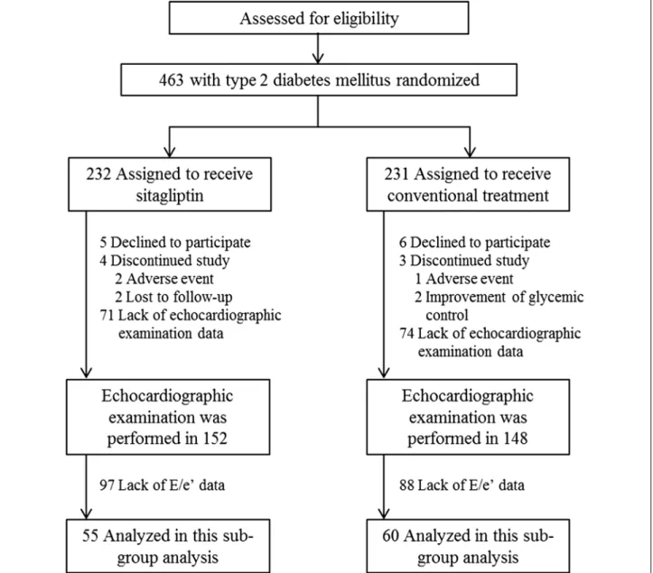
The details of the PROLOGUE study design have been published elsewhere [17]. Briefly, the study was a mul-ticenter, randomized, prospective, open-label, blinded-endpoint trial carried out at 48 institutions in Japan. A total of 463 patients older than 30 years who had T2DM with an HbA1c level of 6.2–9.4% despite conventional treatment with diet, exercise, and/or pharmacologic therapy with oral antidiabetic agents (except incretin-related therapy) for more than 3 months were enrolled in the study between June 2011 and September 2012. Patients with severe heart failure with a New York Heart Association (NYHA) functional classification of III and IV were excluded. The inclusion and exclusion criteria for the study have been published previously [17, 18]. The patients were assigned randomly using a 1:1 ratio to either add-on sitagliptin treatment (sitagliptin group, n = 232) or conventional glucose-lowering treatment (conventional group, n = 231). The primary endpoint of the PROLOGUE study was the change in mean common carotid IMT 24 months after treatment randomization. Echocardiography was performed as an ad hoc exami-nation at baseline and 12 and 24 months after treatment randomization. The ethical committees of each partici-pating institution approved the study protocol, with writ-ten informed consent for participation in the study being obtained from all the subjects.
Study population
Of the 436 participants in the PROLOGUE study, an echocardiographic examination was performed at base-line in 152 patients in the sitagliptin group and 148 patients in the conventional group. The present study
Conclusions: Adding sitagliptin to conventional antidiabetic regimens in patients with T2DM for 24 months attenu-ated the annual exacerbation in the echocardiographic parameter of diastolic dysfunction (E/e′) independent of other clinical variables such as blood pressure and glycemic control.
Trial registration UMIN000004490 (University Hospital Medical Information Network Clinical Trials). https://upload.umin. ac.jp/cgi-open-bin/ctr_e/ctr_view.cgi?recptno=R000005356; registered November 1, 2010
analyzed the data of 115 patients (55 patients in the sit-agliptin group and 60 patients in the conventional group) who had echocardiographic data of the ratio of peak early diastolic transmitral flow (TMF) velocity (E) to peak early diastolic mitral annular velocity (e′) at baseline and at both 12 and 24 months (Fig. 1).
Echocardiographic examination
Echocardiography was performed in a standard man-ner using commercially available ultrasound diagnostic machines with various hemodynamic parameters being measured at each institution. The recordings and meas-urements were performed in accordance with the guide-lines issued by the American Society of Echocardiograph
[19]. TMF velocity was recorded from the apical long-axis or four-chamber view. The ratio of the peak early diastolic (E) and the peak atrial systolic (A) TMF veloci-ties was calculated. The deceleration time (DT) of early TMF velocity was also measured. The mitral annular motion velocity pattern was recorded from the apical four-chamber view with the sample volume located at the lateral or septal side of the mitral annulus using pulsed tissue Doppler echocardiography. The mean peak early diastolic mitral annular velocity (e′) in the septal and lateral side was measured, and the ratio of E to e′ (E/e′) then calculated as a marker of LV filling pressure. In addition to these diastolic parameters, routine echocar-diographic parameters were also measured and included
LV end-diastolic dimensions (LVDd) and LV end-systolic dimensions (LVDs) measured from M-mode or 2-dimen-sional echocardiogram of the LV. Fractional shortening was calculated as (LVDs—LVds)/LVDdx100. The LV ejec-tion fracejec-tion (LVEF) was measured and calculated from the apical two- and four-chamber view using a modified Simpson’s method. LV mass was calculated as reported previously [20]. Relative wall thickness was calculated as two times posterior wall thickness divided by LVDd [21]. All Doppler recordings were performed during an end-expiratory breath hold. The mean values of three consecutive cardiac cycles were used in the analysis. Measurement and interpretation of the echocardiogra-phy was performed locally at each institution. The read-ers were blinded to the patients’ assignment to treatment. Laboratory examination
Blood samples were collected at baseline and after 12 and 24 months. The parameters analyzed are listed in Table 1. The serum levels of N-terminal pro-brain natriu-retic peptide (NT-proBNP) and high-sensitive CRP were measured in a centralized laboratory (SRL Co. Tokyo, Japan) using an electrochemiluminescence immunoassay (ECLIA) and nephelometry, respectively.
Statistical analysis
Data were expressed as mean ± standard deviation for normally distributed variables, median and interquar-tile range for variables with a skewed distribution, and frequencies (%) for categorical variables. All reported probability values were two-sided with a p value <0.05 considered statistically significant. The percentage changes in the variables during the study period were calculated as (values obtained at 12 or 24 months after treatment randomization—the baseline value)/base-line value. The differences between the two groups were assessed, where appropriate, by either the Student’s t test, Mann–Whitney test, or Fisher’s exact test. Variables with a skewed distribution were analyzed in the analysis of covariance after logarithmic conversion. We performed baseline-adjusted and multivariable regression analysis to confirm differences between the two groups. All the anal-yses were conducted using the JMP software program, version 12.1.0 (SAS Institute Inc., Cary, NC, USA).
Results
Clinical characteristics
Table 1 shows a comparison of the clinical characteris-tics at baseline and at 24 months and baseline-adjusted changes after 24 months of glycemic control between the two patient groups. There was no difference in body mass index and blood pressure between the groups
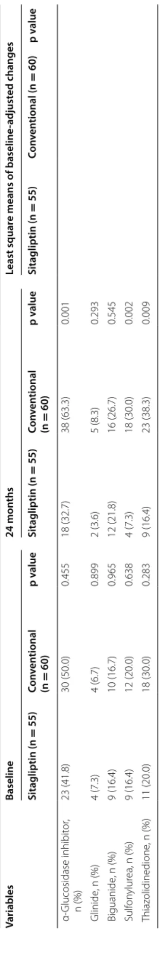
throughout the study, while heart rate was increased in the sitagliptin group at 24 months. Although more than 70% of the subjects had hypertension, blood pres-sure was well controlled in both groups. Other param-eters, such as lipid and renal profiles, were similar in the two groups throughout the study. The incidence of a previous history of CV diseases, including heart fail-ure was not different in the two groups. Although the use of background medications for hypertension, dys-lipidemia, or diabetes at baseline was also comparable in the groups, the incidence of some types of antidia-betic agent increased during the treatment period. This was especially apparent in the conventional group, pos-sibly due to many patients achieving the glycemic con-trol goal (HbA1c <6.2%) set in the PROLOGUE study protocol.
Glycemic control and neurohumoral effects
The levels of fasting plasma glucose, HbA1c, and 1, 5 AG were similar at baseline in the two groups and there were no significant changes in these parameters during the 24 months of treatment between the groups (Table 1). These results indicate similar degrees of improved gly-cemic control had been achieved. The serum levels of NT-proBNP and high-sensitive CRP were also similar at baseline and after 24 months of treatment.
Echocardiographic parameters
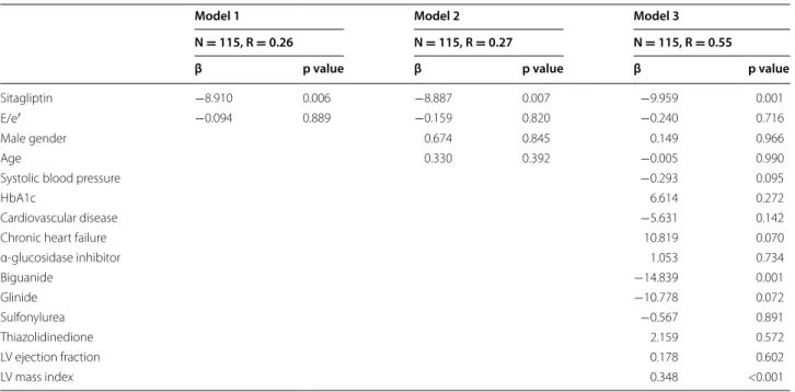
A comparison of echocardiographic parameters at baseline and after 24 months of treatment and base-line-adjusted changes after 24 months of treatment in both groups is shown in Table 2. Although baseline E/e′ was significantly higher in the sitagliptin group than in the conventional group, the baseline-adjusted change in E/e′ during 24 months of treatment was sig-nificantly lower in the sitagliptin group than in the conventional group (Fig. 2a). Analysis of covariance showed this difference remained significant (sitagliptin group, −30.9 ± 9.8%/24 months; conventional group, −11.0 ± 9.0%/24 months; p = 0.001, Table 3), even after adjustment for various confounding factors, such as age, sex, baseline systolic blood pressure, baseline HbA1c, history of CV diseases, history of heart failure, baseline medications for diabetes, baseline E/A, baseline LVEF, and baseline LV mass index. Other parameters relevant to diastolic function such as e′, E/A, and DT were simi-lar in the two groups during the 24 months of treatment (Table 2; Fig. 2b). There were also no significant differ-ences in parameters of cardiac structure and systolic function at baseline and 24 months, or baseline-adjusted changes after 24 months of treatment between the two groups (Table 2; Fig. 2c, d).
Table 1 C linic al char ac teristics b et w een the t w o gr oups Variables Baseline 24 mon ths Least squar e means of baseline -adjust ed changes Sitagliptin (n = 55) Con ven tional (n = 60) p v alue Sitagliptin (n = 55) Con ven tional (n = 60) p v alue Sitagliptin (n = 55) Con ven tional (n = 60) p v alue A ge , y ear 69 ± 8 69 ± 9 0.973 G ender (male/f emale) 38/17 39/21 0.641
Body mass index, k
g/m 2 25.9 ± 3.3 24.8 ± 3.9 0.098 25.5 ± 3.4 24.6 ± 3.8 0.203 − 0.10 ± 0.16 − 0.13 ± 0.15 0.368 Syst olic BP , mmHg 132 ± 17 129 ± 19 0.287 133 ± 16 130 ± 20 0.377 1.70 ± 2.20 (n = 54) 0.34 ± 2.09 0.656 Diast olic BP , mmHg 74 ± 11 71 ± 12 0.090 75 ± 13 (n = 54) 71 ± 11 0.076 1.76 ± 1.47 (n = 54) − 0.54 ± 1.40 0.263 Hear t rat e, beats/min 70 ± 12 67 ± 12 0.196 71 ± 16 67 ± 10 0.129 3.37 ± 1.49 − 2.14 ± 1.43 0.009 Total cholest er ol , mmol/L 4.4 ± 0.8 4.6 ± 0.9 0.43 4.4 ± 0.8 (n = 54) 4.4 ± 1.0 0.690 − 0.08 ± 0.09 (n = 54) − 0.10 ± 0.08 0.899 HDL cholest er ol , mmol/L 1.3 ± 0.3 (n = 55) 1.4 ± 0.4 (n = 58) 0.617 1.3 ± 0.3 (n = 54) 1.4 ± 0.4 0.412 − 0.03 ± 0.03 (n = 54) 0.00 ± 0.03 (n = 58) 0.362 Tr igly cer ides , mmol/L 3.1 [2.2–4.5] (n = 54) 3.1 [2.3–4.2] 0.536 2.9 [2.2–5.0] (n = 54) 2.9 [2.1–3.9] 0.151 0.06 ± 0.06 (n = 53) − 0.08 ± 0.05 0.078 Cr eatinine , μmol/L 71.6 [61.9–86.6] 68.1 [59.2–89.3] 0.496 75.1 [62.8–87.5] (n = 54) 69.0 [61.0–98.1] 0.740 2.65 ± 1.77 (n = 54) 4.42 ± 1.77 0.345 eGFR, mL/min/1.73 m 2 66.6 ± 15.9 67.3 ± 18.4 0.834 65.1 ± 14.1 (n = 54) 67.1 ± 19.7 0.757 − 1.96 ± 1.10 (n = 54) –3.18 ± 1.04 0.419
Fasting plasma glucose
, mmol/L 7.5 ± 1.8 (n = 53) 7.1 ± 1.4 0.135 7.0 ± 1.6 (n = 54) 6.7 ± 1.7 (n = 58) 0.248 − 0.41 ± 0.20 (n = 52) − 0.49 ± 0.19 (n = 58) 0.780 H bA1c , % 7.0 ± 0.6 6.9 ± 0.5 0.737 6.5 ± 0.6 (n = 54) 6.6 ± 0.7 0.412 − 0.47 ± 0.08 (n = 52) − 0.34 ± 0.07 (n = 57) 0.211 1,5A G, µg/mL 13.9 [8.3–20.3] (n = 52) 15.6 [10.7–22.8] (n = 58) 0.086 16.4 [8.7–22.6] 15.1 [9.6–25.0] (n = 59) 0.747 2.55 ± 0.82 (n = 52) 0.95 ± 0.79 (n = 57) 0.165 NT -pr oBNP , pg/mL 111.5 [42.1–240.8] (n = 52) 99.6 [52.1–234.9] (n = 58) 0.848 114.5 [51.8–261.9] 114.1 [60.2–323.8] (n = 59) 0.673 0.09 ± 0.08 (n = 52) 0.16 ± 0.08 (n = 57) 0.535 H igh-sensitiv e CRP , ng/mL 540 [279–1100] (n = 52) 576 [236–1618] (n = 58) 0.952 478 [232–1150] (n = 55) 478 [199–1590] (n = 59) 0.984 − 0.06 ± 0.16 (n = 52) − 0.06 ± 0.15 (n = 57) 0.983 Cur rent smok er , n (%) 8 (17.4) (n = 46) 13 (28.3) (n = 46) 0.321 H yper tension, n (%) 45 (81.8) 46 (76.7) 0.497 D yslipidemia, n (%) 42 (76.4) 42 (70.0) 0.530 Cer ebr ovascular disease , n (%) 8 (14.6) 4 (6.7) 0.226 Car dio vascular disease , n (%) 38 (69.1) 41 (68.3) 0.930 Chr onic hear t failur e, n (%) 3 (5.5) 7 (11.7) 0.326 M edications ACE inhibit or or ARB 37 (67.3) 42 (70.0) 0.753 39 (70.9) 41 (68.3) 0.764 β-block er 12 (21.8) 11 (18.3) 0.641 13 (23.6) 12 (20.0) 0.637 Diur etic 17 (30.9) 12 (20.0) 0.178 16 (29.1) 15 (25.0) 0.621 Statin 42 (76.4) 40 (66.7) 0.251 39 (70.9) 39 (65.0) 0.498
Da ta f or ca tegor ical v ar iables ar e g iv en as number (%); da ta f or c on tinuous v ar iables g iv en as mean ± standar d devia
tion or median [in
ter quar tile r ange]. Skew ed da ta w as calcula ted af ter logar ithmic tr ansla tion. I n the righ t c olumn, v alues ar e sho wn as baseline -adjust ed least squar e mean ± standar d er ror BP blood pr essur e, HDL high-densit y lipopr ot ein, eGFR estima
ted glomerular filtr
ation r at e, 1,5A G 1,5-anh ydr oglucit ol ,1,4-anh ydr o-d -glucit ol , NT -pr oBNP N-t er minal pr o-br ain na tr iur etic peptide , CRP C-r eac tiv e pr ot ein. AC E ang iot ensin-con ver ting enz yme , ARB ang iot ensin r ec ept or blocker Table 1 c on tinued Variables Baseline 24 mon ths Least squar e means of baseline -adjust ed changes Sitagliptin (n = 55) Con ven tional (n = 60) p v alue Sitagliptin (n = 55) Con ven tional (n = 60) p v alue Sitagliptin (n = 55) Con ven tional (n = 60) p v alue α-Glucosidase inhibit or , n (%) 23 (41.8) 30 (50.0) 0.455 18 (32.7) 38 (63.3) 0.001 Glinide , n (%) 4 (7.3) 4 (6.7) 0.899 2 (3.6) 5 (8.3) 0.293 Biguanide , n (%) 9 (16.4) 10 (16.7) 0.965 12 (21.8) 16 (26.7) 0.545 Sulf on ylur ea, n (%) 9 (16.4) 12 (20.0) 0.638 4 (7.3) 18 (30.0) 0.002 T hiaz olidinedione , n (%) 11 (20.0) 18 (30.0) 0.283 9 (16.4) 23 (38.3) 0.009
Table 2 C omparisons of echo car dio gr aphic par amet ers a t baseline , 24 mon ths and baseline -adjust ed changes af ter 24 mon ths of gly cemic c on tr ol Da ta f or ca tegor ical v ar iables ar e g iv en as number (%); da ta f or c on tinuous v ar iables g iv en as mean ± standar d devia tion. I n the r igh t c olumn, v alues ar e sho wn as baseline -adjust ed least squar e mean ± standar d er ror TMF tr ansmitr al flo w v elocit y, E ear ly diast olic v elocit y, e′ ear ly diast olic mitr al annular v elocit y, LV lef t v en tr icular , LA lef t a tr ial Variables Baseline 24 mon ths Least squar e means of baseline -adjust ed changes Sitagliptin (n = 55) Con ven tional (n = 60) p v alue Sitagliptin (n = 55) Con ven tional (n = 60) p v alue Sitagliptin (n = 55) Con ven tional (n = 60) p v alue TMF-E, cm/s 74.2 ± 32.6 69.3 ± 31.0 0.417 73.1 ± 33.4 73.6 ± 34.3 0.939 − 0.85 ± 2.37 4.04 ± 2.27 0.139 e′ , cm/s 6.35 ± 1.72 7.03 ± 2.13 0.065 6.60 ± 2.19 6.76 ± 2.11 0.684 0.13 ± 0.23 − 0.16 ± 0.22 0.370 E/e ′ 12.17 ± 5.20 10.34 ± 4.18 0.040 12.19 ± 6.93 12.06 ± 7.06 0.922 − 0.18 ± 0.55 1.91 ± 0.53 0.008 TMF-A, cm/s 80.1 ± 20.3 (n = 48) 83.4 ± 21.3 (n = 54) 0.424 82.4 ± 19.3 (n = 48) 88.2 ± 22.9 (n = 55) 0.168 3.06 ± 2.10 (n = 47) 5.73 ± 1.96 (n = 54) 0.354 E/A 0.91 ± 0.52(n = 48) 0.79 ± 0.24 (n = 54) 0.125 0.86 ± 0.35 (n = 48) 0.81 ± 0.31 (n = 54) 0.418 − 0.03 ± 0.04 (n = 48) − 0.04 ± 0.04 (n = 54) 0.825 D eceleration time , msec 233.5 ± 65.8 (n = 50) 237.6 ± 67.7 (n = 55) 0.751 238.1 ± 78.1 (n = 52) 240.6 ± 64.6 (n = 58) 0.859 9.53 ± 9.06 (n = 49) 5.38 ± 8.63 (n = 54) 0.741 LV end-diast olic dimension, mm 48.5 ± 5.6 48.6 ± 5.9 0.912 47.5 ± 5.7 47.7 ± 5.3 0.836 − 1.04 ± 0.51 − 0.91 ± 0.48 0.855 LV end-syst olic dimension, mm 31.7 ± 6.1 32.6 ± 7.2 0.462 31.5 ± 7.1 31.6 ± 6.9 0.935 − 0.30 ± 0.60 − 0.94 ± 0.58 0.447 Frac tional shor tening , % 35.1 ± 6.7 33.6 ± 7.9 0.279 33.9 ± 13.3 34.4 ± 8.5 0.787 − 1.05 ± 1.31 0.64 ± 1.25 0.355 LV ejec tion frac tion, % 63.6 ± 9.6 60.8 ± 10.8 0.153 62.5 ± 9.3 61.9 ± 9.8 0.721 − 0.63 ± 0.85 (n = 54) 0.67 ± 0.81 0.273 LV mass , g 173.0 ± 66.6 160.2 ± 56.3 0.266 159.9 ± 49.1 159.2 ± 52.9 0.940 − 10.29 ± 4.94 − 3.50 ± 4.73 0.324 LV mass index, g/m 2 101.6 ± 35.0 96.2 ± 30.6 0.383 94.8 ± 26.3 95.6 ± 28.3 0.881 − 5.50 ± 2.81 − 1.80 ± 2.68 0.343 Relativ e wall thick ness 0.40 ± 0.09 0.38 ± 0.08 0.230 0.41 ± 0.08 0.39 ± 0.08 0.400 0.01 ± 0.01 0.00 ± 0.01 0.886 LA dimension, mm 40.8 ± 6.9 (n = 54) 40.0 ± 7.6 (n = 58) 0.572 39.7 ± 6.9 40.3 ± 6.9 (n = 59) 0.652 − 0.87 ± 0.55 (n = 54) 0.25 ± 0.54 (n = 58) 0.151
-10% -5% 0% 5% 10% -10% 0% 10% 20% -10% 0% 10% 20%
30% %change of E/e’ %change of e’
Leas
t square mean
Leas
t square mean
*
%change of LVEF %change of LVMI
Leas t square mean Leas t squa re mean : Conven onal group (n=55) : Sitaglip n group (n=60) -10% -5% 0% 5% 10% 0M 12M 24M 0M 12M 24M 0M 12M 24M 0M 12M 24M
Least square mean ± standard error
a b
d c
Fig. 2 Percentage changes in E/e′, e′, LVEF, and LVMI at 12 and 24 months in the two treatment groups. Each graph shows sex-, age- and baseline-adjusted least square means (±standard error) at 12 and 24 months. The %change values were calculated as (24 or 12 month data-baseline)/base-line. E/e′ at 24 months shows significant difference between the two groups. E peak early diastolic transmitral flow velocity, e′ peak early diastolic mitral annular velocity, LVEF left ventricular ejection fraction, LVMI left ventricular mass index. *p = 0.002 vs. sitagliptin group
Table 3 Factors associated with change in E/e′ from baseline to 24 months
Model 1 means ANCOVA adjusted for baseline E/e′. Model 2 were adjusted for Model 1 and sex, age. Model 3 were adjusted for Model 2 and systolic blood pressure, HbA1c, cardiovascular disease, chronic heart failure, α-glucosidase inhibitor, bigunaide, glinide, sulfonylurea, thiazolidinedione, LV ejection fraction, LV mass index, whose data were obtained at baseline examination
Abbreviations, see Tables 1 and 2
Model 1 Model 2 Model 3
N = 115, R = 0.26 N = 115, R = 0.27 N = 115, R = 0.55
β p value β p value β p value
Sitagliptin −8.910 0.006 −8.887 0.007 −9.959 0.001
E/e′ −0.094 0.889 −0.159 0.820 −0.240 0.716
Male gender 0.674 0.845 0.149 0.966
Age 0.330 0.392 −0.005 0.990
Systolic blood pressure −0.293 0.095
HbA1c 6.614 0.272
Cardiovascular disease −5.631 0.142
Chronic heart failure 10.819 0.070
α-glucosidase inhibitor 1.053 0.734 Biguanide −14.839 0.001 Glinide −10.778 0.072 Sulfonylurea −0.567 0.891 Thiazolidinedione 2.159 0.572 LV ejection fraction 0.178 0.602 LV mass index 0.348 <0.001
Discussion
The present study was a subgroup analysis of the PRO-LOGUE trial that focused on the effect of sitagliptin on echocardiographic parameters of diastolic function. The key finding of the study was that addition of sitagliptin to conventional diabetic care significantly attenuated the increase in echocardiographic parameters of diastolic function (E/e′), relative to conventional treatment alone. On the other hand, changes in other parameters such as LV size and LVEF did not differ between the two groups. We also found no significant differences in the biomark-ers measured during the study. It is known that metabolic disturbances and diabetes are associated closely with cardiac diastolic dysfunction such as diabetic cardiomyo-pathy, and there is also evidence that patients with diabe-tes and an increased E/e′ have higher mortality [22, 23]. Given these results, it appears that sitagliptin treatment may have a protective effect on cardiac diastolic function, leading to improved prognosis independent of glycemic control and blood pressure.
Recently we demonstrated a possible effect of sitaglip-tin on carotid atherosclerosis [18], endothelial function [24], and arterial stiffness [25] using data of the PRO-LOGUE study. This series of studies did not show ben-eficial effects of sitagliptin treatment on these variables, relative to conventional glucose-lowering treatment with the exception of incretin-related agents, with better gly-cemic control being observed in the sitagliptin treatment group. In contrast, there is another report that additional administration of DPP-4 inhibitors, including sitagliptin, to conventional antidiabetic regimes significantly attenu-ated the progression of carotid IMT [26, 27]. Although the participants’ backgrounds including age, concomitant agents, and severity of diabetes differed between these studies [18], these findings suggest that DPP-4 inhibitors at least cause no harm to the vasculature and are useful for glycemic control in the usual clinical settings. The findings are also consistent with the results of a large CV outcome trial [13]. This led us to investigate the effect of sitagliptin on cardiac function and biomarkers in the cur-rent sub-group analysis of the PROLOGUE study data.
DPP-4 inhibitors promote glucose-dependent insulin secretion and suppress glucagon secretion by inhibiting the activity of an enzyme which inactivates endogenous incretin like glucagon-like peptide-1 (GLP-1) and gas-tric inhibitory polypeptide. This leads to improved post-prandial hyperglycemia similar to that seen with the normal physiological response. DPP-4 inhibitors do not cause weight gain and a single administration is unlikely to induce hypoglycemia. It is therefore relatively easy to use DPP-4 inhibitors safely in the elderly and patients with renal dysfunction. To date, the three randomized
clinical trials mentioned above have reported non-infe-riority, relative to placebo, for CV outcomes in patients with T2DM with high cardiovascular risk or established CV disease [11–13]. In particular, the rate of hospitaliza-tion for heart failure was similar between sitagliptin and placebo treatments in the TECOS trial [13], despite the SAVOR-TIMI 53 trial showing that saxagliptin, another DPP-4 inhibitor, significantly increased the hospitali-zation rate [12]. However, because these trials did not fully investigate CV physiological functions and relevant biomarkers, it proved difficult to determine how DPP-4 inhibitors affected cardiac function. Mechanistic studies using these surrogate markers are therefore required to determine the possible actions of DPP-4 inhibitors on the CV system.
Accumulated evidence suggests that patients with TD2M often exhibit LV diastolic dysfunction and heart failure due to underlying metabolic derangement, such as insulin resistance, independent of hypertension and cor-onary artery disease (CAD) [28–30]. Recent studies have also demonstrated that DPP-4 activity correlates with cardiac systolic and diastolic dysfunction and remodeling via several molecular pathways, such as increased inflam-mation and altered angiogenesis [31–34]. Experimental studies have shown that sitagliptin improved survival rate and cardiac function in an ischemia–reperfusion mice model [35] and reduced infarction size in a myocar-dial infarction mice model [36]. Long-term administra-tion of sitagliptin was also shown to suppress the onset of heart failure in a rat model of heart failure [31]. A meta-analysis of clinical trials also described the advan-tages of DPP-4 inhibitors on risk reduction in CV events and death compared with other antidiabetic agents [37]. Sitagliptin treatment in T2DM patients with CAD also improved parameters of diastolic function and cardiac dysfunction due to post-ischemic stunning during dob-utamine stress echocardiography [38]. In contrast, sit-agliptin treatment did not improve systolic function in T2DM patients with ischemic heart failure [39]. Further-more, Oe et al. [40] reported that sitagliptin treatment in T2DM patients with LV diastolic dysfunction was not associated with improvement in the relevant echocardio-graphic parameters. As a consequence of these different findings the therapeutic effect of DPP-4 inhibitors on cardiac function remains controversial.
In the present study, adding sitagliptin to usual diabe-tes treatment significantly attenuated the annual increase in E/e′, suggesting a preventive effect on LV compliance and diastolic dysfunction. However, sitagliptin treat-ment did not affect other echocardiographic parameters of systolic function and cardiac structures or other clini-cal variables, such as NT-proBNP, blood pressure, and
glycemic control. Comparison with a previous study in which sitagliptin did not improve diastolic dysfunction [40] showed the following differences: (1) all partici-pants in the previous study were diagnosed with diastolic dysfunction at baseline; (2) the treatment period was 6 months vs. 24 months in the PROLOGUE study; (3) the comparator was voglibose vs. any antidiabetic agents except for incretin-related in the PROLOGUE study. While these differences may have affected the results of the studies, the precise mechanisms by which sitaglip-tin suppressed the increase in diastolic parameter values were not confirmed in our study. As reported previ-ously [40], the increased incidence of concomitant use of thiazolidinediones in the conventional group may have enhanced the acceleration of E/e′ values. Nogueira et al. also reported that beneficial effects in LV diastolic func-tion were observed in T2DM patients on insulin treated with sitagliptin, while the effects were not as apparent in T2DM patients treated with insulin only [41]. That study also reported a possible association between the sitaglip-tin-mediated improvement in diastolic dysfunction and increase in plasma GLP-1 levels. However, we did not measure this incretin in the current study. It is thought that GLP-1 has a wide spectrum of CV protective effects [42]. In fact, treatment with a GLP-1 agonist, one of the incretin-related agents, was shown to improve diastolic function beyond and independent of glycemic control [43]. Because there remains clinical caution regarding DPP-4 inhibitor-induced heart failure [44, 45], further experimental and clinical research is required to eluci-date the precise mechanisms by which DPP-4 inhibitors affect diastolic function and heart failure in patients with T2DM.
Limitations
The present study was a sub-analysis of the PROLOGUE study. Because echocardiography was a voluntary meas-urement in the PROLOGUE study and not performed in all participants, the number of patients in this study was small and included only Japanese subjects. Whether or not echocardiography was performed was left to the judgment of each researcher and therefore selection bias could not be fully excluded. The sample size may there-fore be underpowered and accordingly the clinical impli-cations may be limited. In addition, the PROLOGUE study recruited patients with and without history of heart failure at baseline, with patients with a NYHA functional classification of III and IV being excluded. Because most patients had no history of heart failure evident at base-line, we did not determine whether there were differences in the effects of sitagliptin on diastolic function between
patients with or without heart failure. Further studies on a larger number of subjects are needed to assess whether longer-term DPP-4 inhibitor treatment is safe and has beneficial effect on cardiac function in T2DM patients with or without overt heart failure.
Conclusions
Our present sub-group analysis from the PROLOGUE study demonstrated that adding sitagliptin to conven-tional antidiabetic regimens for 24 months in patients with T2DM attenuated the annual exacerbation in the echocardiographic parameter of diastolic dysfunction, E/e′, independent of other clinical variables such as blood pressure and glycemic control. These results suggest that sitagliptin is potentially a beneficial agent for diastolic function in patients with T2DM.
Abbreviations
A: peak atrial systolic transmitral flow velocity; CAD: coronary artery disease; CV: cardiovascular; DPP-4: dipeptidyl peptidase 4; DT: deceleration time; E: peak early diastolic transmitral flow velocity; e′: peak early diastolic mitral annular velocity; EF: ejection fraction; GLP-1: glucagon-like peptide-1; IMT: intima-media thickness; LV: left ventricular; LVDd: left ventricular end-diastolic dimensions; LVDs: left ventricular end-systolic dimensions; NT-proBNP: N-terminal pro-brain natriuretic peptide; NYHA: New York Heart Association; TMF: transmitral flow; T2DM: type 2 diabetes mellitus.
Authors’ contributions
HY and AT wrote the draft of the article, which was critically supervised by MS and KN. HY performed statistical analysis throughout this study. KK, RA and MM confirmed data collection and study selection criteria. HD, MI, HT, MN, HK, YKB, MO, and TM enrolled patients and performed study quality assessment. All authors approved the final version of manuscript.
Author details
1 Department of Cardiovascular Medicine, Tokushima University Hospital, 2-50-1 Kuramoto, Tokushima, Japan. 2 Department of Cardiovascular Medicine, Saga University, 5-5-1 Nabeshima, Saga, Japan. 3 Department of Diabetes Therapeutics and Research Center, Tokushima University, Tokushima, Japan. 4 Department of Cardiovascular Medicine, Juntendo University Graduate School of Medicine, Tokyo, Japan. 5 Department of Cardiology and Nephrol-ogy, Mie University Graduate School of Medicine, Tsu, Japan. 6 Department of Cardiovascular Medicine, Faculty of Medical Sciences, Kyushu University, Fukuoka, Japan. 7 Cardiovascular Center, Japanese Red Cross Nagoya Daini Hospital, Nagoya, Japan. 8 Division of Cardiology, Japanese Red Cross Nagoya Daiichi Hospital, Nagoya, Japan. 9 Department of Cardiology, Nagoya Univer-sity Graduate School of Medicine, Nagoya, Japan. 10 Department of Diabetes, Endocrinology, Metabolism and Rheumatology, Tokyo Medical University, Tokyo, Japan. 11 Clinical Research Center, Saga University, Saga, Japan.
Acknowledgements
The authors thank the participants and staff for their essential contributions to the PROLOGUE study. The PROLOGUE study is a multicenter collaboration. In addition to the listed authors, the PROLOGUE Study Investigators listed in Additional file 1 were involved in this study.
Additional file
Competing interests
HYa received honoraria from MSD, research grant from MSD and Ono. AT, KK, and RA declares no competing interests. MM received honoraria from Astellas, Eli Lilly, Mitsubishi Tanabe, Novo Nordisk, Sanofi, and Takeda, research grant from Astellas, Nihon Unisys, Nikkiso, and Terumo. HD received honoraria from Astellas, AstraZeneca, Bayer, Boehringer Ingelheim, Daiichi Sankyo, GlaxoS-mithKline, Kowa, Medtronic Japan, Mitsubishi Tanabe, Mochida, MSD, Ono, Pfizer, Sanofi, Shionogi, Takeda and Terumo, research funding from Abbott Vascular Japan, Astellas, Bayer, Boehringer Ingelheim, Boston Scientific Japan, Bristol-Myers, Daiichi Sankyo, Kaken, Kowa, MSD, Novartis, Otsuka, Pfizer, Philips Respironics GK, Sanofi, Sanwa Kagaku Kenkyusho, Shionogi, Sumitomo Dainip-pon and Takeda. MI received honoraria from Daiichi Sankyo and Pfizer, research funding from Astellas, Biotronik, Bristol-Myers, Daiichi Sankyo, GlaxoSmithKline, Mitsubishi Tanabe, MSD, Novartis, Otsuka, Pfizer, Shionogi, Sumitomo Dainip-pon and Takeda. HT received honoraria from Bristol-Myers, Daiichi Sankyo, Mit-subishi Tanabe and Ono, research funding from Astellas, Boehringer Ingelheim, Boston Scientific Japan, Daiichi Sankyo, Mitsubishi Tanabe, Otsuka, Takeda and Teijin Pharma. MN declares no competing interests. HK declares no compet-ing interests. YKB received honoraria from AstraZeneca, Mitsubishi Tanabe, MSD, and Takeda, research grant from AstraZeneca, Daiichi Sankyo, Mitsubishi Tanabe, MSD, and Takeda. MO received honoraria from Astellas, Boehringer Ingelheim, Daiichi Sankyo, Eli Lilly, Kowa, Merck, Mitsubishi Tanabe, Novartis, Ono and Sanofi. HYo declares no competing interests. TM received honoraria from Bayer, Boehringer Ingelheim, Daiichi Sankyo, Kowa, Mitsubishi Tanabe, MSD, Pfizer, Sumitomo Dainippon, and Takeda, research grant from Astellas, Boehringer Ingelheim, Daiichi Sankyo, Kowa, Mitsubishi Tanabe, MSD, Novartis, Otsuka, Pfizer, Sanofi, Sumitomo Dainippon, Takeda, and Teijin Pharma. KN received honoraria from Astellas, Boehringer Ingelheim, Daiichi Sankyo, Merck, Mitsubishi Tanabe, Sanofi, and Takeda. MS received honoraria from MSD, research grant from MSD and Ono. KN received research funding from Astellas, Boehringer Ingelheim, Grant-in-Aid for Scientific Research from the Ministry of Education, Culture, Sports, Science and Technology in Japan, Mitsubishi Tanabe, Sanwa Kagaku Kenkyusho, Takeda, and Teijin Pharma.
Availability of data and materials
The datasets analyzed during the current study are available from the corre-sponding authors on reasonable request (yamadah@tokushima-u.ac.jp, node@ cc.saga-u.ac.jp).
Consent for publication
All authors have read and approved the submission of the manuscript; the manuscript has not been published and is not being considered for publica-tion elsewhere, in whole or in part, in any language. If the manuscript is accepted, we approve it for publication in Cardiovascular Diabetology.
Ethics approval and consent to participate
The ethical committees of the participating institutions approved the study protocol. Written informed consent for participation in the study was obtained from all subjects.
Funding
This study is supported by a research grant from the Clinical Research Promotion Foundation (No. 1026). The funding body had no role in study design, data collection and analysis, decision to publish or preparation of the manuscript.
Publisher’s Note
Springer Nature remains neutral with regard to jurisdictional claims in pub-lished maps and institutional affiliations.
Received: 30 January 2017 Accepted: 4 May 2017
References
1. Kannel WB, Hjortland M, Castelli WP. Role of diabetes in congestive heart failure: the Framingham study. Am J Cardiol. 1974;34(1):29–34. 2. Emerging Risk Factors C. Sarwar N, Gao P, Seshasai SR, Gobin R, Kaptoge
S, Di Angelantonio E, Ingelsson E, Lawlor DA, Selvin E et al.: diabetes
mellitus, fasting blood glucose concentration, and risk of vascular disease: a collaborative meta-analysis of 102 prospective studies. Lancet. 2010;375(9733):2215–22.
3. Gilbert RE, Krum H. Heart failure in diabetes: effects of anti-hyperglycae-mic drug therapy. Lancet. 2015;385(9982):2107–17.
4. Bertoni AG, Hundley WG, Massing MW, Bonds DE, Burke GL, Goff DC Jr. Heart failure prevalence, incidence, and mortality in the elderly with diabetes. Diabetes Care. 2004;27(3):699–703.
5. MacDonald MR, Petrie MC, Varyani F, Ostergren J, Michelson EL, Young JB, Solomon SD, Granger CB, Swedberg K, Yusuf S, et al. Impact of diabetes on outcomes in patients with low and preserved ejection fraction heart failure: an analysis of the candesartan in heart failure: assessment of reduction in mortality and morbidity (CHARM) programme. Eur Heart J. 2008;29(11):1377–85.
6. Turnbull FM, Abraira C, Anderson RJ, Byington RP, Chalmers JP, Duckworth WC, Evans GW, Gerstein HC, Holman RR, Moritz TE, et al. Intensive glucose control and macrovascular outcomes in type 2 diabetes. Diabetologia. 2009;52(11):2288–98.
7. Varas-Lorenzo C, Margulis AV, Pladevall M, Riera-Guardia N, Calingaert B, Hazell L, Romio S, Perez-Gutthann S. The risk of heart failure associated with the use of noninsulin blood glucose-lowering drugs: systematic review and meta-analysis of published observational studies. BMC Car-diovasc Disord. 2014;14:129.
8. Komajda M, McMurray JJ, Beck-Nielsen H, Gomis R, Hanefeld M, Pocock SJ, Curtis PS, Jones NP, Home PD. Heart failure events with rosiglitazone in type 2 diabetes: data from the RECORD clinical trial. Eur Heart J. 2010;31(7):824–31.
9. McMurray JJ, Gerstein HC, Holman RR, Pfeffer MA. Heart failure: a car-diovascular outcome in diabetes that can no longer be ignored. Lancet Diabetes Endocrinol. 2014;2(10):843–51.
10. Fitchett DH, Udell JA, Inzucchi SE. Heart failure outcomes in clinical trials of glucose-lowering agents in patients with diabetes. Eur J Heart Fail. 2017;19(1):43–53.
11. White WB, Cannon CP, Heller SR, Nissen SE, Bergenstal RM, Bakris GL, Perez AT, Fleck PR, Mehta CR, Kupfer S, et al. Alogliptin after acute coronary syndrome in patients with type 2 diabetes. N Engl J Med. 2013;369(14):1327–35.
12. Scirica BM, Bhatt DL, Braunwald E, Steg PG, Davidson J, Hirshberg B, Ohman P, Frederich R, Wiviott SD, Hoffman EB, et al. Saxagliptin and cardiovascular outcomes in patients with type 2 diabetes mellitus. N Engl J Med. 2013;369(14):1317–26.
13. Green JB, Bethel MA, Armstrong PW, Buse JB, Engel SS, Garg J, Josse R, Kaufman KD, Koglin J, Korn S, et al. Effect of sitagliptin on cardiovascular outcomes in type 2 diabetes. N Engl J Med. 2015;373(3):232–42. 14. Zannad F, Cannon CP, Cushman WC, Bakris GL, Menon V, Perez AT, Fleck
PR, Mehta CR, Kupfer S, Wilson C, et al. Heart failure and mortality out-comes in patients with type 2 diabetes taking alogliptin versus placebo in EXAMINE: a multicentre, randomised, double-blind trial. Lancet. 2015;385(9982):2067–76.
15. Sivertsen J, Rosenmeier J, Holst JJ, Vilsboll T. The effect of glucagon-like peptide 1 on cardiovascular risk. Nat Rev Cardiol. 2012;9(4):209–22.
16. Wu S, Hopper I, Skiba M, Krum H. Dipeptidyl peptidase-4 inhibitors and cardiovascular outcomes: meta-analysis of randomized clinical trials with 55,141 participants. Cardiovasc Ther. 2014;32(4):147–58.
17. Oyama J, Ishizu T, Sato Y, Kodama K, Bando YK, Murohara T, Node K. Rationale and design of a study to evaluate the effects of sitagliptin on atherosclerosis in patients with diabetes mellitus: PROLOGUE study. Int J Cardiol. 2014;174(2):383–4.
18. Oyama J, Murohara T, Kitakaze M, Ishizu T, Sato Y, Kitagawa K, Kamiya H, Ajioka M, Ishihara M, Dai K, et al. The effect of sitagliptin on carotid artery atherosclerosis in type 2 diabetes: the PROLOGUE randomized controlled trial. PLoS Med. 2016;13(6):e1002051.
19. Lang RM, Bierig M, Devereux RB, Flachskampf FA, Foster E, Pellikka PA, Picard MH, Roman MJ, Seward J, Shanewise JS, et al. Recommendations for chamber quantification: a report from the American Society of Echo-cardiography’s Guidelines and Standards Committee and the Chamber Quantification Writing Group, developed in conjunction with the Euro-pean Association of Echocardiography, a branch of the EuroEuro-pean Society of Cardiology. J Am Soc Echocardiogr Off Publ Am Soc Echocardiogr. 2005;18(12):1440–63.
• We accept pre-submission inquiries
• Our selector tool helps you to find the most relevant journal • We provide round the clock customer support
• Convenient online submission • Thorough peer review
• Inclusion in PubMed and all major indexing services • Maximum visibility for your research
Submit your manuscript at www.biomedcentral.com/submit
Submit your next manuscript to BioMed Central
and we will help you at every step:
20. Devereux RB, Alonso DR, Lutas EM, Gottlieb GJ, Campo E, Sachs I, Reichek N. Echocardiographic assessment of left ventricular hypertrophy: com-parison to necropsy findings. Am J Cardiol. 1986;57(6):450–8. 21. Lang RM, Bierig M, Devereux RB, Flachskampf FA, Foster E, Pellikka PA,
Picard MH, Roman MJ, Seward J, Shanewise JS, et al. Recommendations for chamber quantification: a report from the American Society of Echo-cardiography’s Guidelines and Standards Committee and the Chamber Quantification Writing Group, developed in conjunction with the Euro-pean Association of Echocardiography, a branch of the EuroEuro-pean Society of Cardiology. J Am Soc Echocardiogr Off Publ Am Soc Echocardiogr. 2005;18(12):1440–63.
22. Seferovic PM, Paulus WJ. Clinical diabetic cardiomyopathy: a two-faced disease with restrictive and dilated phenotypes. Eur Heart J. 2015; 36(27):1718–1727, 1727a–1727c.
23. From AM, Scott CG, Chen HH. Changes in diastolic dysfunction in diabe-tes mellitus over time. Am J Cardiol. 2009;103(10):1463–6.
24. Maruhashi T, Higashi Y, Kihara Y, Yamada H, Sata M, Ueda S, Odawara M, Terauchi Y, Dai K, Ohno J, et al. Long-term effect of sitagliptin on endothe-lial function in type 2 diabetes: a sub-analysis of the PROLOGUE study. Cardiovasc Diabetol. 2016;15(1):134.
25. Tomiyama H, Miwa T, Kan K, Matsuhisa M, Kamiya H, Nanasato M, Kitano T, Sano H, Ohno J, Iida M, et al. Impact of glycemic control with sitagliptin on the 2-year progression of arterial stiffness: a sub-analysis of the PRO-LOGUE study. Cardiovasc Diabetol. 2016;15(1):150.
26. Mita T, Katakami N, Shiraiwa T, Yoshii H, Onuma T, Kuribayashi N, Osonoi T, Kaneto H, Kosugi K, Umayahara Y, et al. Sitagliptin attenuates the progression of carotid intima-media thickening in insulin-treated patients with type 2 diabetes: the sitagliptin preventive study of intima-media thickness evaluation (SPIKE): a randomized controlled trial. Diabetes Care. 2016;39(3):455–64.
27. Mita T, Katakami N, Yoshii H, Onuma T, Kaneto H, Osonoi T, Shiraiwa T, Kosugi K, Umayahara Y, Yamamoto T, et al. Alogliptin, a dipeptidyl pepti-dase 4 inhibitor, prevents the progression of carotid atherosclerosis in patients with type 2 diabetes: the study of preventive effects of alogliptin on diabetic atherosclerosis (SPEAD-A). Diabetes Care. 2016;39(1):139–48. 28. Fontes-Carvalho R, Ladeiras-Lopes R, Bettencourt P, Leite-Moreira A,
Azevedo A. Diastolic dysfunction in the diabetic continuum: association with insulin resistance, metabolic syndrome and type 2 diabetes. Cardio-vasc Diabetol. 2015;14:4.
29. Bugger H, Abel ED. Molecular mechanisms of diabetic cardiomyopathy. Diabetologia. 2014;57(4):660–71.
30. Fuentes-Antras J, Picatoste B, Ramirez E, Egido J, Tunon J, Lorenzo O. Tar-geting metabolic disturbance in the diabetic heart. Cardiovasc Diabetol. 2015;14:17.
31. dos Santos L, Salles TA, Arruda-Junior DF, Campos LC, Pereira AC, Barreto AL, Antonio EL, Mansur AJ, Tucci PJ, Krieger JE, et al. Circulating dipeptidyl peptidase IV activity correlates with cardiac dysfunction in human and experimental heart failure. Circ Heart Fail. 2013;6(5):1029–38.
32. de Almeida Salles T, Zogbi C, de Lima TM, de Godoi Carneiro C, Garcez AT, Barbeiro HV, Antonio EL, Dos Santos L, da Costa Pereira A, Tucci PJ, et al. The contributions of dipeptidyl peptidase IV to inflammation in heart failure. Ame J Physiol Heart Circ Physiol. 2016;310(11):H1760–72.
33. Shigeta T, Aoyama M, Bando YK, Monji A, Mitsui T, Takatsu M, Cheng XW, Okumura T, Hirashiki A, Nagata K, et al. Dipeptidyl peptidase-4 modulates left ventricular dysfunction in chronic heart failure via angiogenesis-dependent and -inangiogenesis-dependent actions. Circulation. 2012;126(15):1838–51. 34. Zaruba MM, Theiss HD, Vallaster M, Mehl U, Brunner S, David R, Fischer R,
Krieg L, Hirsch E, Huber B, et al. Synergy between CD26/DPP-IV inhibition and G-CSF improves cardiac function after acute myocardial infarction. Cell Stem Cell. 2009;4(4):313–23.
35. Sauve M, Ban K, Momen MA, Zhou YQ, Henkelman RM, Husain M, Drucker DJ. Genetic deletion or pharmacological inhibition of dipeptidyl pepti-dase-4 improves cardiovascular outcomes after myocardial infarction in mice. Diabetes. 2010;59(4):1063–73.
36. Ye Y, Keyes KT, Zhang C, Perez-Polo JR, Lin Y, Birnbaum Y. The myocardial infarct size-limiting effect of sitagliptin is PKA-dependent, whereas the protective effect of pioglitazone is partially dependent on PKA. Am J Physiol Heart Circ Physiol. 2010;298(5):H1454–65.
37. Patil HR, Al Badarin FJ, Al Shami HA, Bhatti SK, Lavie CJ, Bell DS, O’Keefe JH. Meta-analysis of effect of dipeptidyl peptidase-4 inhibitors on cardiovas-cular risk in type 2 diabetes mellitus. Am J Cardiol. 2012;110(6):826–33. 38. McCormick LM, Kydd AC, Read PA, Ring LS, Bond SJ, Hoole SP, Dutka DP.
Chronic dipeptidyl peptidase-4 inhibition with sitagliptin is associated with sustained protection against ischemic left ventricular dysfunction in a pilot study of patients with type 2 diabetes mellitus and coronary artery disease. Circ Cardiovasc Imaging. 2014;7(2):274–81.
39. Arturi F, Succurro E, Miceli S, Cloro C, Ruffo M, Maio R, Perticone M, Sesti G, Perticone F. Liraglutide improves cardiac function in patients with type 2 diabetes and chronic heart failure. Endocrine. 2016 Endocrine. 2016 Nov 9. [Epub ahead of print].
40. Oe H, Nakamura K, Kihara H, Shimada K, Fukuda S, Takagi T, Miyoshi T, Hirata K, Yoshikawa J, Ito H. Comparison of effects of sitagliptin and voglibose on left ventricular diastolic dysfunction in patients with type 2 diabetes: results of the 3D trial. Cardiovasc Diabetol. 2015;14:83. 41. Nogueira KC, Furtado M, Fukui RT, Correia MR, Dos Santos RF, Andrade JL.
Rossi da Silva ME: left ventricular diastolic function in patients with type 2 diabetes treated with a dipeptidyl peptidase-4 inhibitor—a pilot study. Diabetol Metab Syndr. 2014;6(1):103.
42. Ussher JR, Drucker DJ. Cardiovascular actions of incretin-based therapies. Circ Res. 2014;114(11):1788–803.
43. Saponaro F, Sonaglioni A, Rossi A, Montefusco L, Lombardo M, Adda G, Arosio M. Improved diastolic function in type 2 diabetes after a six month liraglutide treatment. Diabetes Res Clin Pract. 2016;118:21–8.
44. Li L, Li S, Deng K, Liu J, Vandvik PO, Zhao P, Zhang L, Shen J, Bala MM, Sohani ZN, et al. Dipeptidyl peptidase-4 inhibitors and risk of heart failure in type 2 diabetes: systematic review and meta-analysis of randomised and observational studies. BMJ (Clin Res ed). 2016;352:i610.
45. Rehman MB, Tudrej BV, Soustre J, Buisson M, Archambault P, Pouchain D, Vaillant-Roussel H, Gueyffier F, Faillie JL, Perault-Pochat MC, et al. Efficacy and safety of DPP-4 inhibitors in patients with type 2 diabetes: meta-analysis of placebo-controlled randomized clinical trials. Diabetes Metab. 2017;43(1):48–58.